Testosterone-Induced Metabolic Changes in Seminal Vesicle Epithelial cells Alter Plasma Components to Enhance Sperm Fertility

  1. Graduate School of Integrated Sciences for Life, Hiroshima University, Hiroshima, Japan
  2. Department of Histology, Faculty of Veterinary Medicine, South Valley University, Qena, Egypt
  3. Graduate School of Innovation and Practice for Smart Society, Hiroshima University, Hiroshima, Japan

Peer review process

Revised: This Reviewed Preprint has been revised by the authors in response to the previous round of peer review; the eLife assessment and the public reviews have been updated where necessary by the editors and peer reviewers.

Read more about eLife’s peer review process.

Editors

  • Reviewing Editor
    Jean-Ju Chung
    Yale University, New Haven, United States of America
  • Senior Editor
    Benoit Kornmann
    University of Oxford, Oxford, United Kingdom

Reviewer #1 (Public review):

Summary:

In this revised report, Yamanaka and colleagues investigate a proposed mechanism by which testosterone modulates seminal plasma metabolites in mice. Based on limited evidence in previous versions of the report, the authors softened the claim that oleic acid derived from seminal vesicle epithelium strongly affects linear progressive motility in isolated cauda epididymal sperm in vitro. Though the report still contains somewhat ambiguous references to the strength of the relationship between fatty acids and sperm motility.

Strengths:

Often, reported epidydimal sperm from mice have lower percent progressive motility compared with sperm retrieved from the uterus or by comparison with human ejaculated sperm. The findings in this report may improve in vitro conditions to overcome this problem, as well as add important physiological context to the role of reproductive tract glandular secretions in modulating sperm behaviors. The strongest observations are related to the sensitivity of seminal vesicle epithelial cells to testosterone. The revisions include the addition of methodological detail, modified language to reflect the nuance of some of the measurements, as well as re-performed experiments with more appropriate control groups. The findings are likely to be of general interest to the field by providing context for follow-on studies regarding the relationship between fatty acid beta oxidation and sperm motility pattern.

Weaknesses:

The connection between media fatty acids and sperm motility pattern remains inconclusive.

Reviewer #2 (Public review):

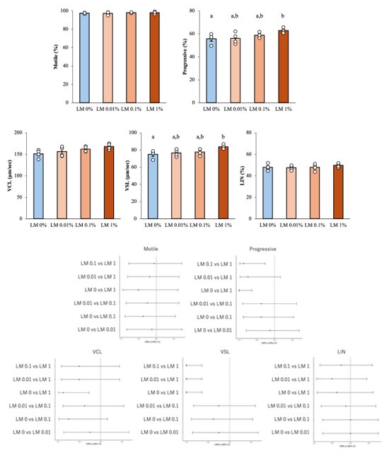
Using a combination of in vivo studies with testosterone-inhibited and aged mice with lower testosterone levels as well as isolated mouse and human seminal vesicle epithelial cells the authors show that testosterone induces an increase in glucose uptake. They find that testosterone induces a difference in gene expression with a focus on metabolic enzymes. Specifically, they identify increased expression of enzymes regulating cholesterol and fatty acid synthesis, leading to increased production of 18:1 oleic acid. The revised version strengthens the role of ACLY as the main regulator of seminal vesicle epithelial cell metabolic programming. The authors propose that fatty acids are secreted by seminal vesicle epithelial cells and are taken up by sperm, positively affecting sperm function. A lipid mixture mimicking the lipids secreted by seminal vesicle epithelial cells, however, only has a small and mostly non-significant effect on sperm motility, suggesting the authors were not apply to pinpoint the seminal vesicle fluid component that positively affects sperm function.

Author response:

The following is the authors’ response to the previous reviews

Public Reviews:

Reviewer #1 (Public review):

Summary:

In this revised report, Yamanaka and colleagues investigate a proposed mechanism by which testosterone modulates seminal plasma metabolites in mice. The authors identify oleic acid as a particularly important metabolite, derived from seminal vesicle epithelium, that stimulates linear progressive motility in isolated cauda epidydimal sperm in vitro. The authors provide additional experimental evidence of a testosterone dependent mechanism of oleic acid production by the seminal vesicle epithelium.

Strengths:

Often, reported epidydimal sperm from mice have lower percent progressive motility compared with sperm retrieved from the uterus or by comparison with human ejaculated sperm. The findings in this report may improve in vitro conditions to overcome this problem, as well as add important physiological context to the role of reproductive tract glandular secretions in modulating sperm behaviors. The strongest observations are related to the sensitivity of seminal vesicle epithelial cells to testosterone. The revisions include addition of methodological detail, modified language to reflect the nuance of some of the measurements, as well as re-performed experiments with more appropriate control groups. The findings are likely to be of general interest to the field by providing context for follow-on studies regarding the relationship between fatty acid beta oxidation and sperm motility pattern.

Thank you for summarizing and your positive evaluation of our study.

Weaknesses:

Support for the proposed mechanism is stronger in this revised report than in the previous report, but there are many challenges in measuring sperm metabolism and its direct relationship with motility patterns. This study is no exception and largely relies on correlations between various experiments in lieu of direct testing. Additionally, the discussion is framed from a human pre-clinical perspective, and it should be noted that the reproductive physiology between mice and humans is very different.

Thank you for pointing out the challenges in our paper. We appreciate your comment on the limited evidence supporting the direct relationship between sperm metabolism and motility patterns under current experimental conditions. Based on your and reviewer2’s suggestions, we have decided to remove the experiments and discussion on the “effects of OA on sperm metabolism, motility and fertility (Fig. 7, Supplemental Figure 5A and C-F.)” and the corresponding parts in the Discussion section from the paper. (See also Reviewer 2's main comment) These data mainly show correlations, and did not show direct evidence of causality. Instead, we added a new experiment to the manuscript, in which a lipid mixture that mimics the fatty acid profile secreted testosterone-dependently from seminal vesicle epithelial cells was added to the sperm culture medium (New Supplemental Figure 5, Lines 259-268). In this experiment, motility parameters were measured using CASA. This experiment evaluates the direct effects of lipid exposure on sperm motility. With these revisions, we are able to focus on the metabolic changes caused by testosterone in seminal vesicle epithelial cells, which are the central focus of our research. We have added a short statement agreeing the potential importance of OA and our intention to more rigorously investigate the role of OA in sperm function in subsequent studies (Lines 402-407).

Furthermore, we have revised text, clearly state the limitations of the species difference and clarify that the translational aspects to humans are speculative (Lines 383-384, 395-397, 408-410).

We appreciate your guidance. We believe that these changes will strengthen our research.

Reviewer #2 (Public review):

Using a combination of in vivo studies with testosterone-inhibited and aged mice with lower testosterone levels as well as isolated mouse and human seminal vesicle epithelial cells the authors show that testosterone induces an increase in glucose uptake. They find that testosterone induces a difference in gene expression with a focus on metabolic enzymes. Specifically, they identify increased expression of enzymes regulating cholesterol and fatty acid synthesis, leading to increased production of 18:1 oleic acid. The revised version strengthens the role of ACLY as the main regulator of seminal vesicle epithelial cell metabolic programming. 18:1 oleic acid is secreted by seminal vesicle epithelial cells and taken up by sperm, inducing an increase in mitochondrial respiration. The difference in sperm motility and in vivo fertilization in the presence of 18:1 oleic acid and the absence of testosterone, however, is small. Additional experiments should be included to further support that oleic acid positively affects sperm function.

Thank you very much for carefully reading the manuscript and for your comments. We appreciate your understanding that the role of ACLY in metabolic programming of seminal vesicle epithelial cells has been strengthened in the revised version. On the other hand, we agree with your view that the increase in sperm motility and fertilization rate by oleic acid is minimal under the current experimental conditions. We agree that further evidence is needed to support our conclusion regarding the positive effects of oleic acid on sperm function. Based on your comments and our re-evaluation of the data, we have decided to remove the experiments and discussion on “OA and sperm motility” from the current paper (Fig. 7, Supplemental Figure 5A and C-F). In the revised paper, we have significantly toned down the claims on the previous role of oleic acid and instead focused on the metabolic regulatory mechanisms of seminal vesicle epithelial cells.

We hope that these revisions address your concerns and improve the overall clarity of the manuscript.

Recommendations for the authors:

Note from the reviewing editor: The reviewers agree that the revised manuscript is significantly improved and view the work as important. Both reviewers agree that the evidence for testosterone effects on seminal vesicle epithelial cells to support fatty acid synthesis is strong and suggest that the authors tone down their conclusion of oleic acid effect on sperm motility as the effect is very small. With this minor changes, the evidence to support the conclusion of the study is viewed as solid.

Thank you for recognizing the improvements that we have made to our manuscript and for appreciating the importance of our research. We also appreciate your assessment that the evidence for the effect of testosterone on seminal vesicle epithelial cells that support fatty acid synthesis is solid.

On the other hand, we agree with the two reviewers that the effect of oleic acid on sperm motility is limited and that the relevant data do not measure a direct relationship. Therefore, we have decided to withdraw the data set on the effect of oleic acid on sperm (Fig. 7, Supplemental Figure 5A and C-F) and focus this paper on seminal vesicle epithelial cells (in response to reviewer 2's suggestion). Given that testosterone-induced lipid (Fatty acid) synthesis in seminal vesicle epithelial cells is a key aspect of our study, we have included additional experiments in the revised manuscript to show how lipids affect sperm (New Supplemental Figure5, Lines 259-263).

With these revisions, the manuscript emphasizes the importance of testosterone-dependent fatty acid synthesis in seminal vesicle epithelial cells and the fact that this includes oleic acid. The title has also been partially revised in line with these revisions.

Reviewer #1 (Recommendations for the authors):

Minor Comments:

(1) The authors indicate in the methods that extracellular flux analysis was normalized to cell count. However, the y-axis units in Figs 4, 8, 9 and SFig 9 are not normalized.

(2) The OA label appears to be missing from Fig 7A. Additionally, the scale bar is offset in one of the images and the length of the scale bar does not appear to be mentioned in the figure legend.

Thank you for raising these points. We have corrected.

Fig. 7 has been withdrawn in response to Reviewer 2's suggestion.

Reviewer #2 (Recommendations for the authors):

With the experiments included in their revised version the authors strengthen their conclusions about testosterone-induced metabolic reprogramming in seminal vesicle cells resulting in reduced proliferation. The experiments surrounding ACLY are well-designed and give insights into the underlying molecular mechanisms. For other parts, the manuscript became less clear and it is often hard to follow the author's line of thoughts for their conclusions.

Based on the experiments shown in the manuscript this reviewer is still not convinced that OA positively affects sperm function. The changes in linear motility are minor, blastocyst levels are lower and the authors do not show that OA alone positively affects cleavage rate during AI. Without additional experiments that show a stronger effect on sperm function, the authors should consider focusing the manuscript exclusively on seminal vesicle epithelial cells.

Thank you for your constructive comments on our paper. We thank the reviewer for pointing out that the effect of oleic acid (OA) on sperm function is limited in our current experiments. As reviewer 1 also pointed out, we agree that further experiments and improved methodology are needed to reliably demonstrate the functional effects of OA on sperm. Because the strength of the data on the direct relationship between fatty acids in seminal fluid and improved sperm function is currently insufficient, we have removed the data set for oleic acid and sperm motility (Fig. 7, Supplemental Figure 5A and C-F) and focused on the “the mechanism of metabolic regulation of testosterone in seminal vesicle epithelial cells”. We have consistently narrowed the focus of the paper to the theme of “how testosterone changes energy metabolism in seminal vesicle epithelial cells”. In accordance with this change, the structure of the paper has also been partially revised (red text in the manuscript). With these revisions, the main point of the paper focuses on the mechanism by which testosterone regulates metabolic pathways in the seminal vesicle epithelial cells.

For more detailed revisions, please see the responses to your comments below.

(1) 45-55 still need major revision. It will not become clear to the reader what the authors mean by epididymal maturation. 'Ability to fertilize in in vitro?' Epididymal sperm are moving linearly in the absence of seminal vesicle fluid. Increased progressive motility, hyperactivation, and the ability to undergo the acrosome reaction are induced upon exposure to seminal vesicle fluid. The authors should introduce the concept of capacitation and that capacitation can be induced in vitro by exposure to bicarbonate and a cholesterol acceptor.

Thank you for pointing out the ambiguity of epididymal maturation, the need to clarify the concept of capacitation, and the role of seminal plasma in this context. The revised text explains that epididymal maturation only gives sperm their potential ability to fertilize. It also explains that it is the subsequent capacitation process—inducible in vitro by incubation with bicarbonate and cholesterol acceptors—that gives full fertilization potential. On the other hands, we emphasize that in vivo, seminal plasma, which contains both capacitation-promoting and decapacitation factors, plays a key role in fine-tuning the timing of capacitation, ensuring that sperm acquire fertilization competence at the appropriate moment. We hope that these revisions clarify our intended meaning and strengthen the overall message of the paragraph. (lines 42-54)

“Sperm that have completed spermatogenesis in the testis acquire their potential to fertilize while maturing in the epididymis (5–7). The physiological change of sperm during fertilization process are collectively referred to as “capacitation”. This change includes a large amplitude of flagella (called hyperactivation) and developing the capacity to undergo the acrosome reaction, and can be induced by culturing sperm collected from the epididymis in a medium containing bicarbonate and cholesterol acceptors (8, 9). However, once capacitation is complete, sperm cannot maintain that state for a long time. Therefore, even if epididymal sperm that have not been exposed to seminal plasma are artificially inseminated into the cervix or uterus, the fertilization rate remains low (10–12). That is because, in vivo, during ejaculation, exposure of epididymal sperm to seminal plasma masks the unintended capacitation as they pass through the female reproductive tract and ensures fertilization of sperm that reach the oviduct (13). In other words, seminal plasma plays an important role in fine-tuning the timing of sperm capacitation and in maintaining the sustained sperm motility needed to reach the oviduct.”

(2) 81: Similar as in their rebuttal the authors should further elute on the connection between fructose, citrate, and testosterone. That still does not become clear. Based on the author's explanation in the rebuttal, why are citrate and fructose levels higher when the animals are castrated?

We thank you for the opportunity to clarify our statement regarding the relationship between fructose, citrate, and testosterone. Our original explanation was intended to reflect the fact that testosterone from the testes has a stimulating effect on the accessory reproductive glands, and to report that the concentrations of fructose and citric acid were higher in the non-castrated (control) animals than in the castrated animals. In castrated animals, the absence of testosterone leads to decreased activity of these glands and, consequently, lower levels of these metabolites. To make this clear, we have revised the manuscript as follows. (lines 76-82)

“Several specific factors produced by the male accessory glands that contribute to seminal plasma and impact male fertility have been elucidated. For example, surgical removal of seminal vesicles in male mice and rats was associated with infertility (17, 22, 23). The observations that fructose (24) and citric acid (25) concentrations in seminal plasma of control mice and rats are higher than in castrated animals suggest that the specific metabolism of the accessory glands might be affected by testosterone derived from the testes, which activate intracellular androgen receptors (AR; NR3C4) required for gene regulation of transcription.”

(3) 111: This reviewer does not understand the author's obsession with reporting linear motility. Sperm are moving linearly when isolated from the epididymis. Again, increase of progressive motility is a well-defined hallmark of capacitation and primarily used in the field when discussing changes in sperm motility during capacitation. This reviewer is assuming that the changes in progressive vs linear motility in Fig. 7 are not significant because the data is more scattered. The % increase seems to be approximately the same. The same is true for Fig. 8. The increase in LIN is so small and not dose-dependent that this reviewer is not comfortable making that one of the main conclusions of the manuscript.

Our claim is based on the observation that seminal vesicle secretions significantly improve the linear motility (VSL and LIN) of sperm even in an environment that does not contain capacitation-inducing factors such as BSA. We interpret this as a survival strategy for sperm to pass through the female reproductive tract efficiently. Therefore, we believe that this does not mean that the meaning of “progressive motility” in the context of conventional capacitation is the same as that of progressive motility observed in seminal plasma.

However, the reviewer's point that the current data set does not sufficiently support what the minor increase in linear motility caused by oleic acid means is agreed with. Therefore, we have decided to withdraw the dataset on the effect of oleic acid on sperm motility (Fig. 7, Supplemental Figure 5A and C-F) and have revised the conclusion. (Lines 406-410)

(4) 128: For the mitochondrial membrane potential measurements the authors should mention that they included antimycin as a control. The manuscript would benefit from including scatter plots with unloaded controls to support their gating strategy. In its current stage, the gating between low and high membrane potential seems arbitrary.

Thank you for pointing this out. We have included an explanation of antimycin as a control in the main text (Lines 920-921). In addition, we have added some reference scatter plots and also added an explanation of the gating strategy between low and high membrane potentials (Supplemental Figure 1C and D, Lines 1101-1104). We hope this change will make the manuscript clearer.

(5) 190: What do the authors mean by: 'However, there was no difference in the Oligomycin-sensitive ECAR, indicating that testosterone may increase glucose metabolism but does not enhance the expression of a group of enzymes involved in the glycolytic pathway.'

Our original intention was to state that testosterone probably increases basal glycolytic flux via increased glucose uptake (as supported by the GLUT4 translocation data), but does not increase maximal glycolytic capacity, as indicated by the lack of difference in oligomycin-sensitive ECAR.

However, as Reviewer 1 previously pointed out, we agree that the assay conditions themselves, such as the use of oligomycin to inhibit oxidative mitochondria, may create non-physiological conditions and not fully reflect the energy distribution in vivo. Under these conditions, there is a possibility that the flow of glycolysis will increase artificially as a compensatory reaction, and parameters such as “maximum glycolytic capacity” should have been interpreted with caution.

Therefore, we have revised the manuscript to clarify that our data are a single-time point under defined experimental conditions and do not necessarily provide direct insight into changes in expression or activity of individual glycolytic enzymes.

“These data indicate that testosterone enhances glucose utilization. This leads to the interpretation that testosterone increases the flow of glycolysis by increasing glucose uptake and alters metabolic flux distribution.” (Lines 186-188)

(6) 205: Could the authors elaborate further on how they came to this conclusion: 'These results suggest that testosterone does not reduce transient enzyme activity in mitochondria but rather weakens the metabolic pathway of the mitochondrial TCA cycle and/or the electron transport chain due to the changes in gene expression patterns in seminal vesicle epithelial cells.' Based on their results at this point the authors have no insights about changes in enzyme activity or gene expression that might explain the phenotype.

Our statement is based on the following observations. In testosterone-treated cells, the addition of glucose increased ECAR, suggesting an increase in glycolytic flux due to an increase in glucose uptake. On the other hand, mitochondrial respiratory parameters (basal respiration, oligomycin-sensitive respiration, FCCP-uncoupled respiration, and reserve respiratory capacity) were significantly decreased under testosterone treatment.

From these results, it was speculated that testosterone promotes the redistribution of metabolic flux, directing it away from mitochondrial oxidative phosphorylation and towards the glycolytic pathway and, possibly, lipid synthesis. However, as the reviewers correctly point out, at this point, we have not directly measured changes in the activity or expression of individual enzymes in the TCA cycle or ETC. Therefore, in the next experiment, we extracted mRNA from the cells and performed gene expression analysis using real-time PCR. To make this clear, we have revised the manuscript as follows.

“Overall, these data indicate that testosterone promotes the redistribution of metabolic flux. In other words, testosterone increased glycolysis in seminal vesicle epithelial cells while decreasing mitochondrial respiration. To determine whether these changes were accompanied by changes in gene expression of specific metabolic-related enzymes, we analyzed gene expression levels.” (Lines 201-205)

(7) 219: Characterizing ACLY as an enzyme of the ETC is misleading. ACLY is a cytosolic enzyme that connects the TCA cycle with fatty acid synthesis.

We would like to thank you for pointing out that the description of the function of ACLY could be misunderstood. We agree that characterizing ACLY as an enzyme of the ETC could be misleading. Therefore, we have revised the sentence to clearly indicate that ACLY is a cytosolic enzyme that links the TCA cycle with fatty acid synthesis. The revised text is as follows:

"Interestingly, testosterone significantly increased the expression of Acly, which encodes a cytoplasmic enzyme that converts citrate transported from the TCA cycle into acetyl-CoA, a substrate that is essential for fatty acid synthesis." (lines216-218)

(8) 228: Which results support that ETC proteins were upregulated by flutamide?

We appreciate the reviewer for this point. In preliminary experiments, we analyzed ETC protein expression using real-time qPCR. Our data show that treatment with flutamide significantly upregulates the expression of genes involved in mitochondrial ETC, such as mtND6, while decreasing the expression of the lipogenic genes Acly and Acc. These additional data are now presented in Supplementary Figure S3B. (lines 223-226)

(9) 245: Aren't the authors showing in Fig. 5 that glut4 expression is reduced in seminal vesicle epithelial cells upon testosterone treatment? How does that fit into the author's hypothesis?

Thank you for pointing this out. We have already responded to a similar comment from Reviewer 3 in a previous revision. Please refer to our response to Reviewer 3 in a previous version.

(10) 285: Based on the author's results OA increases the oocyte cleavage rate but then reduces the rate of blastocyst to cleaved oocyte. Doesn't that mean OA affects negatively early development?

We thank the reviewer for the insightful comment. The one-hour pre-treatment is designed to reflect the transient exposure of sperm to the seminal plasma during ejaculation. In this context, it is unlikely that such a short exposure would impair the overall developmental potential of the embryo. However, although pre-conditioning with oleic acid does not ultimately affect the development of the offspring, it may lead to a decrease in the blastocyst rate at a certain point (approximately 96-120 hours after fertilization). We agree that additional research is needed to demonstrate this.

Therefore, because the experiments related to the effects of oleic acid on sperm and fertilization are currently incomplete, we have decided to withdraw them for future research.

(11) 305: What happens to pyruvate and lactate levels when ACLY expression is reduced?

We appreciate the reviewer’s question regarding the fate of pyruvate and lactate when ACLY expression is reduced. In the absence of testosterone (Ctrl), the expression level of ACLY decreases. At this time, the concentration of pyruvate in the culture medium increased compared to that of testosterone (Testo; Fig. 4D,E). This is probably a reflection of the fact that when the expression of ACLY is suppressed, the rate at which the products of the glycolytic pathway are converted to the fat-producing pathway (i.e., the conversion of citrate to acetyl-CoA) decreases.

On the other hand, lactate levels did not change significantly. This suggests that the flow of lactate production via lactate dehydrogenase is relatively constant, independent of metabolic reprogramming by ACLY.

Therefore, our data suggest that a decrease in ACLY expression leads to a decrease in pyruvate demand, while lactate production is maintained. We interpret these findings as supporting the idea that ACLY is important for directing the carbon produced by the glycolytic pathway to lipid synthesis (by transporting citrate from the mitochondria).

We hope that this explanation clarifies the interpretation of the data.

Minor revision:

189: ECAR: extracellular acidification rate. Please correct.

We have corrected this. (Lines 184-185)

199: Pyruvate is not synthesized, it is metabolized from PEP. Please correct.

The following corrections have been made. “pyruvate is metabolized from phosphoenolpyruvic acid through glycolysis”. (Lines 194-195)

In addition, minor revisions were made to improve the clarity of the overall text.

  1. Howard Hughes Medical Institute
  2. Wellcome Trust
  3. Max-Planck-Gesellschaft
  4. Knut and Alice Wallenberg Foundation