Knockout of cyclin dependent kinases 8 and 19 leads to depletion of cyclin C and suppresses spermatogenesis and male fertility in mice

  1. Center for Precision Genome Editing and Genetic Technologies for Biomedicine, Institute of Gene Biology, Russian Academy of Sciences, 34/5 Vavilov Street, 119334 Moscow, Russia
  2. Blokhin National Medical Research Center of Oncology, 24 Kashirskoye Shosse, 115522 Moscow, Russia
  3. Life Sciences Research Center, Moscow Institute of Physics and Technology, Dolgoprudniy, Russia
  4. Belozersky Institute of Physico-Chemical Biology, Lomonosov Moscow State University, 119992 Moscow, Russia
  5. Institute of Mitoengineering MSU, 119992 Moscow, Russia
  6. Institute of Gene Biology, Russian Academy of Sciences, 34/5 Vavilov Street, 119334 Moscow, Russia
  7. Endocrinology Research Centre, 117292 Moscow, Russia
  8. Department of Drug Discovery and Biomedical Sciences, University of South Carolina, 715 Sumter Street, Columbia, SC 29208, USA
  9. Senex Biotechnology, Inc., 715 Sumter Street, Columbia, SC 29208, USA

Peer review process

Not revised: This Reviewed Preprint includes the authors’ original preprint (without revision), an eLife assessment, public reviews, and a provisional response from the authors.

Read more about eLife’s peer review process.

Editors

  • Reviewing Editor
    Wei Yan
    Washington State University, Pullman, United States of America
  • Senior Editor
    Wei Yan
    Washington State University, Pullman, United States of America

Reviewer #1 (Public Review):

Summary:

In this paper, Bruter and colleagues report the effects of inducible deletion of the genes encoding the two paralogous kinases of the Mediator complex in adult mice. The physiological roles of these two kinases, CDK8 and CDK19, are currently rather poorly understood; although conserved in all eukaryotes, and among the most highly conserved kinases in vertebrates, individual knockouts of genes encoding CDK8 homologues in different species have revealed generally rather mild and specific effects, in contrast to Mediator itself. Here, the authors provide evidence that neither CDK8 nor CDK19 are required for adult homeostasis but they are functionally redundant for maintenance of reproductive tissue morphology and fertility in males.

Strengths:

The morphological data on the atrophy of the male reproductive system and the arrest of spermatocyte meiosis are solid and are reinforced by single-cell transcriptomics data, which is a challenging technique to implement in vivo. The main findings are important and will be of interest to scientists in the fields of transcription and developmental biology.

Weaknesses:

There are several major weaknesses.

The first is that data on the general health of mice with single and double knockouts is not shown, nor is there any data on effects in any other tissues. This gives the impression that the only phenotype is in the male reproductive system, which would be misleading if there were phenotypes in other tissues that are not reported. Furthermore, data for the genitourinary system in single knockouts are very sparse; data are described for fertility in Figure 1H, ploidy, and cell number in Figures 2B and C, plasma testosterone and luteinizing hormone levels in Figures 5C and 5D, and morphology of testis and prostate tissue for single Cdk8 knockout in Supplementary Figure 1C (although in this case the images do not appear very comparable between control and CDK8 KO, thus perhaps wider fields should be shown), but, for example, there is no analysis of different meiotic stages or of gene expression in single knockouts. It is worth mentioning that single knockouts seem to show a corresponding upregulation of the level of the paralogue kinase, indicating that any lack of phenotypes might be due to feedback compensation, which would be an interesting finding if confirmed; this has not been mentioned.

The second major weakness is that the correlation between double knockout and reduced expression of genes involved in steroid hormone biosynthesis is portrayed as a causal mechanism for the phenotypes observed. While this is a possibility, there are no experiments performed to provide evidence that this is the case. Furthermore, there is no evidence showing that CDK8 and/or CDK19 are directly responsible for the transcription of the genes concerned.

Finally, the authors propose that the phenotypes are independent of the kinase activity of CDK8 or CDK19 because treatment of mice for a month with an inhibitor does not recapitulate the effects of the knockout, and nor does expression of two steroidogenic genes change in cultured Leydig cells upon treatment with an inhibitor. However, there are no controls for effective target inhibition shown.

Reviewer #2 (Public Review):

Summary:

The authors tried to test the hypothesis that Cdk8 and Cdk19 stabilize the cytoplasmic CcNC protein, the partner protein of the Mediator complex including CDK8/19 and Mediator protein via a kinase-independent function by generating induced double knockout of Cdk8/19. However, the evidence presented suffers from a lack of focus and rigor and does not support their claims.

Strengths:

This is the first comprehensive report on the effect of a double knockout of CDK8 and CDK19 in mice on male fertility, hormones, and single-cell testicular cellular expression. The inducible knockout mice led to male sterility with severe spermatogenic defects, and the authors attempted to use this animal model to test the kinase-independent function of CDK8/19, previously reported for humans. Single-cell RNA-seq of knockout testis presented a high resolution of molecular defects of all the major cell types in the testes of the inducible double knockout mice. The authors also have several interesting findings such as reentry into cell cycles by Sertoli cells, and loss of Testosterone in induced dko that could be investigated further.

Weaknesses:

The claim of reproductive defects in the induced double knockout of CDK8/19 resulted from the loss of CCNC via a kinase-independent mechanism is interesting but was not supported by the data presented. While the construction and analysis of the systemic induced knockout model of Cdk8 in Cdk19KO mice is not trivial, the analysis and data are weakened by the systemic effect of Cdk8 loss, making it difficult to separate the systemic effect from the local testis effect.

The analysis of male sterile phenotype is also inadequate with poor image quality, especially testis HE sections. The male reproductive tract picture is also small and difficult to evaluate. The mice crossing scheme is unusual as you have three mice to cross to produce genotypes, while we could understand that it is possible to produce pups of desired genotypes with different mating schemes, such a vague crossing scheme is not desirable and of poor genetics practice. Also using TAM-treated wild type as control is ok, but a better control will be TAM-treated ERT2-cre; CDK8f/f or TAM-treated ERT2 Cre CDK19/19 KO, so as to minimize the impact from the well-recognized effect of TAM.

While the authors proposed that the inducible loss of CDK8 in the CDK19 knockout background is responsible for spermatogenic defects, it was not clear in which cells CDK8/19 genes are interested and which cell types might have a major role in spermatogenesis. The authors also put forward the evidence that reduction/loss of Testosterone might be the main cause of spermatogenic defects, which is consistent with the expression change in genes involved in steroigenesis pathway in Leydig cells of inducible double knockout. However it is not clear how the loss of Testosterone contributed to the loss of CcnC protein.

The authors should clarify or present the data on where CDK8 and CDK19 as well as CcnC are expressed so as to help the readers understand which tissues both CDK might be functioning in and cause the loss of CcnC. It should be easier to test the hypothesis of CDK8/19 stabilizing CcnC protein using double knock-out primary cells, instead of the whole testis.

Since CDK8KO and CDK19KO both have significantly reduced fertility in comparison with wildtype, it might be important to measure the sperm quantity and motility among CDK8 KO, CDK19KO, and induced DKO to evaluate spermatogenesis based on their sperm production.

Some data for the inducible knockout efficiency of Cdk8 were presented in Supplemental Figure 1, but there is no legend for the supplemental figures, it was not clear which band represented the deletion band, and which tissues were examined. Tail or testis? It seems that two months after the injection of Tam, all the Cdk8 were completely deleted, indicating extremely efficient deletion of Tam induction by two months post administration. Were the complete deletion of Cdk8 happening even earlier? An examination of time points of induced loss would be useful and instructional as to when is the best time to examine phenotypes.

The authors found that Sertoli cells re-entered the cell cycle in the inducible double knockout but stopped short of careful characterization other than increased expression of cell cycle genes.

Overall this work suffered from a lack of focus and rigor in the analysis and lack of sufficient evidence to support their main conclusions.

Minor:

Dko should be appropriately named iDKO (induced dKO).

"suppress spermatogenesis and male fertility" in the title does not fit the evidence presented.

"DKO males, had an understized and dedifferentiated reproductive system?" what is the evidence for "undifferentiated"?

We performed necropsy ? not the right wording here.

Colchicine-lke apoptotic bodies ? what does this mean? Not clear.

Images throughout the manuscript suffer from poor resolution and are often blurry and hard to evaluate.

To pinpoint the meiotic stage defect of iDKO, it is better to use the meiotic chromosome spread approach.

Author response:

Reviewer #1

The first is that data on the general health of mice with single and double knockouts is not shown, nor is there any data on effects in any other tissues. This gives the impression that the only phenotype is in the male reproductive system, which would be misleading if there were phenotypes in other tissues that are not reported.

We thank the reviewer for helpful and constructive suggestions that we plan to implement in the revision. We agree with this point and we will add a statement that the effect on the urogenital system was not the only observed phenotype, although it was the most striking histological feature that we found. We did notice some other physiological differences that we are examining in detail and determining their mechanisms, for future publications.

Furthermore, data for the genitourinary system in single knockouts are very sparse; data are described for fertility in Figure 1H, ploidy, and cell number in Figures 2B and C, plasma testosterone and luteinizing hormone levels in Figures 5C and 5D, and morphology of testis and prostate tissue for single Cdk8 knockout in Supplementary Figure 1C (although in this case the images do not appear very comparable between control and CDK8 KO, thus perhaps wider fields should be shown), but, for example, there is no analysis of different meiotic stages or of gene expression in single knockouts. It is worth mentioning that single knockouts seem to show a corresponding upregulation of the level of the paralogue kinase, indicating that any lack of phenotypes might be due to feedback compensation, which would be an interesting finding if confirmed; this has not been mentioned.

We agree that a description of the single KO could be beneficial, but we expect no big differences with the WT or Cre-Ert. We found neither histological differences nor changes in cell counts or ratios of cell types. Our ethical committee also has concerns about sacrificing mice without major phenotypic changes, without a well formulated hypothesis about the observed effects. We plan to add histological pictures to the next version of the article.

We thank the reviewer for raising an important point about the paralog upregulation. Indeed, our data on primary cells (supplementary 1B) suggests the upregulation of CDK19 in CDK8KO and vice versa. We will point this out in disc We plan to examine the data for the testis as soon as more tissues are available.

The second major weakness is that the correlation between double knockout and reduced expression of genes involved in steroid hormone biosynthesis is portrayed as a causal mechanism for the phenotypes observed. While this is a possibility, there are no experiments performed to provide evidence that this is the case. Furthermore, there is no evidence showing that CDK8 and/or CDK19 are directly responsible for the transcription of the genes concerned.

We agree with the reviewer that the effects on CDK8/CDK19/CCNC could lead to the observed transcriptional changes in multiple indirect steps. There are, however, major technical challenges in examining the binding of transcription factors in the tissue, especially in Leydig cells which are a relatively minor population. We will clarify it in the revision, and strengthen this point in the discussion.

Finally, the authors propose that the phenotypes are independent of the kinase activity of CDK8 or CDK19 because treatment of mice for a month with an inhibitor does not recapitulate the effects of the knockout, and nor does expression of two steroidogenic genes change in cultured Leydig cells upon treatment with an inhibitor. However, there are no controls for effective target inhibition shown.

We thank the reviewer for raising this concern, which we will address in the revision. This study used the same CDK8/19 inhibitor (SNX631-6) as in the recently published study on prostate cancer (doi: 10.1172/JCI176709). That study describes the inhibitor, its target engagement in cell-free and cell-based assays, its anticancer potency, and its transcriptomic effects in vivo, the same dosage strength as in the present study, which phenocopy the effects of CDK8/19 knockdown. Additional data will be included in the revision.

Reviewer #2

The claim of reproductive defects in the induced double knockout of CDK8/19 resulted from the loss of CCNC via a kinase-independent mechanism is interesting but was not supported by the data presented. While the construction and analysis of the systemic induced knockout model of Cdk8 in Cdk19KO mice is not trivial, the analysis and data are weakened by the systemic effect of Cdk8 loss, making it difficult to separate the systemic effect from the local testis effect.

We agree with the reviewer that the effects on the testis could be due to the systemic loss of CDK8 rather than specifically in the testis, and we will clarify it in the revision. We will also clarify that although our results are suggestive that the effects of CDK8/19 knockout are kinase-independent, and that the loss of Cyclin C is a likely explanation for the kinase independence but we do not claim that it is the mechanism.

The analysis of male sterile phenotype is also inadequate with poor image quality, especially testis HE sections. The male reproductive tract picture is also small and difficult to evaluate.

Unfortunately, during the submission process through Biorxiv the quality of the image worsened. We uploaded the high resolution pictures for the journal but probably they were not presented for the reviewer. We will re-send the high resolution images.

The mice crossing scheme is unusual as you have three mice to cross to produce genotypes, while we could understand that it is possible to produce pups of desired genotypes with different mating schemes, such a vague crossing scheme is not desirable and of poor genetics practice.

We thank the reviewer for this suggestion. Indeed, our scheme is not a representation of the actual breeding scheme but just a brief explanation of lineages used for the acquisition of the triple transgenic mice. We will include the full crossing scheme into the revision.

Also using TAM-treated wild type as control is ok, but a better control will be TAM-treated ERT2-cre; CDK8f/f or TAM-treated ERT2 Cre CDK19/19 KO, so as to minimize the impact from the well-recognized effect of TAM.

We used TAM-treated ERT2-cre for most of the experiments, and did not observe any major histological or physiological differences with the WT+TAM. We will make sure to present them in the revision.

While the authors proposed that the inducible loss of CDK8 in the CDK19 knockout background is responsible for spermatogenic defects, it was not clear in which cells CDK8/19 genes are interested and which cell types might have a major role in spermatogenesis. The authors also put forward the evidence that reduction/loss of Testosterone might be the main cause of spermatogenic defects, which is consistent with the expression change in genes involved in steroigenesis pathway in Leydig cells of inducible double knockout. However it is not clear how the loss of Testosterone contributed to the loss of CcnC protein.

We agree with the reviewer that the spermatogenic defects could be caused by the effects on gene expression in tissues other than Leydig cells. Nevertheless, this is our primary hypothesis since these changes resemble the effects of chemical castration in rats (https://www.ncbi.nlm.nih.gov/pmc/articles/PMC3408499/), and in SCARKO mice (https://www.ncbi.nlm.nih.gov/pmc/articles/PMC3968405/).

Our hypothesis is actually the reversed scenario proposed by the reviewer. We think that the loss of steroidogenic gene expression is caused by the loss of CDK8/19 and Cyclin C in Leydig cells. This, in turn, leads to a drop of testosterone levels. We will expand this explanation for clarity.

The authors should clarify or present the data on where CDK8 and CDK19 as well as CcnC are expressed so as to help the readers understand which tissues both CDK might be functioning in and cause the loss of CcnC. It should be easier to test the hypothesis of CDK8/19 stabilizing CcnC protein using double knock-out primary cells, instead of the whole testis.

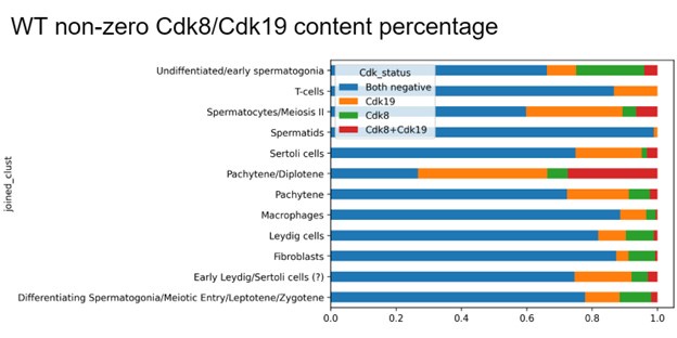
The stabilizing effect of Cdk8/19 on CcnC has been previously discovered and reported in cell culture (doi: 10.1093/nar/gkad538.), and here we have confirmed it at the level of whole tissue. Due to a limited sensitivity of single cell sequencing (only ~5,000 transcripts are sequenced from total of average 500,000 transcripts per cell, so the low expressed transcripts are not sequenced in all cells) it is challenging to firmly establish CDK8/19 positive and -negative tissues from single cell data because both transcripts are minor. This image will be included in the next version. We plan to resolve this matter using two approaches. First, we will try immunohistochemistry. If this method is not sufficiently sensitive we will analyze published single cell sequencing data from mouse databases and re-analyze our data. So far the former approach was challenging for us due to the absence of anti-mouse antibodies which are specific for CDK8 and CDK19 and work on tissue sections. We and others could not produce a tissue-specific staining, with the currently available commercially available antibodies. The only published specific antibody is currently not available.

Since CDK8KO and CDK19KO have significantly reduced fertility compared to the wild type, it might be important to measure the sperm quantity and motility among CDK8 KO, CDK19KO, and induced DKO to evaluate spermatogenesis based on their sperm production.

We agree that this is an interesting question. We did not do spermograms for single KOs but we don’t think that a decreased sperm count would explain CDK8KO infertility as the vasectomized males are able to produce copulative plugs in females whereas CDK8KO males do not, suggesting the absence of mating behavior as a reason for low fertility in the latter genotype.

Some data for the inducible knockout efficiency of Cdk8 were presented in Supplemental Figure 1, but there is no legend for the supplemental figures, it was not clear which band represented the deletion band, and which tissues were examined. Tail or testis?

We apologize for the accidental loss of supplementary figure legends, which will be presented in the next version. The efficiency of CDK8 KO in different tissues was previously examined by us in https://www.ncbi.nlm.nih.gov/gene/264064. The western blot in the MS represents deletion data for the testis.

It seems that two months after the injection of Tam, all the Cdk8 were completely deleted, indicating extremely efficient deletion of Tam induction by two months post administration. Were the complete deletion of Cdk8 happening even earlier?

The complete deletion of CDK8 occurs within a week or even as early as 2-3 days in culture, and at least after at two weeks in vivo. We chose the two mo. period to prevent the effect of tamoxifen on gene expression. We examined other time points (Figure 6) and registered the beginning of effects at 2 weeks and maximum effect by one mo.

The authors found that Sertoli cells re-entered the cell cycle in the inducible double knockout but stopped short of careful characterization other than increased expression of cell cycle genes.

We agree with the reviewer, and we will add Ki67 (or equivalent) staining along with Sertoli cell markers.

Dko should be appropriately named iDKO (induced dKO).

We will make the corresponding change.

We performed necropsy ? not the right wording here. Colchicine-lke apoptotic bodies ? what does this mean? Not clear.

We will amend the next version to address these minor points, and we thank the reviewer for careful reading of the manuscript.

Images throughout the manuscript suffer from poor resolution and are often blurry and hard to evaluate.

As mentioned above, we had a problem with image quality during the submission through Biorxiv and we will provide high resolution images in the next version.

To pinpoint the meiotic stage defect of iDKO, it is better to use the meiotic chromosome spread approach.

Unfortunately, meiotic spreads would not be feasible or informative, due to a low number of surviving cells in iDKO and the fact that there were evidently no cells in stages after SYCP3+.

  1. Howard Hughes Medical Institute
  2. Wellcome Trust
  3. Max-Planck-Gesellschaft
  4. Knut and Alice Wallenberg Foundation