Synaptic plasticity and cognitive function are disrupted in the absence of Lrp4
- Skirball Institute of Biomolecular Medicine, NYU Medical Center, United States
Figures
Figure 1
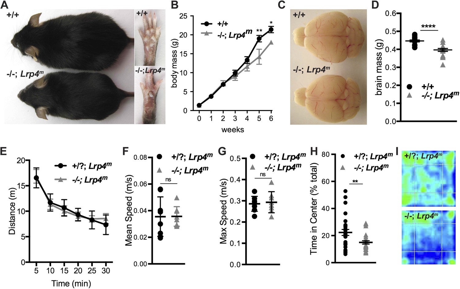
Restoring Lrp4 expression selectively in muscle of Lrp4 mutant mice rescues neonatal lethality.
(A) Lrp4−/−; Lrp4m mice are fertile and live a normal lifespan. (B) The body mass of six-week old Lrp4−/−; Lrp4m mice is reduced by 16% (wild-type, 21.4 ± 0.8 g, n = 10; Lrp4−/−; Lrp4m, 18.0 ± 0.5 g, n = 5). (C) The gross morphology of the adult brain is similar in wild-type and Lrp4−/−; Lrp4m mice. (D) The size of the adult brain is reduced by 11% in Lrp4−/−; Lrp4m mice (wild-type, 0.45 ± 0.005 g, n = 17; Lrp4−/−; Lrp4m, 0.4 ± 0.008 g, n = 15). (E, F, G) The locomotor activity of Lrp4−/−; Lrp4m mice in an open field test was normal as measured by distance traveled (E), mean velocity (F), and maximum velocity (G) (wild-type, n = 17; Lrp4−/−; Lrp4m, n = 15). (H) Lrp4−/−; Lrp4m mice showed reduced exploratory behavior (wild-type, 22.3 ± 2.2%, n = 29; Lrp4−/−; Lrp4m, 14.9 ± 1.2%, n = 25). (I) Representative heat maps of wild-type and Lrp4−/−; Lrp4m mice during a 30 min open field test.
Figure 2
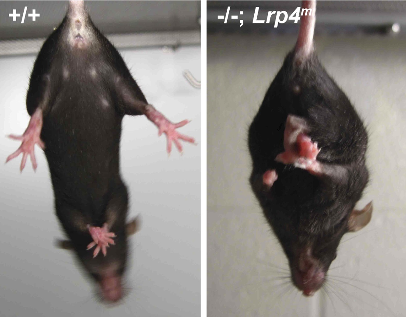
Forelimb and hindlimb clasping in Lrp4−/−; Lrp4m mice.
Wild-type mice splay their limbs when suspended by their tail, whereas Lrp4−/−; Lrp4m mice clasp their limbs.
Figure 3
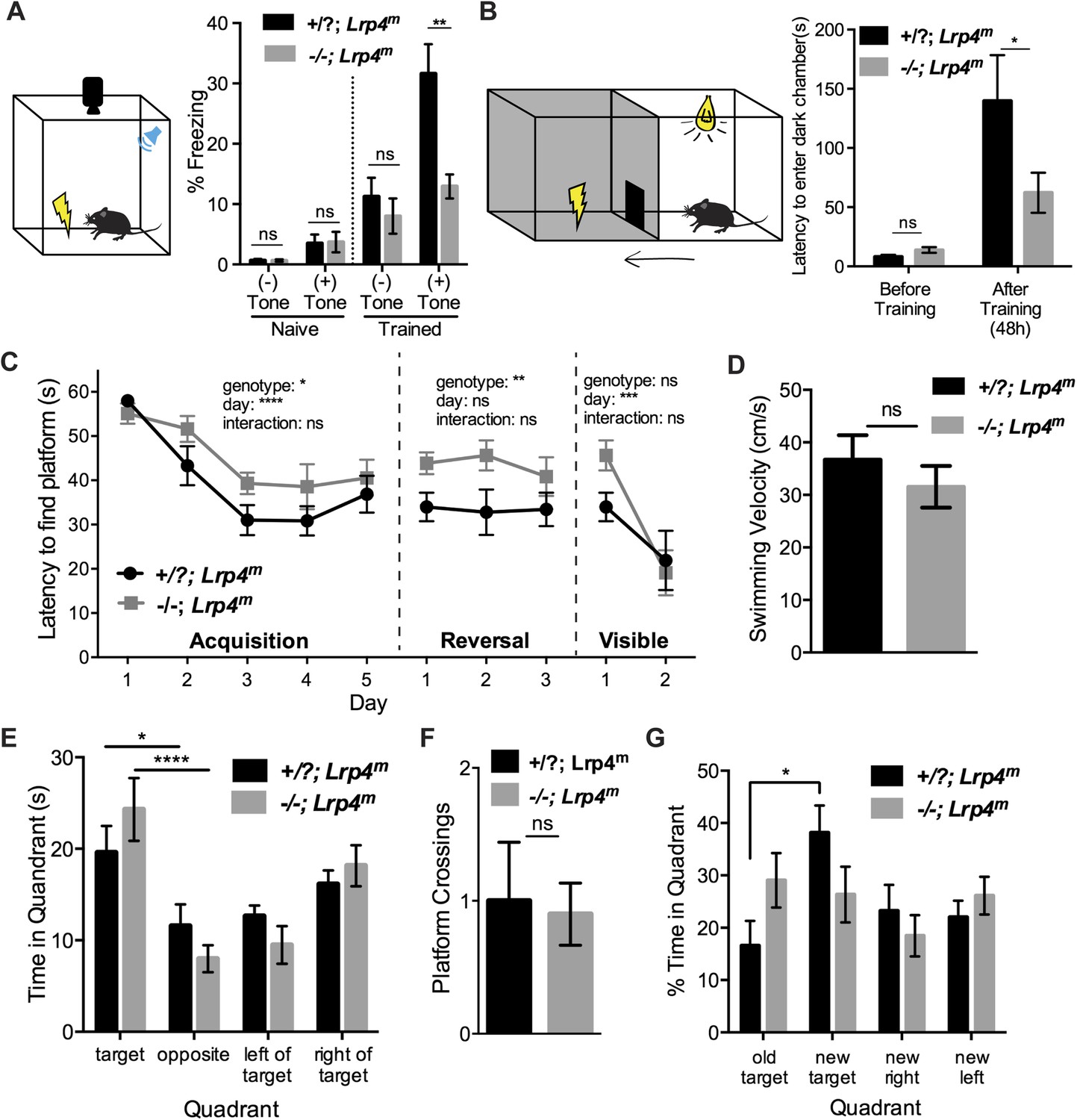
Mice lacking Lrp4 in the CNS display defects in learning and memory.
(A) A schematic representation of the fear-conditioning paradigm. Lrp4−/−; Lrp4m mice exhibit a decrease in freezing behavior, compared to littermate control mice, when presented with an aversive conditioned stimulus (Lrp4−/−; Lrp4m, n = 12; littermate controls, n = 14). (B) A schematic representation of the passive avoidance paradigm. Lrp4−/−; Lrp4m mice were less hesitant to enter a dark chamber associated with an aversive stimulus (Lrp4−/−; Lrp4m, n = 13; littermate controls, n = 17). (C) Lrp4−/−; Lrp4m mice showed spatial learning deficits and reduced cognitive flexibility in the Morris water maze. Both control and Lrp4−/−; Lrp4m mice were able to locate the escape platform during the visible version of the water maze (Lrp4−/−; Lrp4m, n = 8; littermate controls, n = 9). (D) Lrp4−/−; Lrp4m and control mice displayed comparable swimming velocity during the Morris water maze. (E) Lrp4−/−; Lrp4m and control mice spent more time searching in the target quadrant region than other quadrants during the probe trial. (F) Lrp4−/−; Lrp4m and control mice crossed the platform site with similar frequency during the probe trial. (G) Lrp4−/−; Lrp4m mice spent less time in the new target quadrant during reversal training. See also Figure 4.
Figure 4
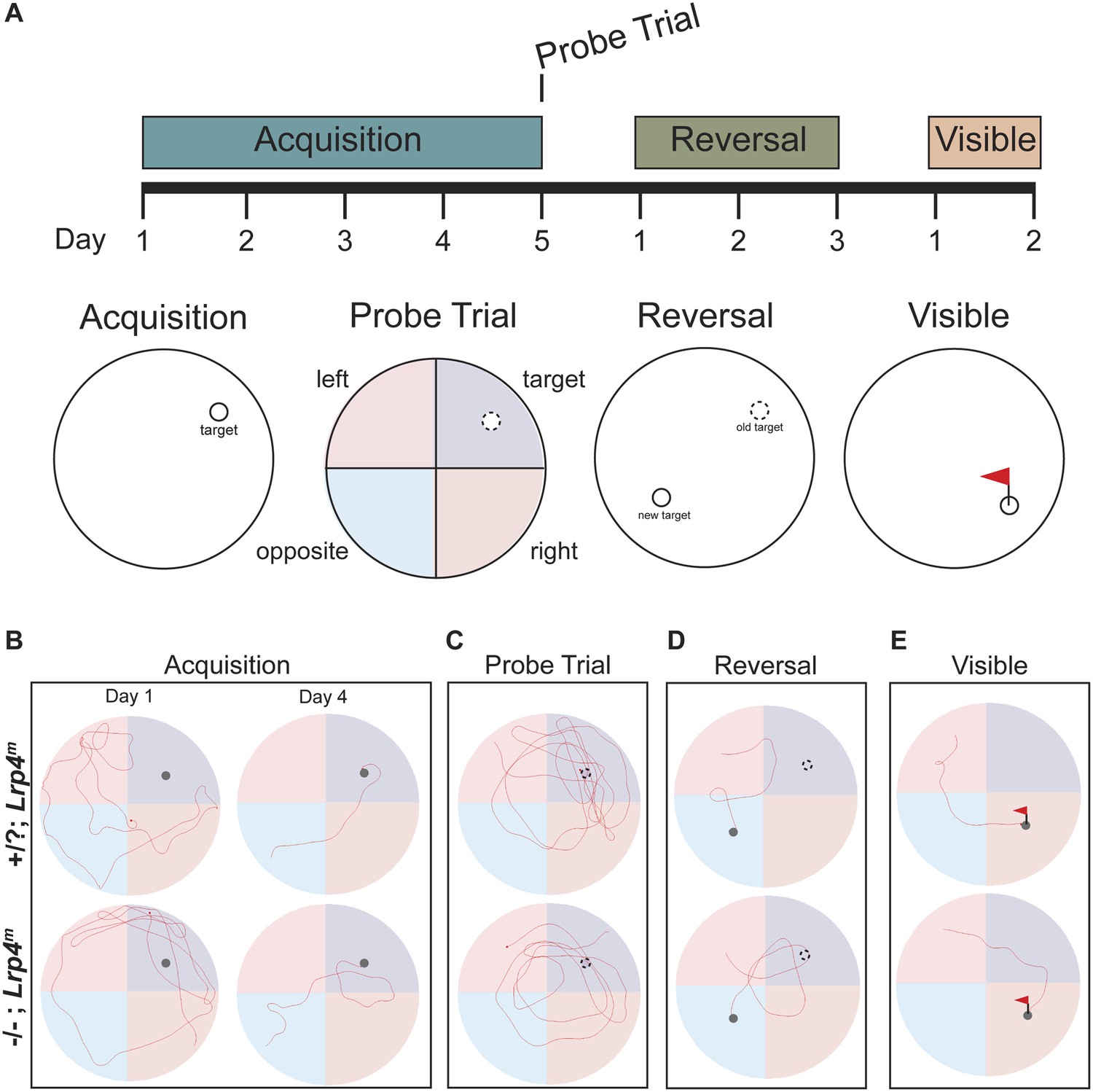
Mice lacking Lrp4 in the CNS display defects in learning and memory in the Morris water maze.
(A) A schematic representation of the Morris water maze training protocol. Mice were trained for 5 days to locate a hidden platform. A probe trial was performed on the fifth day, when the platform was removed. The hidden platform was moved to the opposite quadrant during reversal training. A flag was placed on the hidden platform during the visible training phase. Representative trajectories of control and Lrp4−/−; Lrp4m mice during the acquisition (B), probe (C), reversal (D), and visible (E) tests of the Morris water maze trial.
Figure 5

Lrp4 is required for normal synaptic transmission.
(A) The panel shows the configuration of whole-cell voltage-clamp recordings made from acute hippocampal slices of young, adult mice. Postsynaptic responses in CA1 pyramidal neurons were measured following stimulation of Schaffer collaterals (SC). (B) Representative traces from CA1 neurons to paired stimuli show that paired-pulsed facilitation is normal in Lrp4−/−; Lrp4m mice (wild-type, n = 9; Lrp4−/−; Lrp4m, n = 6). (C) Representative traces of spontaneous miniature excitatory postsynaptic currents (mEPSC) of wild-type or Lrp4 mutant CA1 neurons. (D) mEPSC frequency is reduced in Lrp4 mutant CA1 neurons (wild-type, n = 12; Lrp4−/−; Lrp4m, n = 12. (E, F) mEPSC ampitudes of Lrp4 mutant CA1 neurons are comparable to wild-type (wild-type, n = 12; Lrp4−/−; Lrp4m, n = 12). (G, H) Representative traces from individual CA1 neurons following a TBS delivered to neurons, which were voltage-clamped at −70 mV, show LTP from wild-type but not Lrp4 mutant neurons (upper panel). Excitation, measured as the amplitude of an EPSC (pA), is shown below, and input resistance (Ri) is shown in the bottom panel. There is considerable variability in the baseline EPSC amplitudes in slices from wild-type and Lrp4−/−; Lrp4m mice. However, there was no significant difference in the baseline EPSC amplitudes between wild-type and mutant mice (wild-type, 52.6 ± 9.7 pA, n = 21; Lrp4−/−; Lrp4m, 44.9 ± 4.9 pA, n = 27, p = 0.44). Further, there was not a significant correlation between the magnitude of LTP and initial synaptic strength (wild-type, R2 = 0.05, n = 21, p = 0.35; Lrp4−/−; Lrp4m, R2 = 0.09, n = 21, p = 0.13). (I) Representative traces from individual CA1 neurons during TBS show a reduction in the integral of the summed EPCSs recorded during a single TBS in Lrp4-deficient neurons, top panel. Induction volume, quantified as the integral of the postsynaptic response, is shown in the bottom panel (wild-type, n = 21; Lrp4−/−; Lrp4m; n = 32) (J, K) Representative responses from individual CA1 neurons following a TBS delivered to neurons, which were voltage-clamped at 0 mV, show a restoration of LTP in Lrp4 mutant neurons (upper panel). Excitation, measured as the amplitude of an EPSC (pA), is shown in the middle panel, and input resistance (Ri) is shown in the bottom panel. (L) Averaged data from control cells (n = 21) and Lrp4 mutant (n = 32) neurons shows that the response from wild-type neurons is potentiated 1.5-fold, whereas Lrp4 mutant CA1 neurons potentiate little, if at all 30 min following a TBS delivered at Vc = −70 mV. (M) Depolarization of Lrp4 mutant CA1 neurons during TBS restores LTP (wild-type, n = 11; Lrp4−/−; Lrp4m, n = 9). (N) Quantitation of potentiation at 10-20 min after TBS demonstrates a lack of LTP in Lrp4 mutant CA1 neurons, which is restored upon depolarization (Vc = −70 mV: wild-type, n = 21; Lrp4−/−; Lrp4m, n = 32; Vc = 0 mV: wild-type, n = 11; Lrp4−/−; Lrp4m, n = 9).
Figure 6
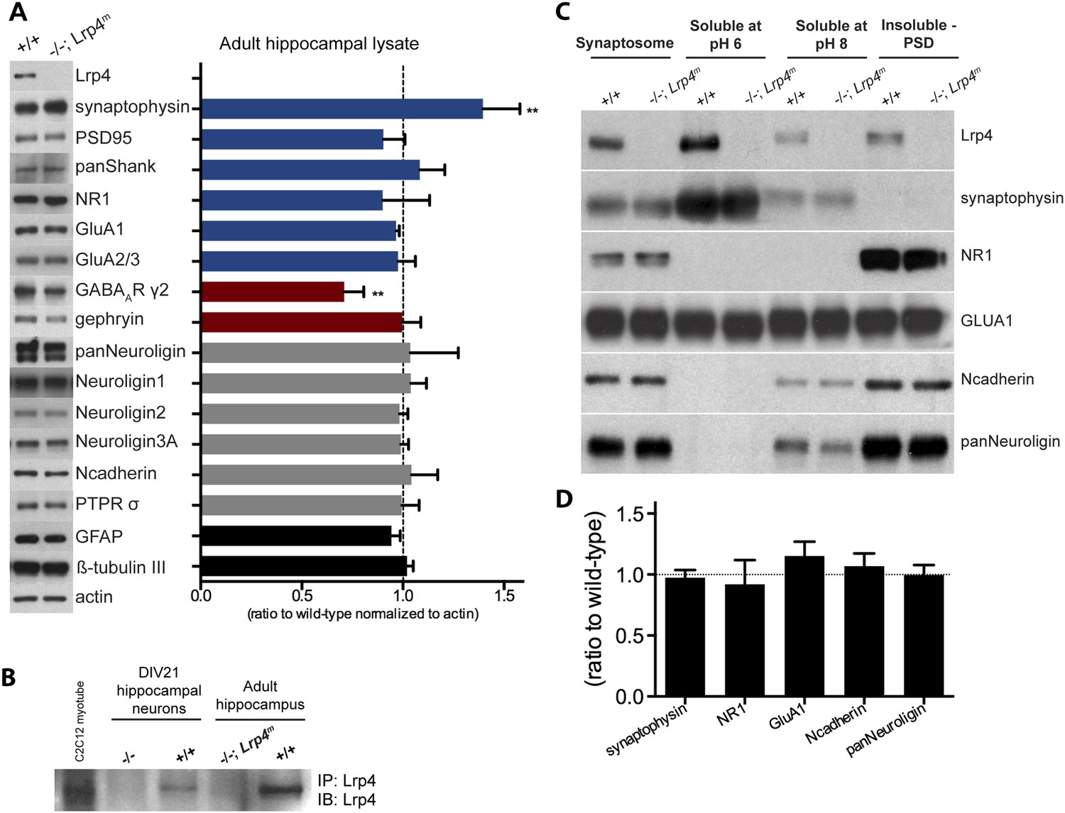
Lrp4 is enriched in synaptic membranes.
(A) Quantitative analysis of proteins in hippocampal lysates from wild-type and Lrp4−/−; Lrp4m mice. The expression level of each protein, normalized to actin, was determined and assigned a value of 1.0 in wild-type mice. The graph shows the ratio of values in Lrp4−/−; Lrp4m mice compared to wild-type mice. Expression of most proteins is not dependent upon Lrp4, but expression of the synaptic vesicle-associated protein, Synaptophysin, was modestly elevated (30%) and expression of the GABAARγ2 subunit was modestly decreased (20%) in Lrp4 mutant hippocampi. (B) Lrp4 expression is detected in cultured hippocampal neurons, grown in cell culture for 21 days, and in hippocampal tissue from wild-type but not from Lrp4−/−; Lrp4m mice. (C) Lrp4 co-isolates with synaptosomes, the pH 6-solubilzed fraction, which is enriched for Synaptophysin, a presynaptic marker, and the postsynaptic density (PSD) fraction, which is highly enriched for NR1. (D) The expression levels of presynaptic and postsynaptic proteins present in the synaptosomal fraction (C) are not altered in Lrp4−/−; Lrp4m mutant hippocampi.
Figure 7
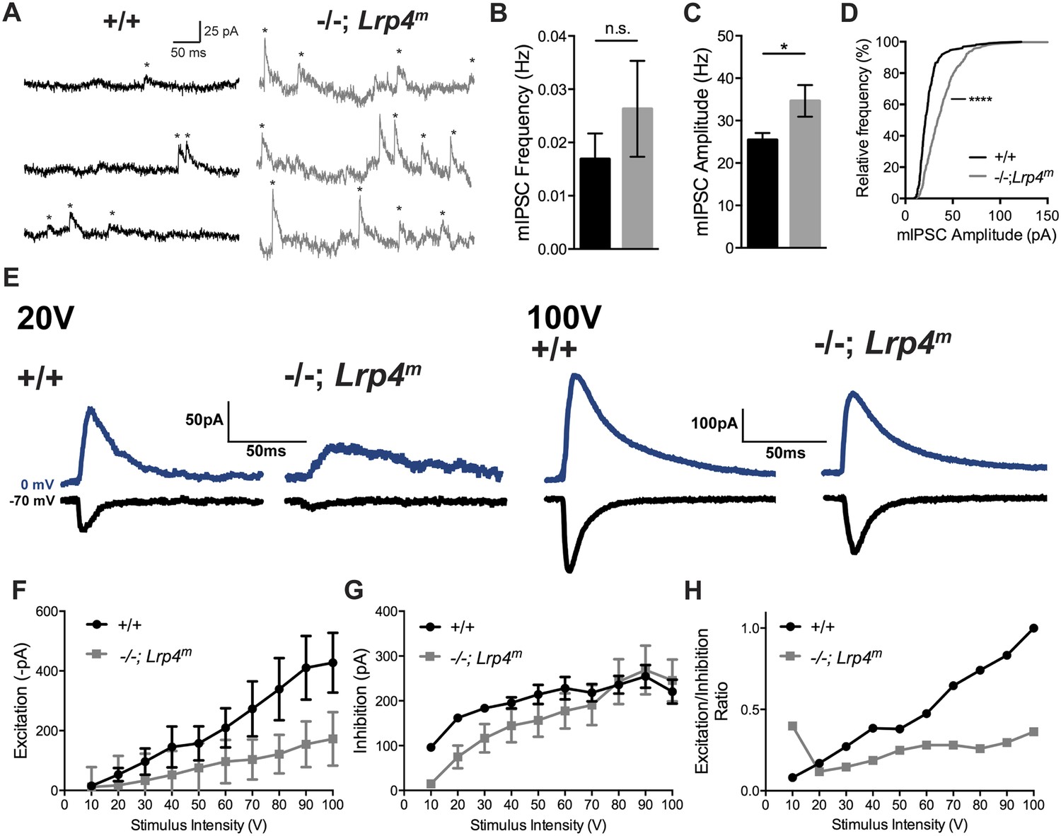
The strength of inhibition is unchanged in CA1 neurons from Lrp4−/−; Lrp4m mice.
(A) Representative traces of spontaneous miniature inhibitory postsynaptic currents (mIPSC) from CA1 neurons from wild-type or Lrp4−/−; Lrp4m mice, which were voltage-clamped at 0 mV. (B) The mIPSC frequency is similar in Lrp4 mutant and wild-type CA1 neurons (wild-type, 0.017 ± 0.005 /sec, n = 7; Lrp4−/−; Lrp4m, 0.026 ± 0.009 /sec, n = 6). (C, D) mIPSC amplitudes are increased in CA1 neurons from Lrp4−/−; Lrp4m mice (wild-type, 25.4 ± 1.6 pA, n = 7; Lrp4−/−; Lrp4m, 34.7 ± 3.7 pA, n = 6). (E) Representative traces of excitation (black) and inhibition (blue) at varied stimulus intensities. (F) Excitation is decreased in CA1 neurons from Lrp4−/−; Lrp4m mice. (G) Inhibition is similar in CA1 neurons from wild-type and Lrp4−/−; Lrp4m mice. (H) The E/I ratio is decreased in CA1 neurons from Lrp4−/−; Lrp4m mice.
Figure 8
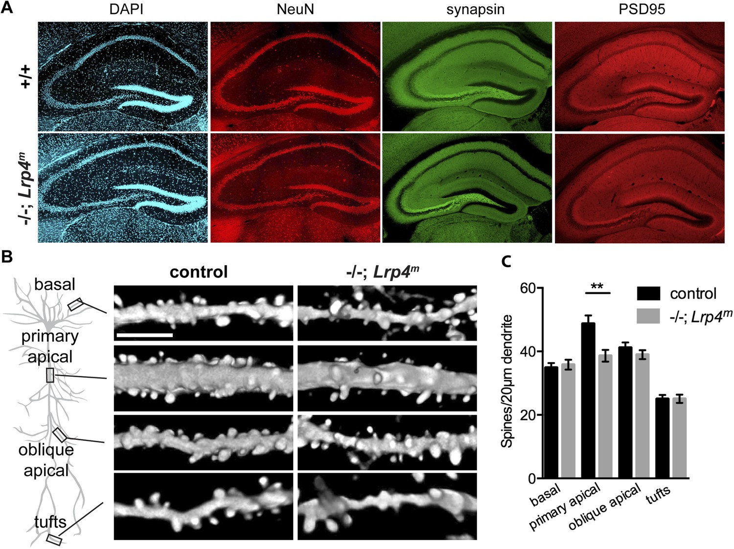
A loss of Lrp4 decreases spine density in primary apical dendrites of CA1 neurons.
(A) The organization of the hippocampus appears normal as assessed by staining sections of the adult hippocampus for DNA (DAPI), neuronal nuclei (NeuN), presynaptic terminals (synapsin) or excitatory postsynaptic membranes (PSD95). (B) Representative images of dendrites of Thy1-YFP labeled CA1 Lrp4 mutant pyramidal neurons near (basal and apical) and far (oblique apical and tufts) from the cell body. Scale bar: 5 μm. (C) The spine density is reduced selectively at primary apical dendrites (basal: wild-type, n = 45; Lrp4−/−; Lrp4m, n = 45; primary apical: wild-type, n = 43; Lrp4−/−; Lrp4m, n = 46; oblique apical: wild-type, n = 42; Lrp4−/−; Lrp4m, n = 46; tufts: wild-type, n = 42; Lrp4−/−; Lrp4m, n = 45).
Download links
A two-part list of links to download the article, or parts of the article, in various formats.
Downloads (link to download the article as PDF)
Open citations (links to open the citations from this article in various online reference manager services)
Cite this article (links to download the citations from this article in formats compatible with various reference manager tools)
Synaptic plasticity and cognitive function are disrupted in the absence of Lrp4
eLife 3:e04287.
https://doi.org/10.7554/eLife.04287


