Ultrastructural analysis of adult mouse neocortex comparing aldehyde perfusion with cryo fixation
- Ecole Polytechnique Fédérale de Lausanne (EPFL), Switzerland
Figures
Figure 1 with 2 supplements
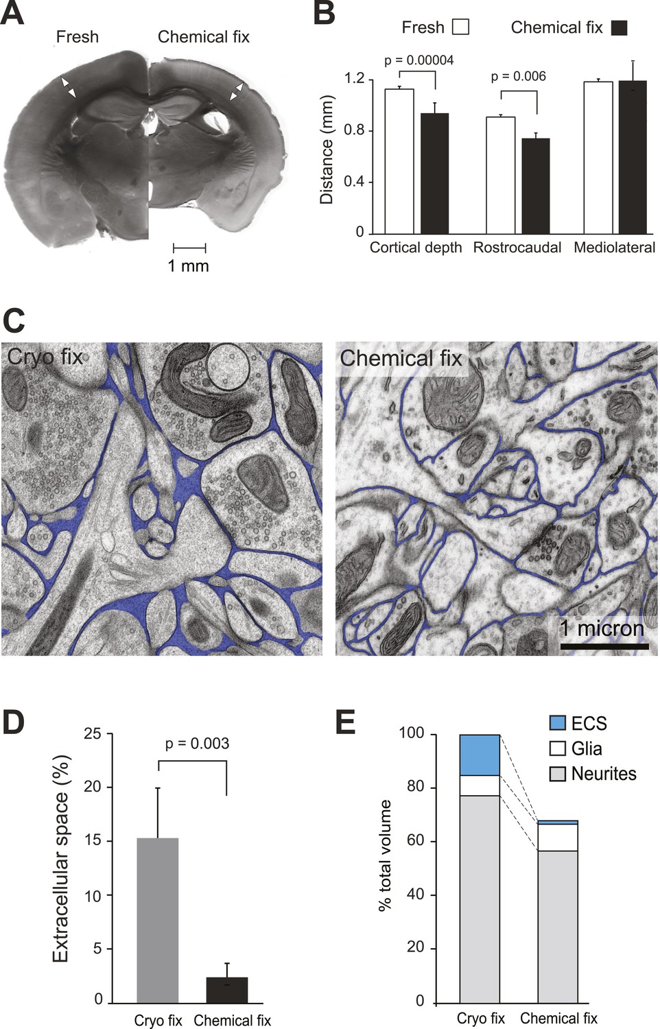
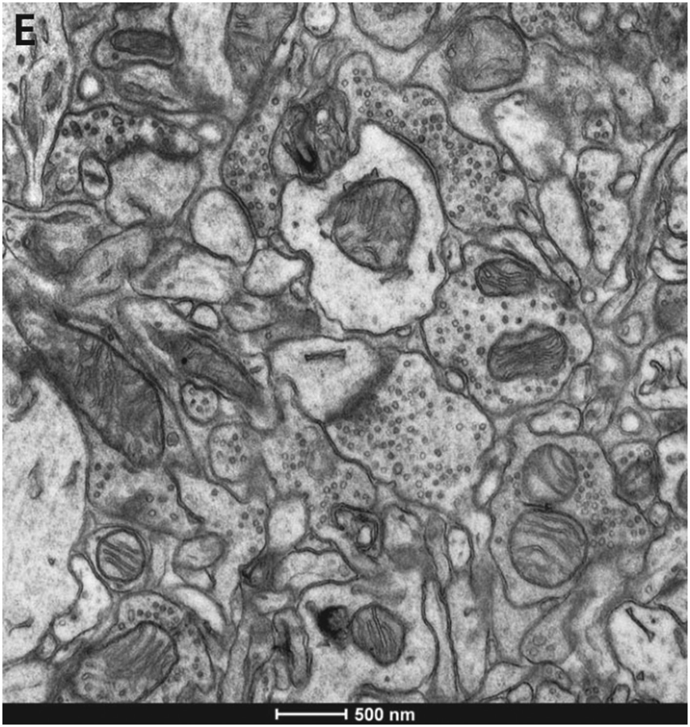
Chemical fixation reduces cortical volume and extracellular space.
(A) Coronal sections of fresh (left) and chemically fixed (right) adult mouse brains. Double-headed arrows overlaying the somatosensory cortex of each section show the position at which the cortical thickness was measured. (B) Measurements of cortical thickness show a 16% reduction after chemical fixation (p = 0.00004, unpaired Student's t-test, left). Measurements across tangential sections show 18% shrinkage in the rostrocaudal axis (p = 0.006, one way ANOVA), but not in the mediolateral axis (p = 0.942, one way ANOVA, right). (C) TEM of cryo fixed (left) and chemically fixed (right) neuropil from the adult mouse cerebral cortex show reduction in the extracellular space (pseudo-coloured in blue) after chemical fixation. (D) Measurements of the volume fraction of extracellular space from serial section analysis showed a six-fold difference between the two fixation techniques (p = 0.003, one way ANOVA). (E) Measurements of volumes occupied by extracellular space, neurites, and glia, from serial section transmission electron microscopy sections showed how the different compartments are altered by chemical fixation. Volume occupied by astrocytic processes was significantly increased after chemical fixation (p = 0.01, one way ANOVA). However, there was no change in the volume occupied by axons and dendrites (p = 0.074, one way ANOVA). As the volume of the cortex is reduced by 31% after chemical fixation, these percentages are shown in the bar chart in which the total volume of chemically fixed neuropil is 69% of the cryo-fixed value (100%).
-
Figure 1—source data 1
Data values and statistics underlying Figure 1B, D, E.
- https://doi.org/10.7554/eLife.05793.004
Figure 1—figure supplement 1
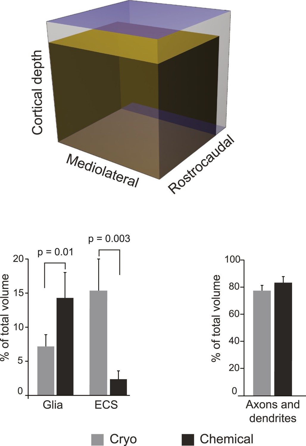
Schematic scale model (upper image) representing the fresh somatosensory cortex (outer cube) and chemically fixed cortex (inner cube), showing the extent to which the two fixations change the volume of this brain region.
Graph (lower left) showing the percentage of neuropil volume occupied by astrocytic (marked ‘Glia’) elements and ECS, in the cryo and chemically fixed neuropil. Graph (lower right) shows the percentage occupied by axons and dendrites. Data are represented as mean ± SD. One way ANOVA assessed statistical significance.
Figure 1—figure supplement 2
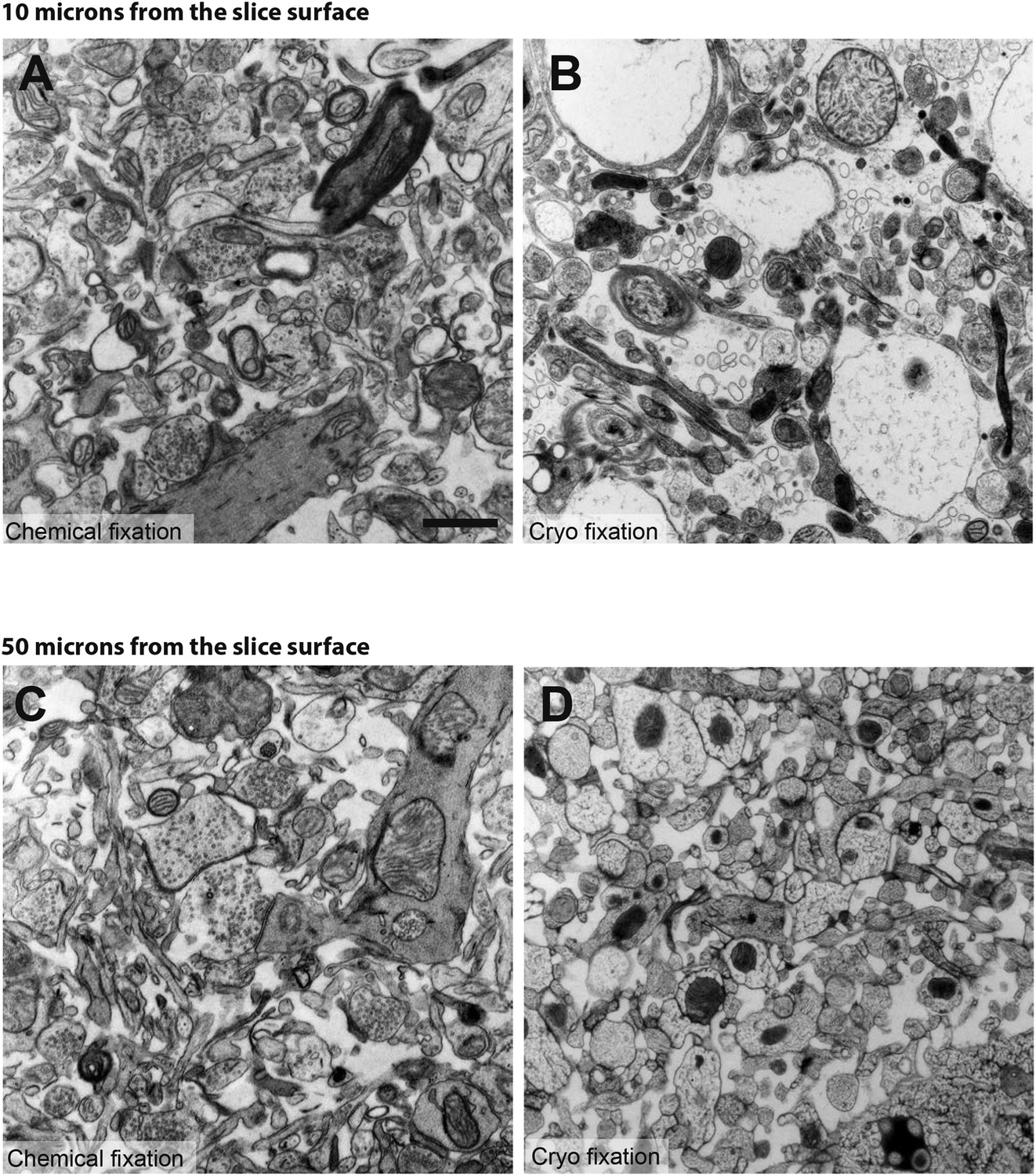
Comparison between chemical fixation (left hand images; A and C) and cryo fixation (right hand images; B and D) of acute brain slices shows that both fixation conditions are able to reveal significant amounts of extracellular space.
However, this can only be clearly seen within 10 microns of the slice surface (B). Deeper into the cryo-fixed slice, the cellular elements appear disrupted with a fine latticed patterning indicating damage caused by ice crystal formation (D). This is not apparent at the same depth in the chemical fixed slice (C). Scale bar is 1 micron.
Figure 2
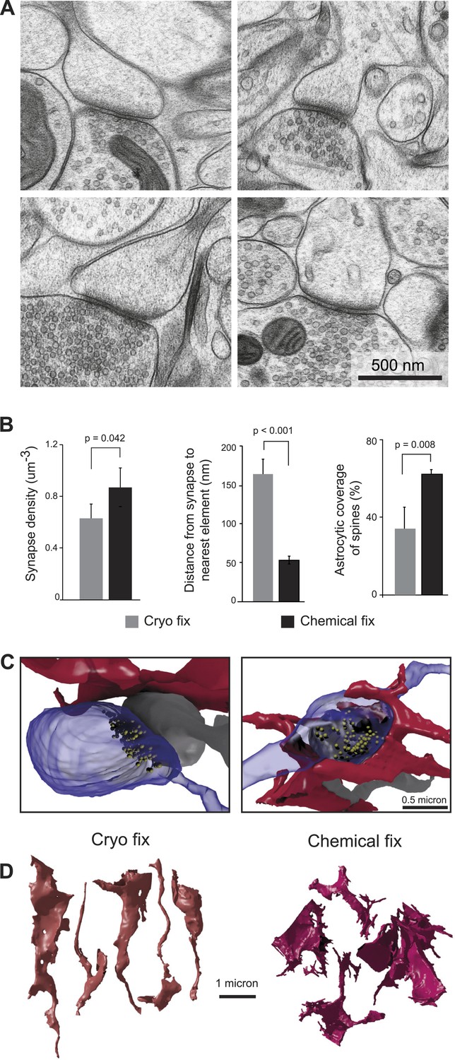
Cryo fixation reveals a larger peri-synaptic space and reduced astrocytic coverage.
(A) Cryo-fixed neuropil shows synaptic contacts with large amounts of surrounding extracellular space. (B) Synaptic density measurements show the chemically fixed neuropil to have 38% more synapses (left graph, p < 0.05, one way ANOVA). Dendritic spine synapses (presumed glutamatergic) show greater distances between the edge of the contact zone and the nearest membrane compared with chemical fixation (middle graph, p < 0.001, unpaired Student's t-test). Cryo-fixed synapses show less astrocytic coverage (right graph, p < 0.01; one way ANOVA). (C) Reconstructions from serial electron microscope images, of axonal boutons (blue) synapsing with dendritic spines (grey), show the astrocytic processes in the near vicinity (red). In the cryo-fixed synapse (left), the astrocytic process is not squeezed close to the synaptic contact (indicated with vesicles in yellow). In the chemically fixed example (right), the astrocyte tightly surrounds the synapse, where the vesicle-filled axonal bouton contacts the spine behind it. (D) Astrocytic processes reconstructed from serial FIBSEM images using the ilastik software (www.ilastik.org) show that chemically fixed astrocytic processes (right) have a more elaborate morphology with small processes extending from the flattened lamellae compared with the less complex structure of cryo-fixed astrocytes (left).
-
Figure 2—source data 1
Data values and statistics underlying Figure 2B.
- https://doi.org/10.7554/eLife.05793.008
Figure 3
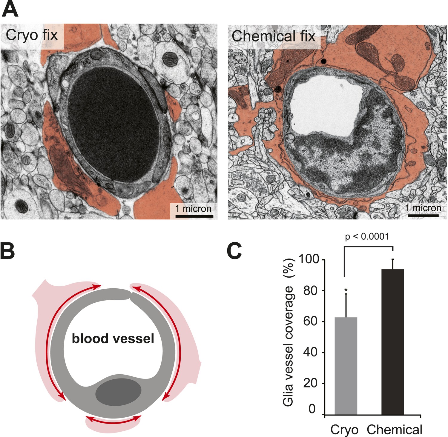
Cryo-fixed capillaries show less astrocytic coverage.
(A) Electron micrographs of transversely sectioned capillaries show the astrocytic endfeet pseudo-coloured in orange. Cryo-fixed example shows a darkly stained erythrocyte within the vessel lumen. (B) Schematic diagram indicates the coverage measured. (C) Chemically fixed tissue contains capillaries with more glial coverage (p < 0.0001, n = 11 vessels cryo, n = 69 vessels perfused, unpaired Student's t-test).
-
Figure 3—source data 1
Data values and statistics underlying Figure 3C.
- https://doi.org/10.7554/eLife.05793.010
Figure 4 with 1 supplement

Vesicles of symmetric synapses are distorted by chemical fixation.
(A) Cryo-fixed synapses on a dendritic shaft (left image) and on a dendritic spine (middle image) show similar rounded vesicles. A chemically fixed, high-pressure frozen and cryo-substituted (right hand image) synapse on a dendritic shaft, however, shows typical features of a symmetric (presumed GABAergic) synapse with ovoid vesicles. (B) Measurements of the short (x) and long (y) diameters of synaptic vesicles. Synapses in cryo-fixed tissue cannot be classified according to the symmetry of pre- and post-synaptic densities and all synaptic vesicles were round. Asymmetric synapses in chemically fixed tissue show similarly shaped vesicles, as do the vesicles at asymmetric synapses of chemically fixed tissue that is then high-pressure frozen and freeze substituted in resin. The symmetric synapses, seen in chemically fixed tissue, show vesicles with characteristic ovoid shapes irrespective of how they were resin embedded.
-
Figure 4—source data 1
Statistics underlying Figure 4B.
- https://doi.org/10.7554/eLife.05793.012
Figure 4—figure supplement 1
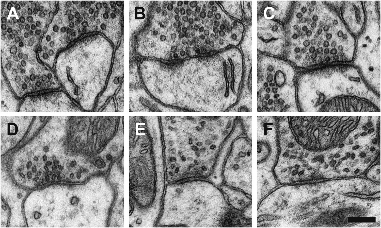
Examples of glutamatergic synapses (A, B, C), situated on dendritic spines, with round clear vesicles; and presumed GABAergic synapses (D, E, F) on dendritic shafts showing flattened, dark vesicles.
Scale bar is 200 microns.
Figure 5 with 1 supplement
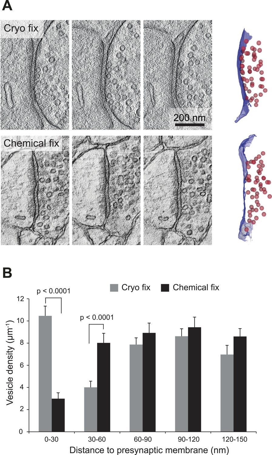
Cryo fixation preserves larger numbers of vesicles at the pre-synaptic membrane.
(A) Electron tomography of a 200-nm thick section shows a cryo-fixed (upper) synapse with a large number of vesicles close to the presynaptic membrane in comparison with a similar chemically fixed synapse (lower). In each case three sample images are shown from complete tomographic series. Three-dimensional reconstructions of this region (right hand images) show all the vesicles (red) in relation to the presynaptic membrane (blue). (B) Measurements of the distance of vesicles from the presynaptic membrane show that more vesicles are arranged closer (0–30 nm) to the synapse after cryo fixation (p < 0.0001, unpaired Student's t-test).
-
Figure 5—source data 1
Data values and statistics underlying Figure 5B.
- https://doi.org/10.7554/eLife.05793.015
Figure 5—figure supplement 1
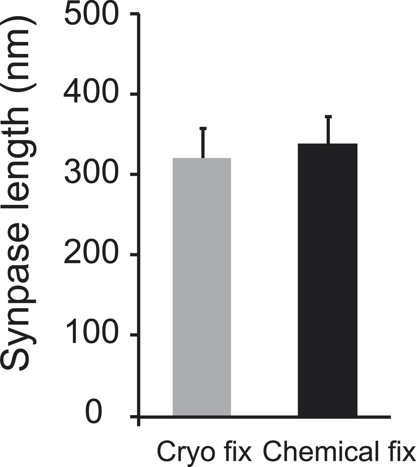
Synapses found on dendritic spines were the same length.
Data are represented as mean ± SD. Unpaired Student's t-test assessed statistical significance; p = 0.561.
Author response image 1
Electron micrograph showing the neuropil from an acute slice chemically fixed immediately after it was vibratome sectioned. The image is taken 50 micons from the slice surface.
Download links
A two-part list of links to download the article, or parts of the article, in various formats.
Downloads (link to download the article as PDF)
Open citations (links to open the citations from this article in various online reference manager services)
Cite this article (links to download the citations from this article in formats compatible with various reference manager tools)
Ultrastructural analysis of adult mouse neocortex comparing aldehyde perfusion with cryo fixation
eLife 4:e05793.
https://doi.org/10.7554/eLife.05793


