Complement 3a receptor 1 on macrophages and Kupffer cells is not required for the pathogenesis of metabolic dysfunction-associated steatotic liver disease
- Division of Cardiology, Department of Medicine, Cardiovascular Research Institute, Weill Center for Metabolic Health, Weill Cornell Medicine, United States
Figures
Figure 1 with 1 supplement
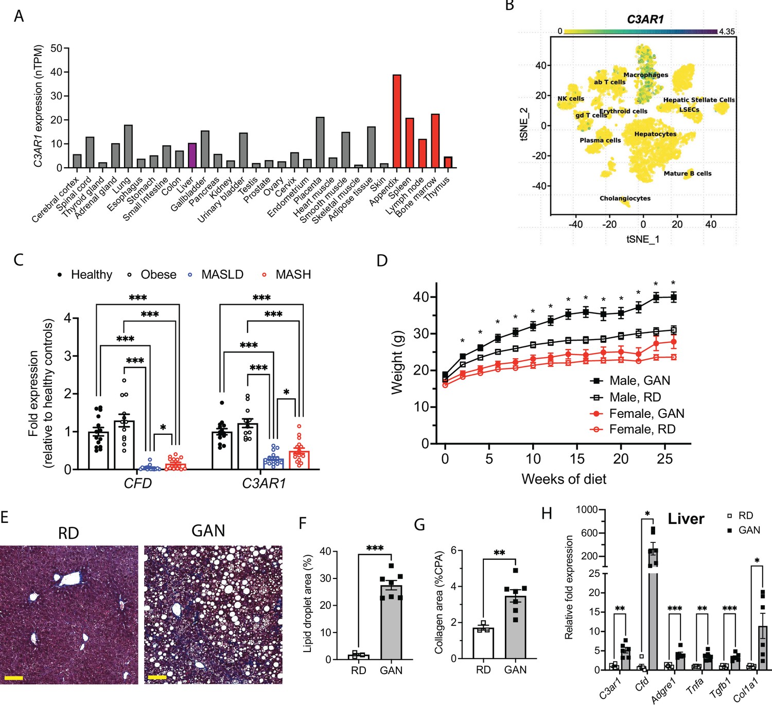
C3AR1 is found in macrophages, is modulated by MASLD/MASH in humans, and is induced by a murine dietary model of MASH.
(A) Relative C3AR1 human tissue expression level by tissue, derived from deep sequencing of the mRNA combined dataset (HPA and GTEx) in the Human Protein Atlas, shown as normalized transcripts per million (nTPM). Liver is highlighted in purple and immunologic tissues are highlighted in red. (B) Single-cell RNA sequencing distribution of C3AR1 expression in human liver (tSNE, t-distributed Stochastic Neighbor Embedding). (C) Analysis of CFD and C3AR1 expression from liver biopsy samples in patients with MASH, MASLD, obesity without MASLD, and age-matched healthy controls (n=12–16 per group, Welch t test with Holm-Šídák correction for multiple comparisons). (D) Weight curve in male and female C3ar1flox/flox control mice placed on GAN high-fat diet compared to regular diet (RD) controls (males, n=7; females, n=6). (E) Representative liver section staining by Masson’s Trichrome in male control mice on RD or GAN diet for 28 weeks (scale bar = 100 mm). (F) Lipid droplet area quantification in liver sections from male control mice, excluding vessel lumens (RD, n=3; GAN, n=7). (G) Collagen area quantification in liver sections of male control mice (RD, n=3; GAN, n=7). (H) Gene expression of key macrophage or fibrosis genes in male control mice on GAN or RD (n=6 per group). Unpaired two-tailed Student’s t test (Except 1 C as above). Annotations: *, p<0.05; **, p<0.01; ***, <0.001. Error bars represent standard error of the mean.
-
Figure 1—source data 1
Source data for Figure 1A, C, D, F, G and H.
Source data for Figure 1—figure supplement 1B-D.
- https://cdn.elifesciences.org/articles/100708/elife-100708-fig1-data1-v1.xlsx
Figure 1—figure supplement 1
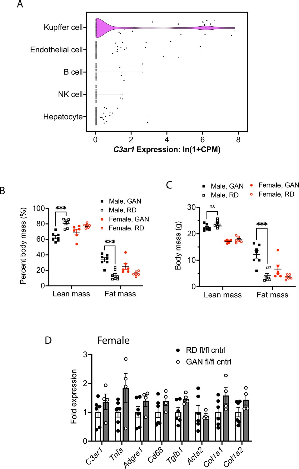
C3ar1 is expressed in liver, primarily in Kupffer cells.
GAN diet induces fat accumulation without induction of C3ar1 in female mice. (A) Single cell RNA sequencing analysis of C3ar1 expression in mouse liver tissue (derived from Tabula Muris). (B) Percent lean and fat mass of C3ar1flox/flox control mice after 20 weeks of GAN or RD (n=6–7 per group). (C) Absolute lean and fat mass of control mice after 20 weeks of GAN or RD (n=6–7 per group). (D) Relative gene expression in female control mice after 30 weeks on RD (n=4–6 per group). Unpaired two-tailed Student’s t test: ***, p<0.001. Error bars represent standard error of the mean.
Figure 2 with 1 supplement
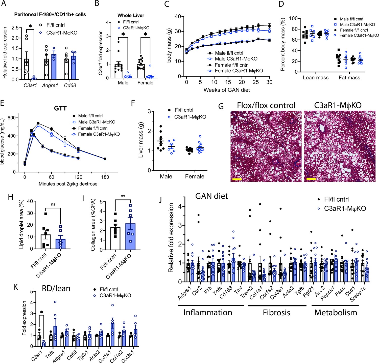
C3aR1 deletion in all macrophages does not affect weight gain, glucose homeostasis, liver steatosis or fibrosis.
(A) Expression of C3ar1 in peritoneal F4/80+/CD68+ cells from C3ar1flox/flox control (n=6) or C3aR1-MφKO male mice (n=3). (B) Expression of C3ar1 in whole liver from control or C3aR1-MφKO mice (n=11–12 per male group, n=13–14 per female group). (C) Body mass curve of control or C3aR1-MφKO mice on GAN high-fat diet starting at 5 weeks of age (n=11–12 per male group, n=14 per female group). (D) Body composition analysis by EchoMRI in control or C3aR1-MφKO mice after 30 weeks GAN diet (n=6–9 per male group, n=9–13 per female group). (E) Glucose tolerance test in control or C3aR1-MφKO mice with 14 hr fast after 28 weeks GAN diet (n=6–9 per male group, n=9–14 per female group). (F) Liver mass in control or C3aR1-MφKO male mice at time of euthanasia after 30 weeks GAN diet (n=6–9 per male group, n=9–14 per female group). (H) Representative liver section staining by Masson’s Trichrome in male control or C3aR1-MφKO mice (scale bar = 100 mm). (I) Lipid droplet area in liver sections from male control or C3aR1-MφKO mice, excluding vessel lumens (n=6–7 per group). (J) Collagen area in liver sections from male control or C3aR1-MφKO mice (n=6–7 per group). (J,K) Relative mRNA expression of key markers for inflammation, fibrosis, and liver metabolism in liver from male control or C3aR1-MφKO mice after 30 weeks of either GAN (J) diet (n=11–12 per group) or regular (K) diet (n=3–5 per group). Unpaired two-tailed Student’s t test: Student’s t test: *, p<0.05. Error bars represent standard error of the mean.
-
Figure 2—source data 1
Source data for Figure 2A-F, H-K.
Source data for Figure 2—figure supplement 1A-D.
- https://cdn.elifesciences.org/articles/100708/elife-100708-fig2-data1-v1.xlsx
Figure 2—figure supplement 1
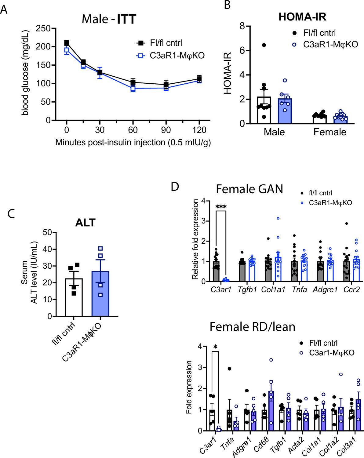
C3aR1 deletion in macrophages does not affect insulin-glucose axis, circulating marker of liver injury, or expression of key genes in female mice.
(A) Insulin tolerance test in C3ar1flox/flox control or C3aR1-MφKO male mice with 14 hr fast after 29 weeks GAN diet (n=6–9 per group). (B) HOMA-IR measurement of insulin resistance in control or C3aR1-MφKO mice with 6 hr fast after 27 weeks GAN diet (n=6–9 per male group, n=9–13 per female group). (C) Serum alanine aminotransferase levels in control or C3aR1-MφKO male mice after 30 weeks GAN diet (n=4 per group). (D) Relative gene expression in control or C3aR1-MφKO female mice after 30 weeks of either GAN (n=13–14 per group) or RD (n=5–6 per group). Unpaired two-tailed Student’s t test: *, p<0.05; ***, p<0.001. Error bars represent standard error of the mean.
Figure 3 with 1 supplement
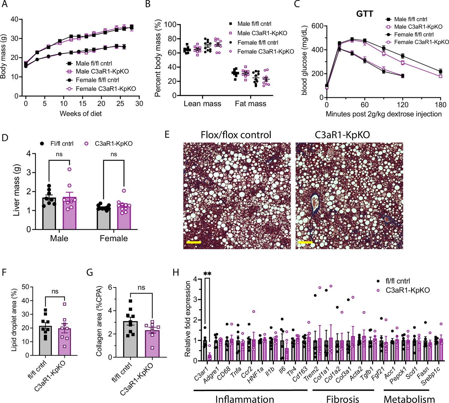
C3aR1 deletion in Kupffer cells does not affect weight gain, glucose homeostasis, liver steatosis or fibrosis.
(A) Body mass curve on GAN diet in C3ar1flox/flox control or C3aR1-KpKO mice beginning at 5 weeks of age (n=8–10 per group). (B) Body composition analysis by EchoMRI in control or C3aR1-KpKO mice after 28 weeks GAN diet (n=8–10). (C) Glucose tolerance test in control or C3aR1-KpKO mice with 14 hr fast after 26 weeks GAN diet (n=8–10). (D) Liver mass in control or C3aR1-KpKO male mice at time of euthanasia after 30 weeks GAN diet (n=8–10). (E) Representative liver section staining by Masson’s Trichrome in control or C3aR1-KpKO male mice (scale bar = 100 mm). (F) Lipid droplet area quantified on liver sections of control or C3aR1-KpKO male mice, excluding vessel lumens (n=8–9). (G) Collagen area quantified on whole liver section of control or C3aR1-KpKO male mice (n=8–9). (H) Relative gene expression in male control or C3aR1-KpKO mice after 30 weeks GAN diet (n=5–6). Unpaired two-tailed Student’s t test: **, p<0.01. Error bars represent standard error of the mean.
-
Figure 3—source data 1
Source data for Figure 3A–D and F–H.
Source data for Figure 3—figure supplement 1A.
- https://cdn.elifesciences.org/articles/100708/elife-100708-fig3-data1-v1.xlsx
Figure 3—figure supplement 1
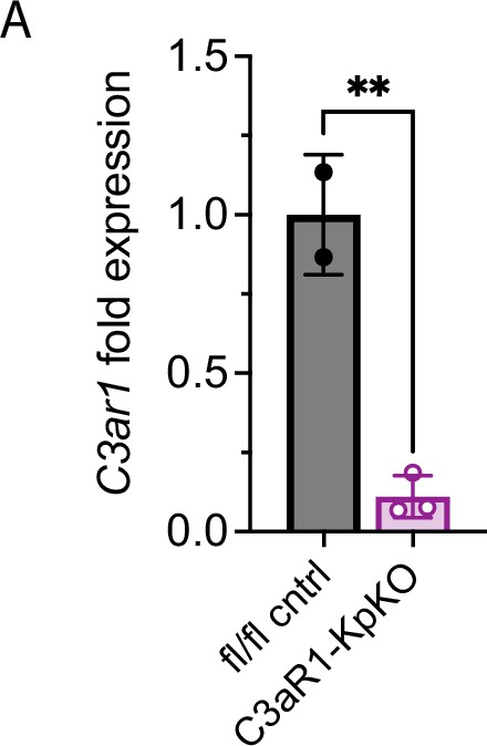
C3ar1 expression in female mice with Kupffer cell-specific deletion of C3ar1.
(A) Relative C3ar1 expression in C3ar1flox/flox control or C3aR1-KpKO female mice after 30 weeks RD (n=2–3 per group). Unpaired two-tailed Student’s t test: **, p<0.01. Error bars represent standard error of the mean.
Additional files
-
Supplementary file 1
Table containing forward and reverse primer sequences for the gene targets used in quantitative PCR experiments performed in this study.
- https://cdn.elifesciences.org/articles/100708/elife-100708-supp1-v1.xlsx
-
MDAR checklist
- https://cdn.elifesciences.org/articles/100708/elife-100708-mdarchecklist1-v1.docx
Download links
A two-part list of links to download the article, or parts of the article, in various formats.
Downloads (link to download the article as PDF)
Open citations (links to open the citations from this article in various online reference manager services)
Cite this article (links to download the citations from this article in formats compatible with various reference manager tools)
Complement 3a receptor 1 on macrophages and Kupffer cells is not required for the pathogenesis of metabolic dysfunction-associated steatotic liver disease
eLife 13:RP100708.
https://doi.org/10.7554/eLife.100708.3


