T3SS translocon induces pyroptosis by direct interaction with NLRC4/NAIP inflammasome
- CAS and Shandong Province Key Laboratory of Experimental Marine Biology, Institute of Oceanology; CAS Center for Ocean Mega-Science, Chinese Academy of Sciences, China
- Tsinghua University-Peking University Joint Center for Life Sciences, School of Basic Medical Sciences, Tsinghua University, China
- Laboratory for Marine Biology and Biotechnology, Qingdao Marine Science and Technology Center, China
- College of Marine Sciences, University of Chinese Academy of Sciences, China
- NHC Key Laboratory of Tropical Disease Control, School of Tropical Medicine, Hainan Medical University, China
Figures
Figure 1 with 1 supplement
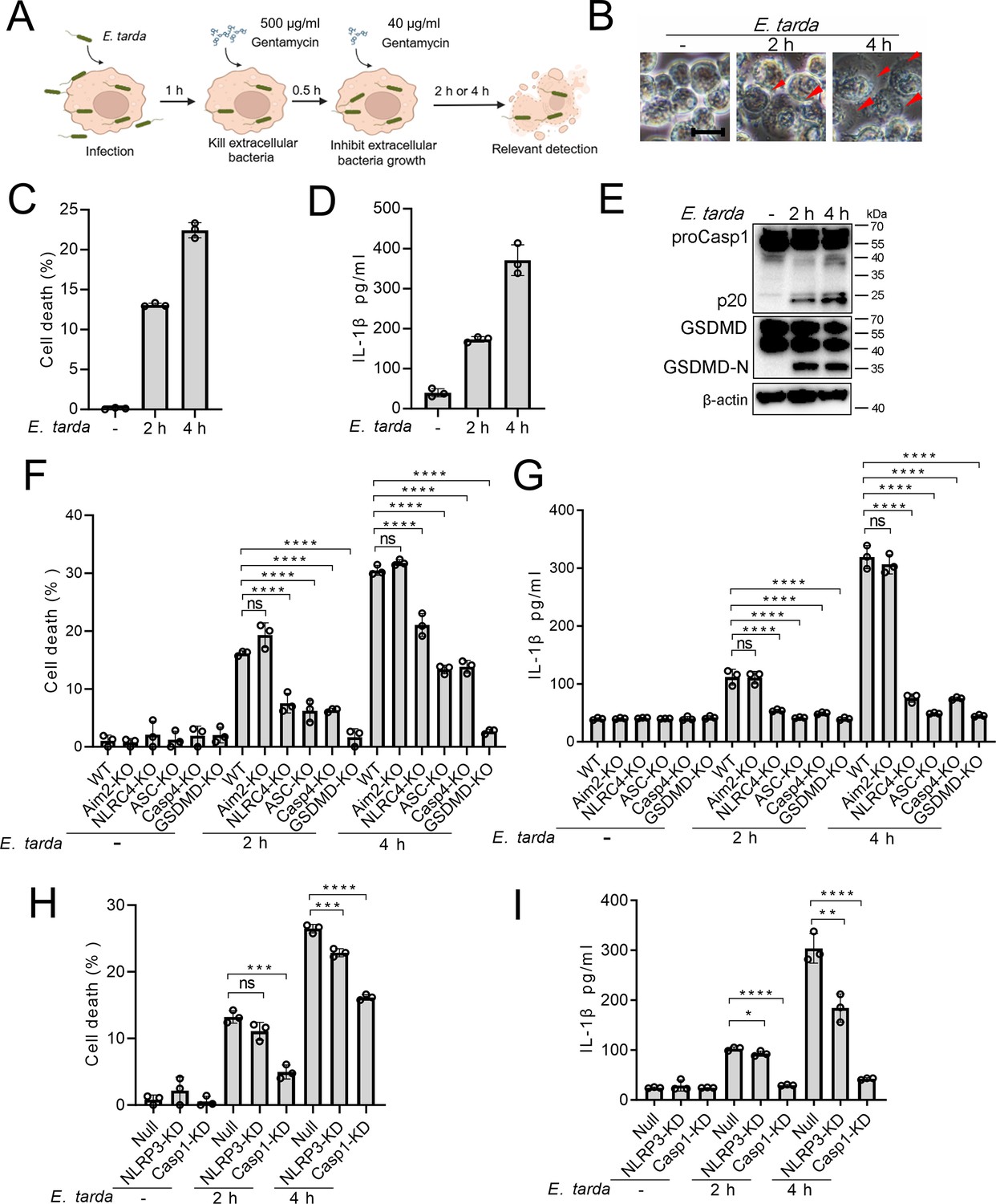
The ability of Edwardsiella tarda to induce pyroptosis in human macrophages.
(A) The schematic of experimental design. (B–E) Differentiated THP-1 (dTHP-1) cells were infected with E. tarda for the indicated hours and then subjected to microscopy (B), measurement of cell death (C), IL-1β release (D), and Western blot (E) using antibodies against Casp1, GSDMD, and β-actin (loading control). In (B), red arrowheads indicate pyroptotic cells; scale bar, 10 μm. (F–I) dTHP-cells in the form of wild-type (WT), knockout (KO) variants (Aim2-KO, NLRC4-KO, ASC-KO, Casp4-KO, and GSDMD-KO), and knockdown (KD) variants (NLRP3-KD and Casp1-KD) were infected with or without E. tarda for 2 or 4 hr, and then assessed for cell death (F, H) and IL-1β release (G, I). For panels C, D, and F-I, data were the means of triplicate assays and shown as means ± SD. ns, not significant, ***p<0.001, ****p<0.0001, one-way ANOVA with Dunnett’s multiple-comparison test.
-
Figure 1—source data 1
PDF file containing the original blots for Figure 1E.
- https://cdn.elifesciences.org/articles/100820/elife-100820-fig1-data1-v1.pdf
-
Figure 1—source data 2
Original files for blots are displayed in Figure 1E.
- https://cdn.elifesciences.org/articles/100820/elife-100820-fig1-data2-v1.zip
-
Figure 1—source data 3
The numerical source data corresponds to Figure 1.
- https://cdn.elifesciences.org/articles/100820/elife-100820-fig1-data3-v1.xlsx
Figure 1—figure supplement 1
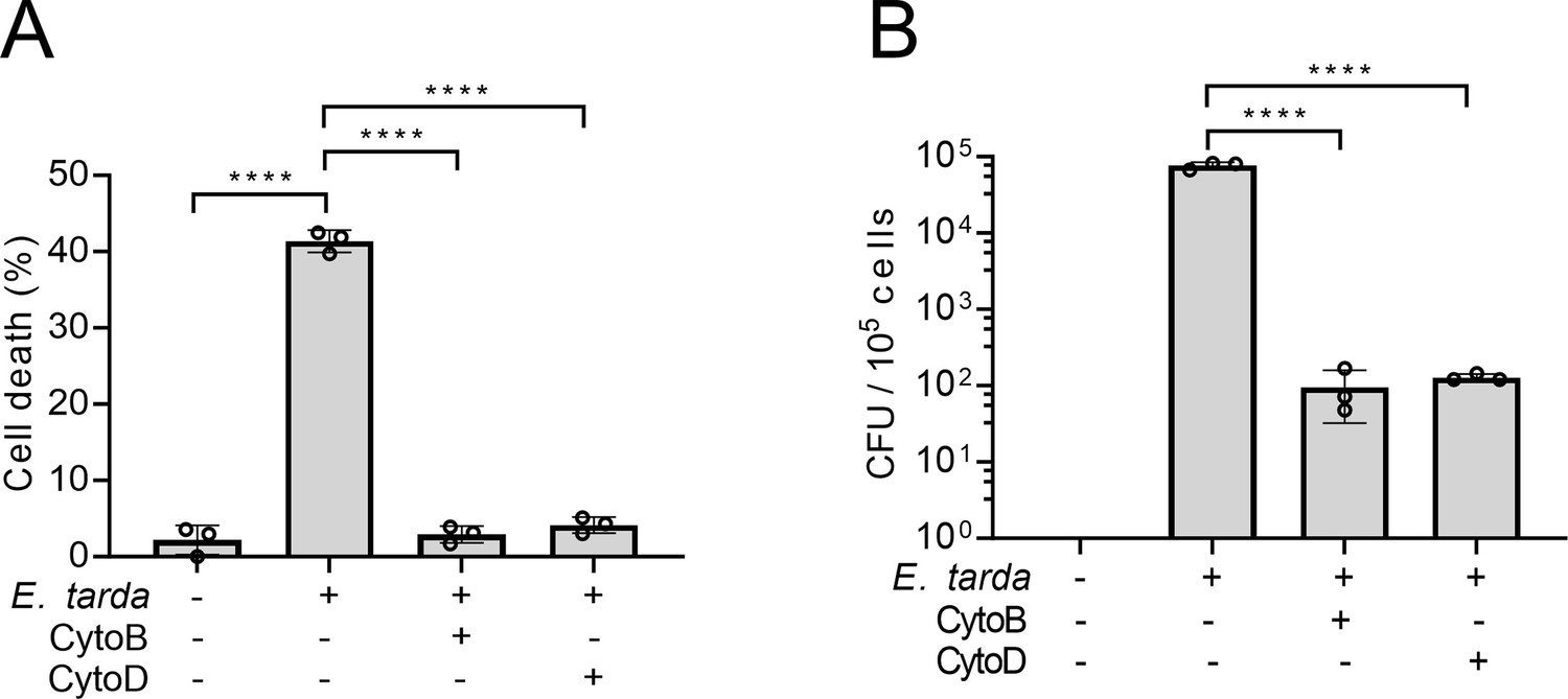
The effect of cytochalasin B (CytoB) and cytochalasin D (CytoD) on the ability of Edwardsiella tarda to induce cell death.
(A) Differentiated THP-1 (dTHP-1) cells pretreated with or without DMSO, CytoB, or CytoD were infected with or without E. tarda for 2 hr and then determined for cell death. (B) dTHP-1 cells pretreated with or without DMSO, CytoB, or CytoD were infected with or without E. tarda for 1 hr. The extracellular bacteria were killed by gentamycin. The intracellular bacteria were quantified by plate count. Data are the means of triplicate assays and are shown as means ± SD. ****p<0.0001, one-way ANOVA with Dunnett’s multiple-comparison test.
-
Figure 1—figure supplement 1—source data 1
The numerical source data corresponds to Figure 1—figure supplement 1.
- https://cdn.elifesciences.org/articles/100820/elife-100820-fig1-figsupp1-data1-v1.xlsx
Figure 2 with 2 supplements
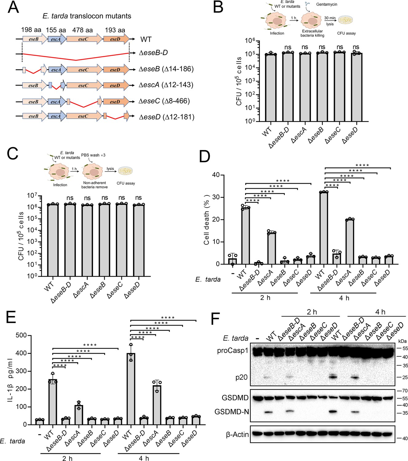
The importance of the translocon for Edwardsiella tarda-induced pyroptosis.
(A) A diagram showing the in-frame deletion (red curved line) of eseB-D, escA, eseB, eseC, and eseD. (B, C) Differentiated THP-1 (dTHP-1) cells were infected with wild-type (WT) or mutant E. tarda for 1 hr. The intracellular bacteria (B) and the total bacteria associated with the cells (i.e. both the cell-attached and the intracellular bacteria) (C) were determined by plate count. (D–F) dTHP-1 cells were treated with or without E. tarda variants for 2 or 4 hr, and then subjected to cell death analysis (D), IL-1β release measurement (E), and immunoblot (F) using antibodies against Casp1, GSDMD, and β-actin (loading control). For panels B-E, data are the means of triplicate assays and are shown as means ± SD. ns, not significant, ****p<0.0001, one-way ANOVA with Dunnett’s multiple-comparison test.
-
Figure 2—source data 1
PDF file containing the original blots for Figure 2F.
- https://cdn.elifesciences.org/articles/100820/elife-100820-fig2-data1-v1.pdf
-
Figure 2—source data 2
Original files for blots are displayed in Figure 2F.
- https://cdn.elifesciences.org/articles/100820/elife-100820-fig2-data2-v1.zip
-
Figure 2—source data 3
The numerical source data corresponds to Figure 2.
- https://cdn.elifesciences.org/articles/100820/elife-100820-fig2-data3-v1.xlsx
Figure 2—figure supplement 1
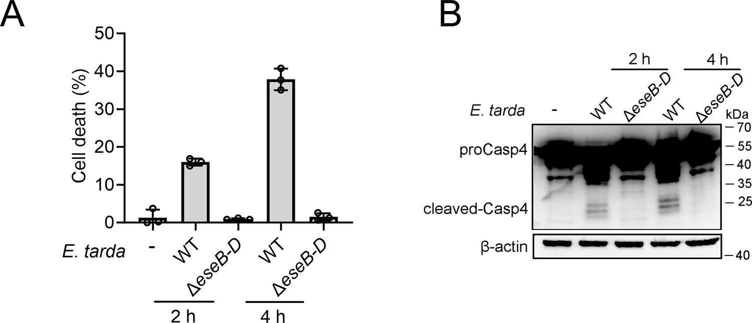
The effect of ΔeseB-D on Casp4 activation.
(A) Differentiated THP-1 (dTHP-1) cells were treated with wild-type (WT) Edwardsiella tarda or the ΔeseB-D mutant for 2 hr or 4 hr. The cells were subjected to cell death analysis (A) and immunoblot with antibodies against Casp4 and β-actin (B). Data in panel A are the means of triplicate assays and are shown as means ± SD.
-
Figure 2—figure supplement 1—source data 1
PDF file containing the original blots for Figure 2—figure supplement 1.
- https://cdn.elifesciences.org/articles/100820/elife-100820-fig2-figsupp1-data1-v1.pdf
-
Figure 2—figure supplement 1—source data 2
Original files for blots are displayed in Figure 2—figure supplement 1.
- https://cdn.elifesciences.org/articles/100820/elife-100820-fig2-figsupp1-data2-v1.zip
-
Figure 2—figure supplement 1—source data 3
The numerical source data corresponds to Figure 2—figure supplement 1.
- https://cdn.elifesciences.org/articles/100820/elife-100820-fig2-figsupp1-data3-v1.xlsx
Figure 2—figure supplement 2
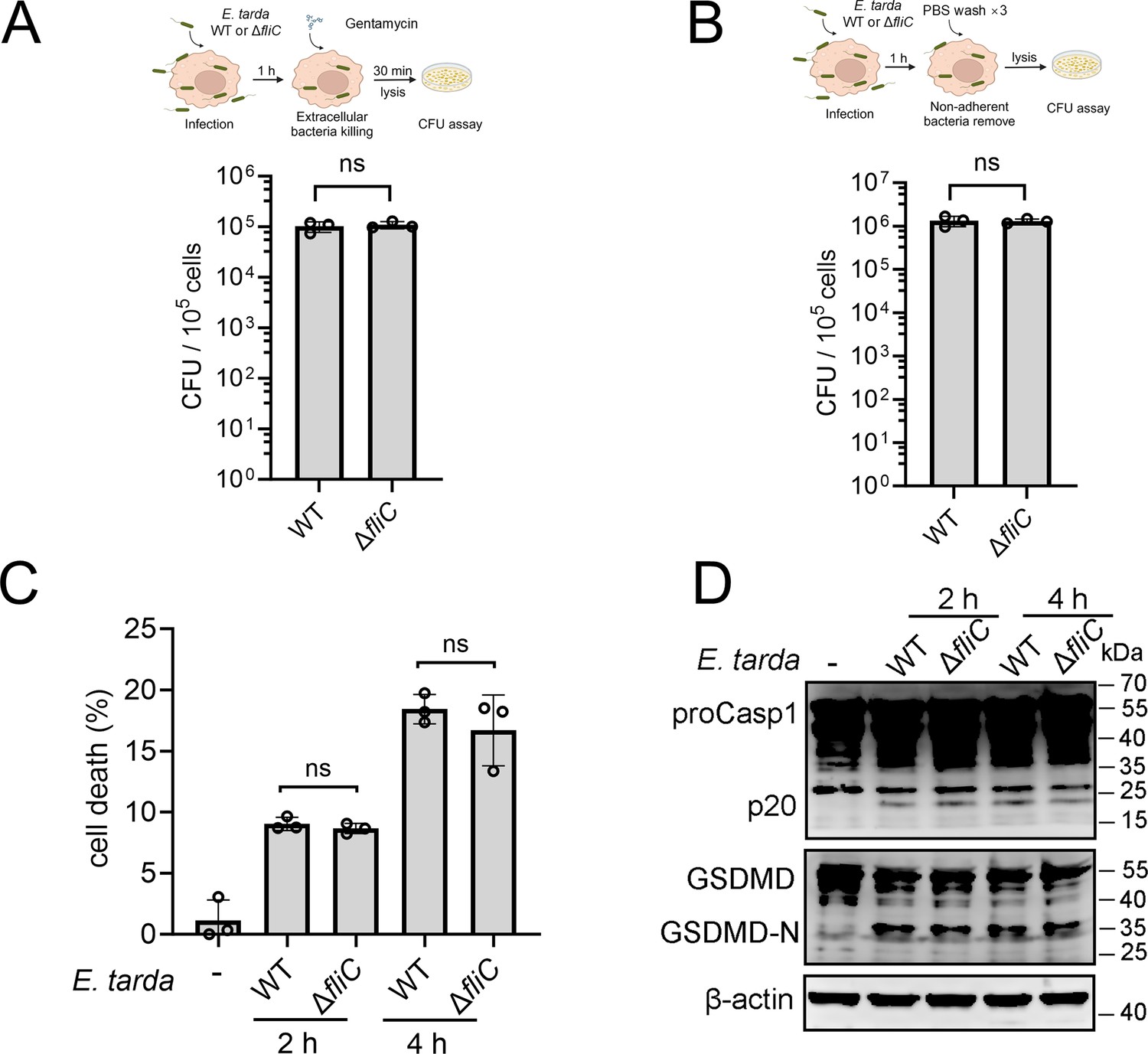
The involvement of flagellin in Edwardsiella tarda-induced pyroptosis of human macrophages.
(A, B) Differentiated THP-1 (dTHP-1) cells were infected with E. tarda wild-type (WT) or ΔfliC for 1 hr. The intracellular bacteria (A) and the total bacteria associated with the cells (B) were determined by plate count. (C, D) dTHP-1 cells were treated with or without E. tarda WT or ΔfliC for 2 or 4 hr, and then subjected to cell death analysis (C) and immunoblotting with antibodies against Casp1, GSDMD, and β-actin (D). Data in panels A-C are the means of triplicate assays and are shown as means ± SD. ns, not significant; p>0.05, Student’s t-test.
-
Figure 2—figure supplement 2—source data 1
PDF file containing the original blots for Figure 2—figure supplement 2.
- https://cdn.elifesciences.org/articles/100820/elife-100820-fig2-figsupp2-data1-v1.pdf
-
Figure 2—figure supplement 2—source data 2
Original files for blots are displayed in Figure 2—figure supplement 2.
- https://cdn.elifesciences.org/articles/100820/elife-100820-fig2-figsupp2-data2-v1.zip
-
Figure 2—figure supplement 2—source data 3
The numerical source data corresponds to Figure 2—figure supplement 2.
- https://cdn.elifesciences.org/articles/100820/elife-100820-fig2-figsupp2-data3-v1.xlsx
Figure 3 with 1 supplement
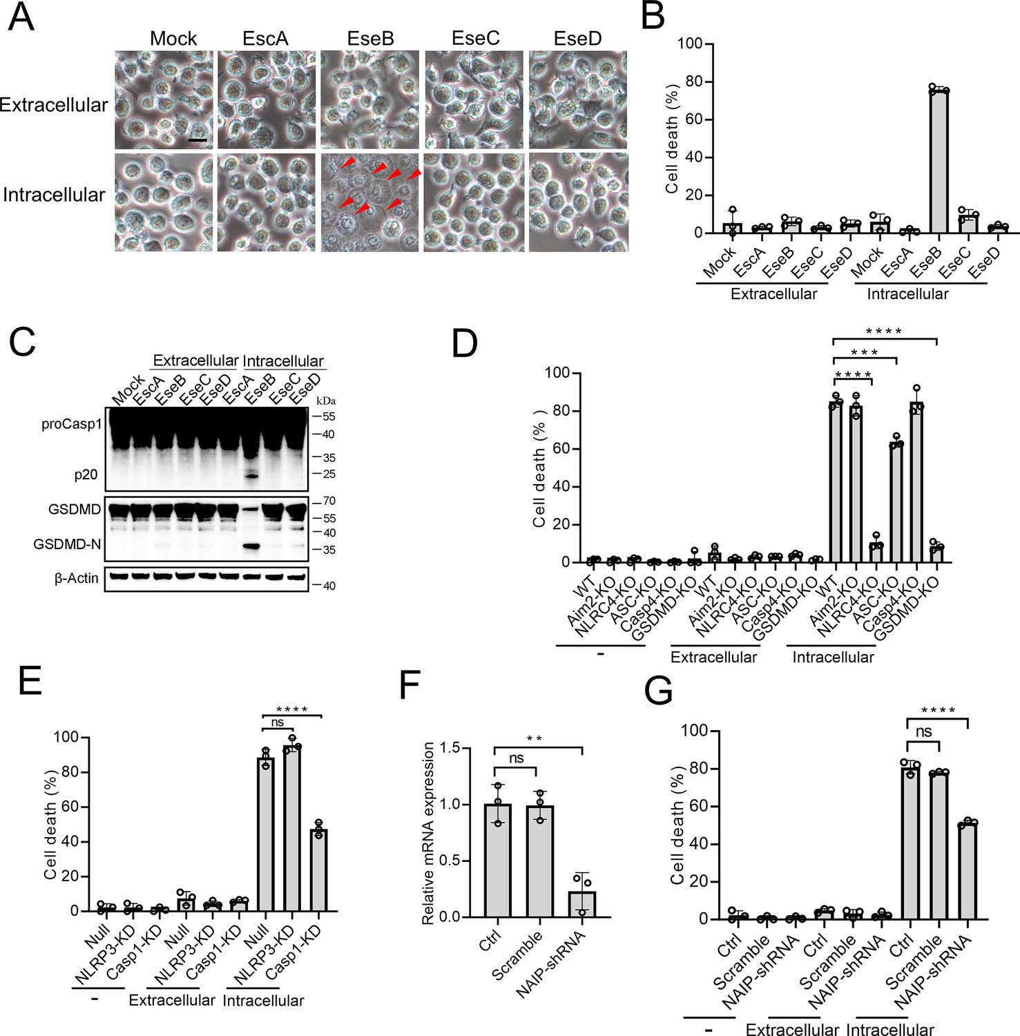
The pyroptotic effect of the translocon proteins and their dependence on the inflammasomes.
(A–C) To determine the extracellular and intracellular effects of EscA, EseB, EseC, and EseD, each of the proteins was added into the culture medium of THP-1 cells (extracellular) or electroporated into THP-1 cells (intracellular). The control cells were mock-treated with PBS. The cells were subjected to microscopy (A), cell death analysis (B), and immunoblot using antibodies against Casp1, GSDMD, and β-actin (loading control) (C). In (A), red arrowheads indicate pyroptotic cells; scale bar, 10 μm. (D) The wild-type (WT) and knockout (KO) THP-1 cells were treated with or without extracellular and intracellular EseB as above and then examined for cell death. (E) The control THP-cells (Null) and the NLRP3/Casp1 knockdown (KD) THP-cells were treated with or without extracellular and intracellular EseB as above and then examined for cells death. (F) THP-1 cell treated with or without (Control, Ctrl) NLR-family apoptosis inhibitory protein (NAIP)-targeting shRNA or scramble RNA (negative control RNA) were examined for NAIP expression by qRT-PCR. (G) THP-1 cells administered with or without NAIP-targeting or scramble RNA were treated or without extracellular and intracellular EseB as above and then examined for cell death. For panels B, and D-F, data are the means of triplicate assays and are shown as means ± SD. ns, not significant, ***p<0.001, ****p<0.0001, one-way ANOVA with Dunnett’s multiple-comparison test.
-
Figure 3—source data 1
PDF file containing the original blots for Figure 3C.
- https://cdn.elifesciences.org/articles/100820/elife-100820-fig3-data1-v1.pdf
-
Figure 3—source data 2
Original files for blots are displayed in Figure 3C.
- https://cdn.elifesciences.org/articles/100820/elife-100820-fig3-data2-v1.zip
-
Figure 3—source data 3
The numerical source data corresponds to Figure 3.
- https://cdn.elifesciences.org/articles/100820/elife-100820-fig3-data3-v1.xlsx
Figure 3—figure supplement 1
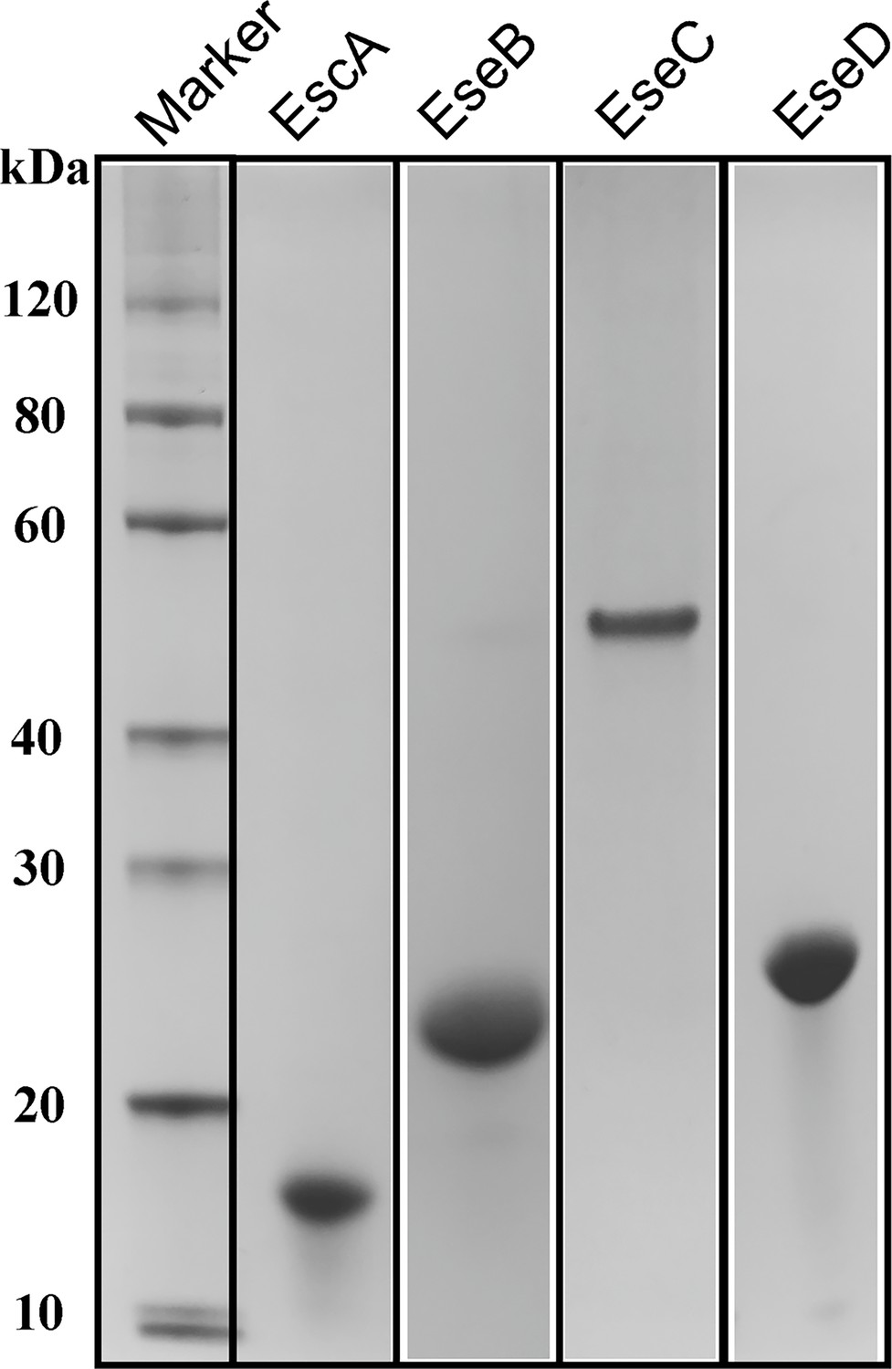
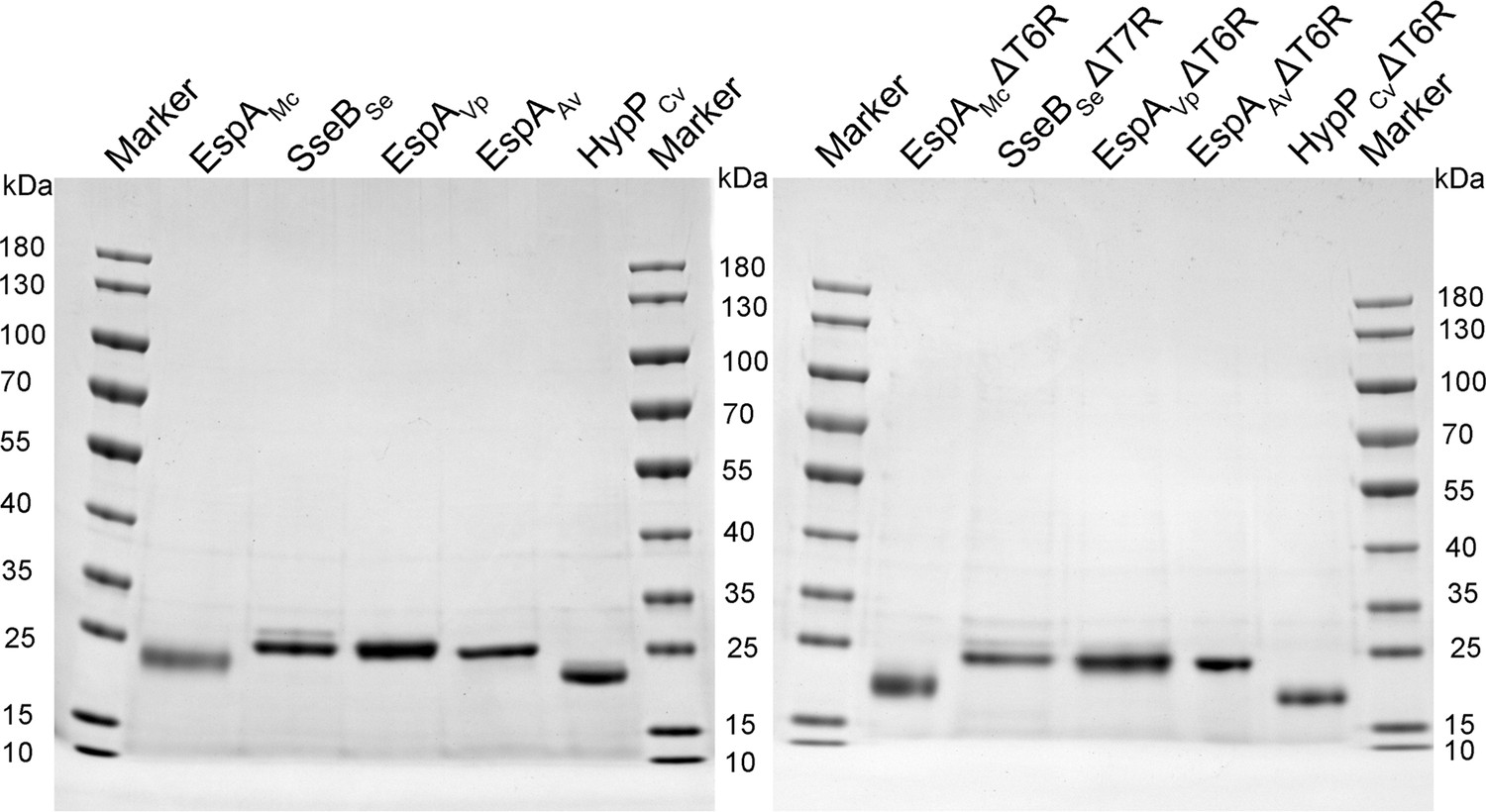
SDS-PAGE analysis of purified recombinant proteins.
The purified recombinant translocon proteins of EscA, EseB, EseC, and EseD were subjected to SDS-PAGE and stained with Coomassie brilliant blue R-250.
-
Figure 3—figure supplement 1—source data 1
PDF file containing the original gels for Figure 3—figure supplement 1.
- https://cdn.elifesciences.org/articles/100820/elife-100820-fig3-figsupp1-data1-v1.pdf
-
Figure 3—figure supplement 1—source data 2
Original files for gels are displayed in Figure 3—figure supplement 1.
- https://cdn.elifesciences.org/articles/100820/elife-100820-fig3-figsupp1-data2-v1.zip
Figure 4 with 2 supplements
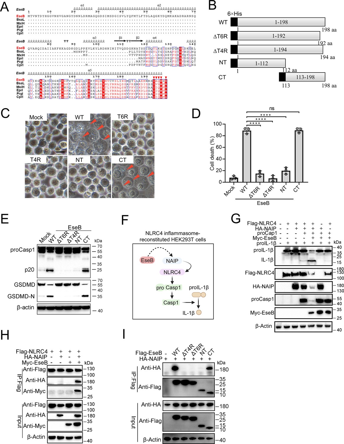
Identification of the functional important region in EseB.
(A) Sequence alignment of EseB and T3SS needle proteins with NLRC4/NAIP-stimulating activity. (B) A diagram showing EseB wild-type (WT) and truncates. (C–E) THP-1 cells were electroporated with or without (Mock) EseB WT or truncated. The cells were subjected to microscopy (C), cell death analysis (D), and immunoblot with antibodies against Casp1, GSDMD, and β-actin (loading control) (E). In (C), red arrowheads indicate pyroptotic cells; scale bar, 10 μm. For panel D, data are the means of triplicate assays and are shown as means ± SD. ns, not significant, ****p<0.0001, one-way ANOVA with Dunnett’s multiple-comparison test. (F) A diagram showing the detection of the activating effect of EseB on NAIP/NLRC4 in NLRC4 inflammasome-reconstituted HEK293T cells by determining proIL-1β cleavage. (G) HEK293T cells were transfected with or without the indicated combination of vectors expressing Flag-tagged NLRC4, HA-tagged NLR-family apoptosis inhibitory protein (NAIP), Myc-tagged EseB, proCasp1, and proIL-1β for 24 hr. The cells were subjected to immunoblot using antibodies against the tags or the proteins with β-actin as a loading control. (H) HEK293T cells were transfected with the indicated combination of vectors expressing Flag-tagged NLRC4, HA-tagged NAIP, and Myc-tagged EseB. The cells were subjected to immunoprecipitation (IP) using antibodies against the tags with β-actin as a loading control. (I) HEK293T cells were transfected with the indicated combination of vectors expressing HA-tagged NAIP and Flag-tagged EseB variants. IP was performed as above.
-
Figure 4—source data 1
PDF file containing the original blots for Figure 4E.
- https://cdn.elifesciences.org/articles/100820/elife-100820-fig4-data1-v1.pdf
-
Figure 4—source data 2
Original files for blots are displayed in Figure 4E.
- https://cdn.elifesciences.org/articles/100820/elife-100820-fig4-data2-v1.zip
-
Figure 4—source data 3
PDF file containing the original blots for Figure 4G.
- https://cdn.elifesciences.org/articles/100820/elife-100820-fig4-data3-v1.pdf
-
Figure 4—source data 4
Original files for blots are displayed in Figure 4G.
- https://cdn.elifesciences.org/articles/100820/elife-100820-fig4-data4-v1.zip
-
Figure 4—source data 5
PDF file containing the original blots for Figure 4H.
- https://cdn.elifesciences.org/articles/100820/elife-100820-fig4-data5-v1.pdf
-
Figure 4—source data 6
Original files for blots are displayed in Figure 4H.
- https://cdn.elifesciences.org/articles/100820/elife-100820-fig4-data6-v1.zip
-
Figure 4—source data 7
PDF file containing the original blots for Figure 4I.
- https://cdn.elifesciences.org/articles/100820/elife-100820-fig4-data7-v1.pdf
-
Figure 4—source data 8
Original files for blots are displayed in Figure 4I.
- https://cdn.elifesciences.org/articles/100820/elife-100820-fig4-data8-v1.zip
-
Figure 4—source data 9
The numerical source data corresponds to Figure 4.
- https://cdn.elifesciences.org/articles/100820/elife-100820-fig4-data9-v1.xlsx
Figure 4—figure supplement 1
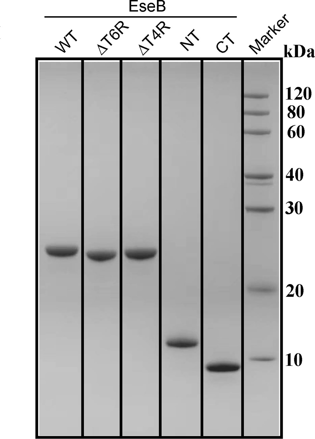
SDS-PAGE analysis of purified recombinant proteins.
The purified recombinant proteins of EseB truncates were subjected to SDS-PAGE and stained with Coomassie brilliant blue R-250. WT, wild-type.
-
Figure 4—figure supplement 1—source data 1
PDF file containing the original gel for Figure 4—figure supplement 1.
- https://cdn.elifesciences.org/articles/100820/elife-100820-fig4-figsupp1-data1-v1.pdf
-
Figure 4—figure supplement 1—source data 2
Original file for gel is displayed in Figure 4—figure supplement 1.
- https://cdn.elifesciences.org/articles/100820/elife-100820-fig4-figsupp1-data2-v1.zip
Figure 4—figure supplement 2
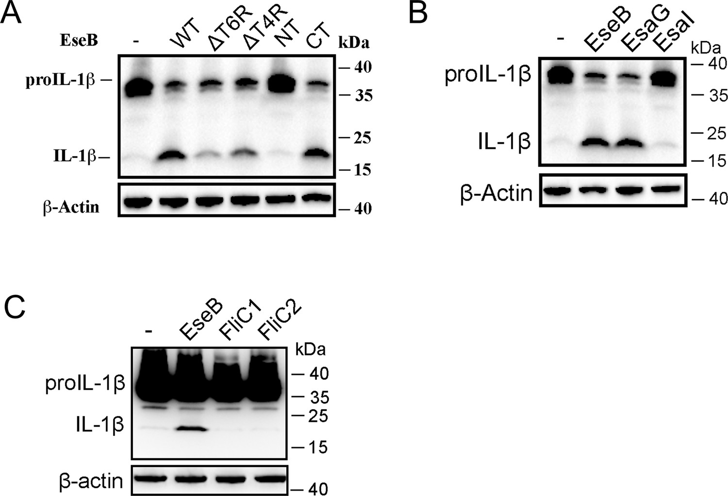
The ability of Edwardsiella tarda EseB, rod (EsaI), needle (EsaG), and flagellin (FliC1/2) to activate NLRC4.
NLRC4 inflammasome-reconstituted HEK293T cells expressing or not expressing wild type (WT) or mutant EseB (A), EsaI/EsaG (B), or FliC1/2 (C) for 24 h were immunoblotted with antibodies against IL-1β and β-actin (loading control).
-
Figure 4—figure supplement 2—source data 1
PDF file containing the original blots for Figure 4—figure supplement 2A.
- https://cdn.elifesciences.org/articles/100820/elife-100820-fig4-figsupp2-data1-v1.pdf
-
Figure 4—figure supplement 2—source data 2
Original files for blots are displayed in Figure 4—figure supplement 2A.
- https://cdn.elifesciences.org/articles/100820/elife-100820-fig4-figsupp2-data2-v1.zip
-
Figure 4—figure supplement 2—source data 3
PDF file containing the original blots for Figure 4—figure supplement 2B.
- https://cdn.elifesciences.org/articles/100820/elife-100820-fig4-figsupp2-data3-v1.pdf
-
Figure 4—figure supplement 2—source data 4
Original files for blots are displayed in Figure 4—figure supplement 2B.
- https://cdn.elifesciences.org/articles/100820/elife-100820-fig4-figsupp2-data4-v1.zip
-
Figure 4—figure supplement 2—source data 5
PDF file containing the original blots for Figure 4—figure supplement 2C.
- https://cdn.elifesciences.org/articles/100820/elife-100820-fig4-figsupp2-data5-v1.pdf
-
Figure 4—figure supplement 2—source data 6
Original files for blots are displayed in Figure 4—figure supplement 2C.
- https://cdn.elifesciences.org/articles/100820/elife-100820-fig4-figsupp2-data6-v1.zip
Figure 5 with 3 supplements
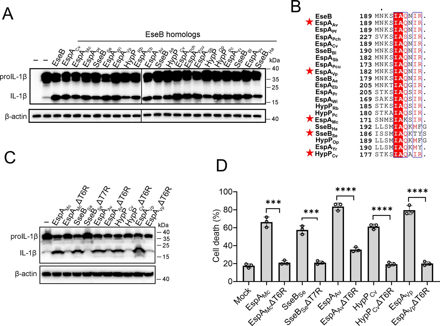
The ability of the EseB homologs to activate the NLRC4/NLR-family apoptosis inhibitory protein (NAIP) inflammasome.
(A) NLRC4 inflammasome-reconstituted HEK293T cells were transfected with or without EseB homologs (Supplementary file 2). The cells were immunoblotted with antibodies against IL-1β and β-actin (loading control). (B) Sequence alignment of the C-terminal regions of the EseB homologs. Red stars indicate the EseB selected for mutation analysis. (C) NLRC4 inflammasome-reconstituted HEK293T cells expressing or not expressing the indicated EseB homologs or their mutants were immunoblotted as panel A. (D) THP-1 cells were electroporated with the indicated EseB homologs or their mutants or PBS (mock) and then determined for cell death. Data are the means of triplicate assays and are shown as means ± SD. ns, not significant, ***p<0.001, ****p<0.0001, Student’s t-test.
-
Figure 5—source data 1
PDF file containing the original blots for Figure 5A.
- https://cdn.elifesciences.org/articles/100820/elife-100820-fig5-data1-v1.pdf
-
Figure 5—source data 2
Original files for blots displayed in Figure 5A.
- https://cdn.elifesciences.org/articles/100820/elife-100820-fig5-data2-v1.zip
-
Figure 5—source data 3
PDF file containing the original blots for Figure 5C.
- https://cdn.elifesciences.org/articles/100820/elife-100820-fig5-data3-v1.pdf
-
Figure 5—source data 4
Original files for blots are displayed in Figure 5C.
- https://cdn.elifesciences.org/articles/100820/elife-100820-fig5-data4-v1.zip
-
Figure 5—source data 5
The numerical source data corresponds to Figure 5.
- https://cdn.elifesciences.org/articles/100820/elife-100820-fig5-data5-v1.xlsx
Figure 5—figure supplement 1
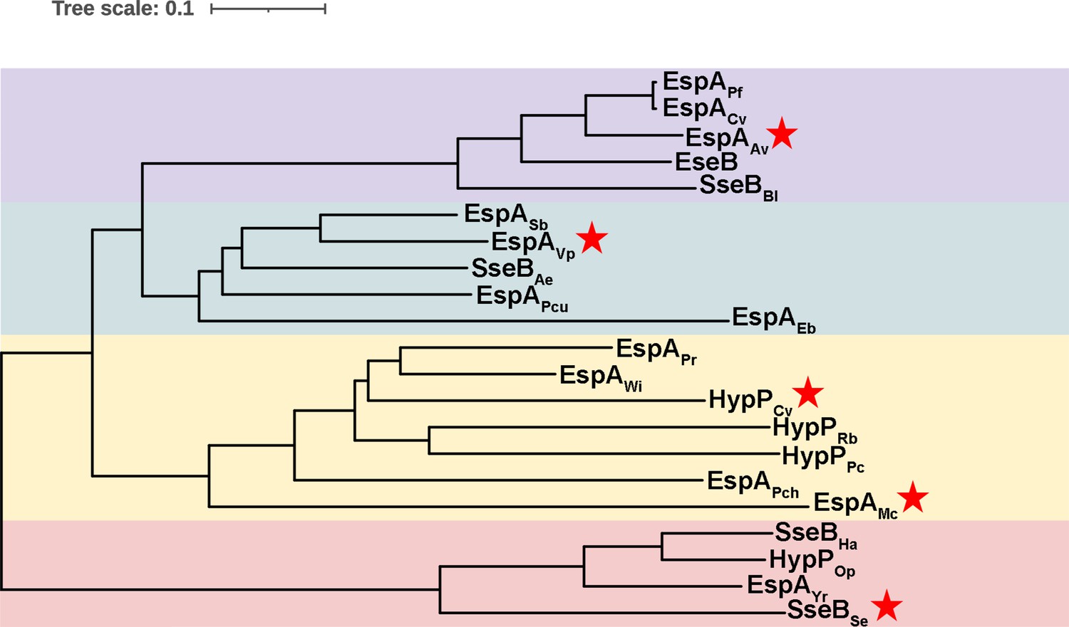
Phylogenetic analysis of EseB homologs.
The phylogenetic tree was generated using ClustalW alignment and the Neighbor-Joining method. Red pentagrams indicate the bacteria whose EseB homologs were selected for mutation analysis.
Figure 5—figure supplement 2
SDS-PAGE analysis of purified recombinant proteins.
Purified recombinant EseB homologues (left) and their mutants (right) were subjected to SDS-PAGE and stained with Coomassie brilliant blue R-250. The full names of the proteins are shown in Supplementary file 2.
-
Figure 5—figure supplement 2—source data 1
PDF file containing the original gels for Figure 5—figure supplement 2.
- https://cdn.elifesciences.org/articles/100820/elife-100820-fig5-figsupp2-data1-v1.pdf
-
Figure 5—figure supplement 2—source data 2
Original files for gels are displayed in Figure 5—figure supplement 2.
- https://cdn.elifesciences.org/articles/100820/elife-100820-fig5-figsupp2-data2-v1.zip
Figure 5—figure supplement 3
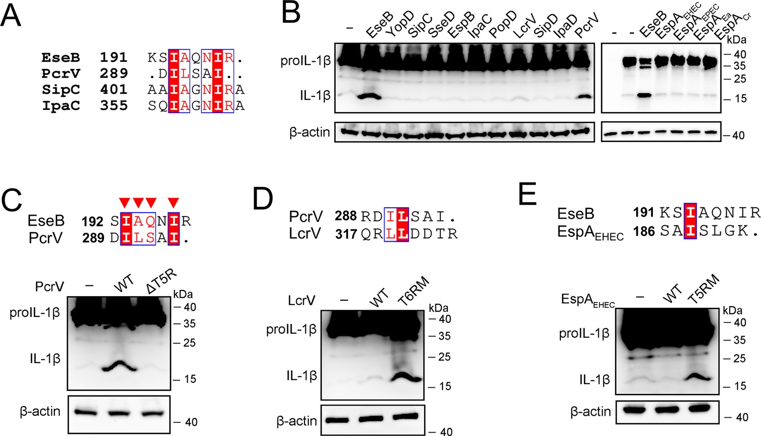
The effects of translocator proteins on NLRC4/NLR-family apoptosis inhibitory protein (NAIP) inflammasome activation.
(A) Sequence alignment of the C-terminal regions of EseB, PcrV, SipC, and IpaC. (B) NLRC4 inflammasome-reconstituted HEK293T cells expressing or not expression (-) the indicated proteins for 24 hr were immunoblotted with antibodies against IL-1β and β-actin (loading control). (C–E) NLRC4 inflammasome-reconstituted HEK293T cells expressing or not expression (-) wild-type (WT) and mutant PcrV (C), LcrV (D), and EspAEHEC (E) were immunoblotted as above. The C-terminal regions of PcrV, LcrV, and EspAEHEC were aligned on top of the panels.
-
Figure 5—figure supplement 3—source data 1
PDF file containing the original blots for Figure 5—figure supplement 3B.
- https://cdn.elifesciences.org/articles/100820/elife-100820-fig5-figsupp3-data1-v1.pdf
-
Figure 5—figure supplement 3—source data 2
Original files for blots are displayed in Figure 5—figure supplement 3B.
- https://cdn.elifesciences.org/articles/100820/elife-100820-fig5-figsupp3-data2-v1.zip
-
Figure 5—figure supplement 3—source data 3
PDF file containing the original blots for Figure 5—figure supplement 3C.
- https://cdn.elifesciences.org/articles/100820/elife-100820-fig5-figsupp3-data3-v1.pdf
-
Figure 5—figure supplement 3—source data 4
Original files for blots are displayed in Figure 5—figure supplement 3C.
- https://cdn.elifesciences.org/articles/100820/elife-100820-fig5-figsupp3-data4-v1.zip
-
Figure 5—figure supplement 3—source data 5
PDF file containing the original blots for Figure 5—figure supplement 3D.
- https://cdn.elifesciences.org/articles/100820/elife-100820-fig5-figsupp3-data5-v1.pdf
-
Figure 5—figure supplement 3—source data 6
Original files for blots are displayed in Figure 5—figure supplement 3D.
- https://cdn.elifesciences.org/articles/100820/elife-100820-fig5-figsupp3-data6-v1.zip
-
Figure 5—figure supplement 3—source data 7
PDF file containing the original blots for Figure 5—figure supplement 3E.
- https://cdn.elifesciences.org/articles/100820/elife-100820-fig5-figsupp3-data7-v1.pdf
-
Figure 5—figure supplement 3—source data 8
Original files for blots are displayed in Figure 5—figure supplement 3E.
- https://cdn.elifesciences.org/articles/100820/elife-100820-fig5-figsupp3-data8-v1.zip
Tables
Key resources table
| Reagent type (species) or resource | Designation | Source or reference | Identifiers | Additional information |
|---|---|---|---|---|
| Gene (Edwardsiella tarda and others) | Translocon genes | GenBank, UniProtKB | Supplementary files 1 and 2 | |
| Strain, strain background (E. tarda) | E. tarda | Li et al., 2022 | ||
| Strain, strain background (E. tarda) | ΔeseB | This paper | In-frame deletion of eseB | |
| Strain, strain background (E. tarda) | ΔescA | This paper | In-frame deletion of escA | |
| Strain, strain background (E. tarda) | ΔeseC | This paper | In-frame deletion of eseC | |
| Strain, strain background (E. tarda) | ΔeseD | This paper | In-frame deletion of eseD | |
| Strain, strain background (E. tarda) | ΔescB-D | This paper | In-frame deletion of eseB-eseD | |
| Strain, strain background (E. tarda) | ΔfliC | This paper | In-frame deletion of fliC1/2 | |
| Strain, strain background (E. coli) | BL21(DE3) | TransGen Biotech | CD601 | |
| Cell line (Homo sapiens) | HEK293T | ATCC | Cat# CRL-3216, RRID:CVCL_0063 | |
| Cell line (H. sapiens) | THP-1 | Cell Resource Center, IBMS, CAMS/PUMC | 1101HUM-PUMC000057, RRID:CVCL_0006 | |
| Cell line (H. sapiens) | THP1-Null | InvivoGen | thp-null | Control cells |
| Cell line (H. sapiens) | THP1-Casp1-KD | InvivoGen | thp-dcasp1 | Casp1 knockdown |
| Cell line (H. sapiens) | THP1-NLRP3-KD | InvivoGen | thp-dnlp | Nlrp3 knockdown |
| Cell line (H. sapiens) | THP1-NLRC4-KO | This paper | Nlrc4 knockout | |
| Cell line (H. sapiens) | THP1-Casp4-KO | This paper | Casp4 knockout | |
| Cell line (H. sapiens) | THP1-Aim2-KO | This paper | Aim2 knockout | |
| Cell line (H. sapiens) | THP1-ASC -KO | Wang et al., 2024 | ASC knockout | |
| Cell line (H. sapiens) | THP1-GSDMD-KO | Zhao et al., 2021 | Gsdmd knockout | |
| Transfected construct (H. sapiens) | shRNA-NAIP | This paper | Lentiviral construct for NAIP knockdown | |
| Transfected construct (H. sapiens) | sgRNA-Aim2, Casp4, NLRC4 | This paper | Lentiviral construct for Aim2, Casp4, NLRC4 gene knockout | |
| Antibody | anti-Caspase-1 (Rabbit polyclonal) | Cell Signaling Technology | Cat# 2225, RRID:AB_2243894 | WB (1:1000) |
| Antibody | anti-GSDMD (Rabbit polyclonal) | Cell Signaling Technology | Cat# 96458, RRID:AB_2894914 | WB (1:1000) |
| Antibody | anti- IL-1β (Rabbit monoclonal) | Cell Signaling Technology | Cat# 12703, RRID:AB_2737350 | WB (1:1000) |
| Antibody | anti-Caspase-4 (Rabbit monoclonal) | Cell Signaling Technology | 42264T | WB (1:1000) |
| Antibody | anti-6×His tag mAb (Rabbit monoclonal) | Abcam | ab213204 | WB (1:1000) |
| Antibody | anti-flag-tag (Rabbit monoclonal) | ABclonal | Cat# AE063, RRID:AB_2771920 | WB (1:1000) |
| Antibody | anti-HA-Tag (Mouse monoclonal) | ABclonal | Cat# AE008, RRID:AB_2770404 | WB (1:1000) |
| Antibody | anti-Myc-Tag (Mouse monoclonal) | ABclonal | Cat# AE010, RRID:AB_2770408 | WB (1:1000) |
| Antibody | anti-β-actin (Mouse monoclonal) | ABclonal | Cat# AC004, RRID:AB_2737399 | WB (1:1000) |
| Antibody | HRP goat anti-mouse IgG | ABclonal | Cat# AS003, RRID:AB_2769851 | WB (1:1000) |
| Antibody | HRP goat anti-rabbit IgG | Abcam | Cat# ab97051, RRID:AB_10679369 | WB (1:1000) |
| Recombinant DNA reagent | pLKO.1 puro | Addgene | 8453, RRID:Addgene_8453 | Negative control lentiviral construct |
| Recombinant DNA reagent | pDM4 | This paper | The suicide plasmid | |
| Recombinant DNA reagent | pET-28a | Novagen | 69864 | |
| Recombinant DNA reagent | pCS2Flag | Addgene | 16331, RRID:Addgene_16331 | |
| Sequence-based reagent | PCR primers | This paper | Supplementary file 1 | |
| Commercial assay or kit | CytoTox 96 Non-Radioactive Cytotoxicity Assay kit | Promega | G1780 | |
| Commercial assay or kit | Human IL-1β ELISA kit | NeoBioscience | EHC002B | |
| Chemical compound, drug | cytochalasin B | Abcam | ab143482 | |
| Chemical compound, drug | cytochalasin D | Invitrogen | PHZ1063 | |
| Software, algorithm | Prism 10 | GraphPad | RRID:SCR_002798 | https://www.graphpad.com/ |
Additional files
-
Supplementary file 1
The primers used for the construction of Edwardsiella tarda translocon mutants.
- https://cdn.elifesciences.org/articles/100820/elife-100820-supp1-v1.docx
-
Supplementary file 2
Translocator proteins used in this study.
- https://cdn.elifesciences.org/articles/100820/elife-100820-supp2-v1.docx
-
MDAR checklist
- https://cdn.elifesciences.org/articles/100820/elife-100820-mdarchecklist1-v1.docx
Download links
A two-part list of links to download the article, or parts of the article, in various formats.
Downloads (link to download the article as PDF)
Open citations (links to open the citations from this article in various online reference manager services)
Cite this article (links to download the citations from this article in formats compatible with various reference manager tools)
T3SS translocon induces pyroptosis by direct interaction with NLRC4/NAIP inflammasome
eLife 13:RP100820.
https://doi.org/10.7554/eLife.100820.3


