Microglia are required for developmental specification of AgRP innervation in the hypothalamus of offspring exposed to maternal high-fat diet during lactation
- Department of Molecular Physiology & Biophysics, Vanderbilt University, United States
Figures
Figure 1
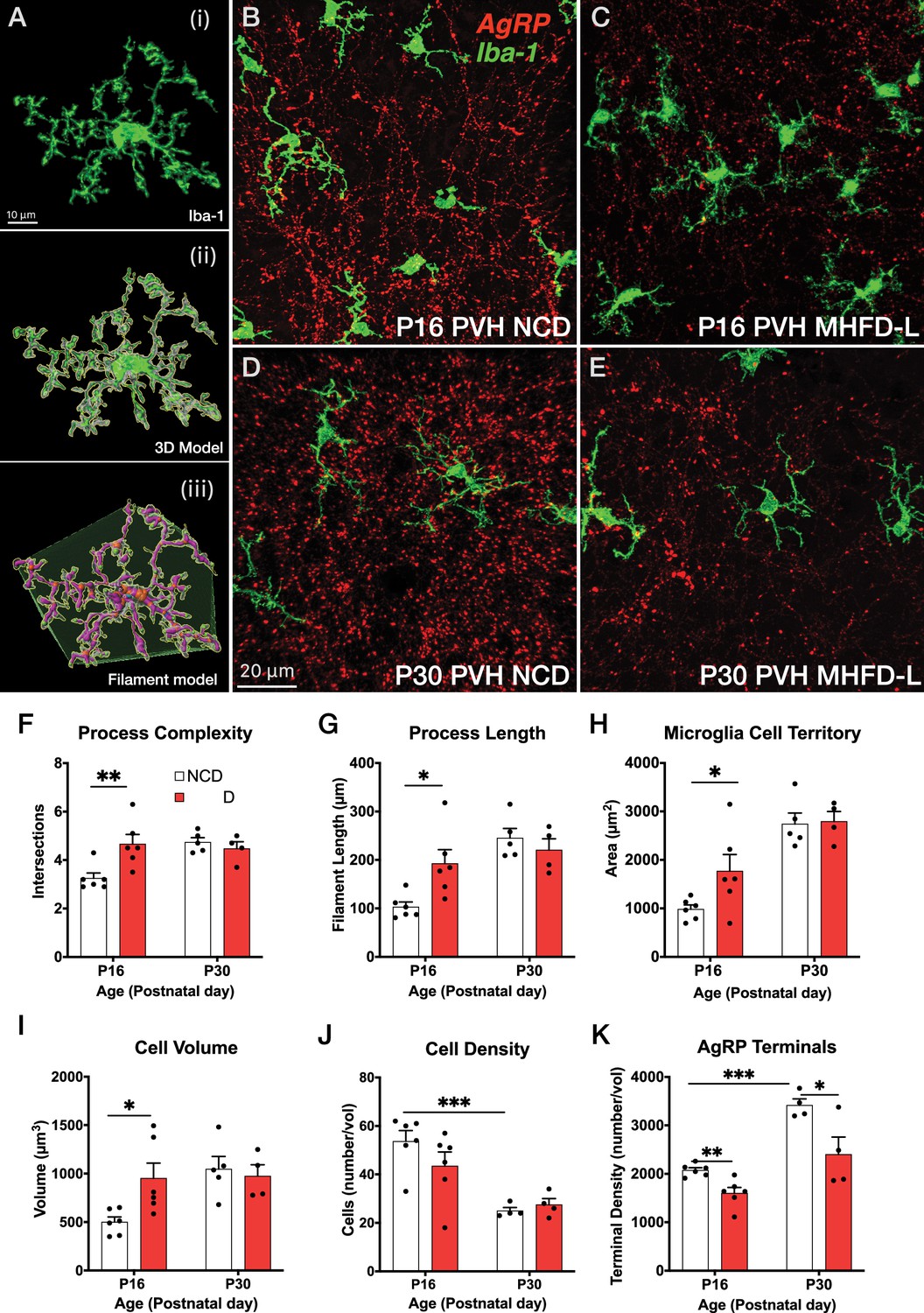
MHFD-L: Microglial morphology in the PVH.
(A) Image analysis pipeline. Fluorescence image of an Iba1-immunostained microglial cell in the PVH (Ai). Confocal images through labeled cells were used to generate 3D reconstructions (Aii), which were then used to create 3D models of microglial cells by using the Filaments tool in Imaris. Polyhedrons were generated around each cell using the Convex Hull function of Imaris to estimate the total tissue ‘territory’ occupied by the microglial cell (Aiii). (B–E) Images of microglial cells (green) and labeled AgRP terminals (red) in the PVH of mice at P16 (B, C) or P30 (D, E) that were raised on NCD (B, D) or MHFD-L (C, E). Graphical comparisons between groups to show that MHFD-L increased microglial ramification complexity (F), process length (G), microglial cell territory (H), and cell volume (I) at P16. The density of microglia in the PVH decreased between P16 and P30, irrespective of diet (J). Density of Agouti-related peptide (AgRP) terminals were decreased in MHFD-L offspring at both P16 and P30 (K). Bars represent the mean ± SEM and each point represents one animal. *p<0.05, **p<0.005. Abbreviations: MHFD-L, maternal HFD during lactation; NCD, normal chow diet; PVH, paraventricular nucleus of the hypothalamus.
Figure 2
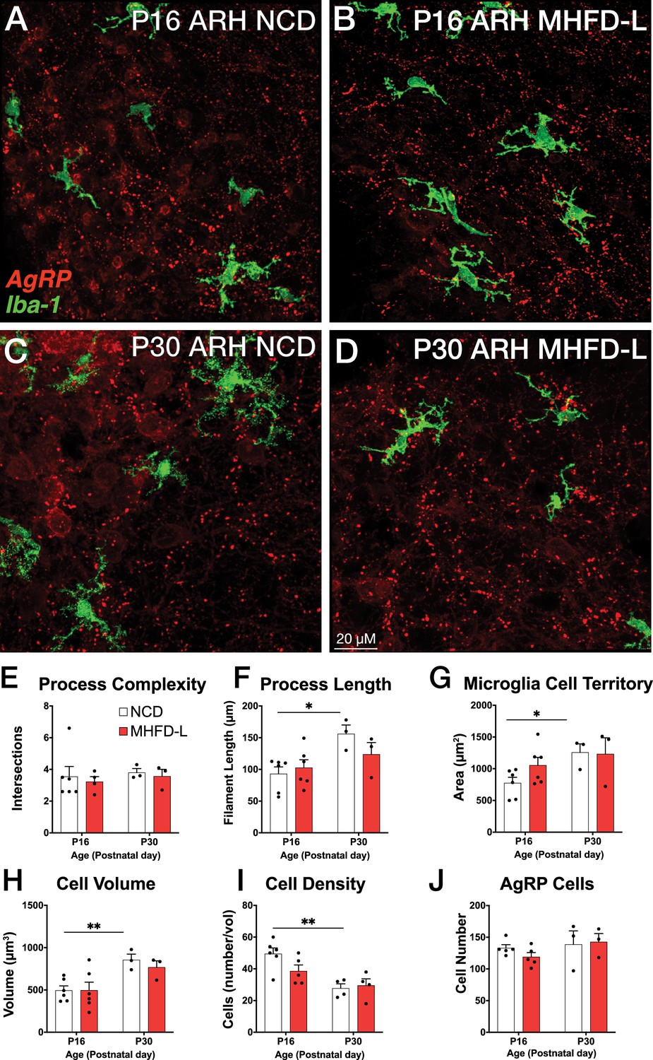
MHFD-L: Microglial morphology in the ARH.
Microglial cells (green) and labeled Agouti-related peptide (AgRP) terminals (red) in the ARH of mice at P16 (A, B) or P30 (C, D) that were exposed to NCD (A, C) or MHFD-L (B, D). Graphical comparisons between groups to show that microglial ramification complexity (E) remained the same, regardless of age or diet. Microglial process length (F), cell territory (G), and cell volume (H) increased between P16 and P30, but were not changed as a result of diet. The density of microglia in the ARH decreased between P16 and P30, irrespective of diet (I). There were no apparent changes in the numbers of AgRP neurons (J). Bars represent the mean ± SEM and each point represents one animal. *p<0.05, **p<0.005. Abbreviations: ARH, arcuate nucleus of the hypothalamus; MHFD-L, maternal HFD during lactation; NCD, normal chow diet.
Figure 3
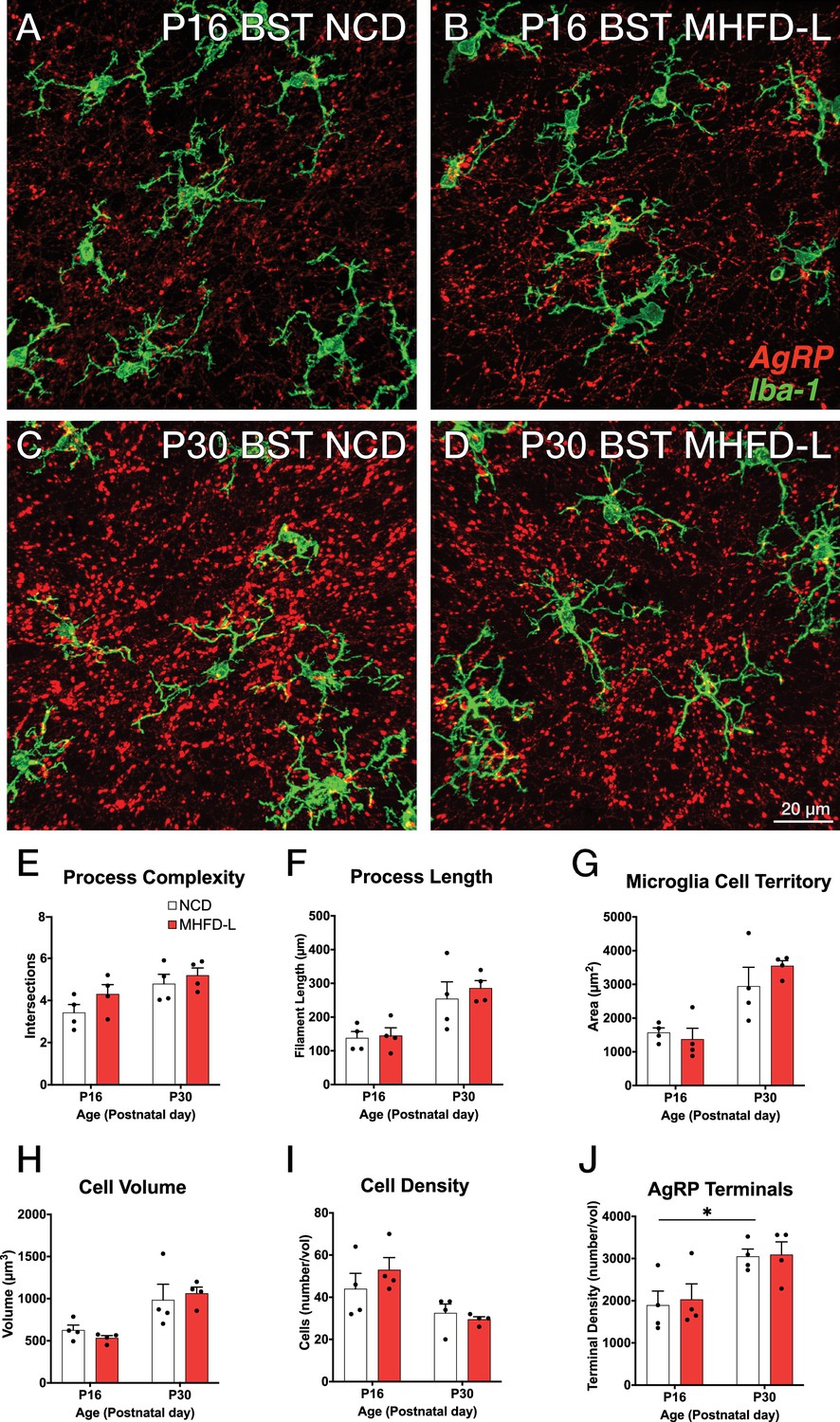
MHFD-L: Microglial morphology in the BST.
Microglial cells (green) and labeled Agouti-related peptide (AgRP) terminals (red) in the BST of mice at P16 (A, B) or P30 (C, D) that were exposed to NCD (A, C) or MHFD-L (B, D). Graphical comparisons between groups to show that microglial ramification complexity (E), process length (F), cell territory (G), and cell volume (H) did not significantly change between P16 and P30, nor were they changed as a result of diet. The density of microglia in the BST decreased between P16 and P30, irrespective of diet (I). The density of AgRP terminals increased between P16 and P30, but there was no effect of maternal diet (J). Bars represent the mean ± SEM and each point represents one animal. *Pp<0.05. Abbreviations: BST, bed nucleus of the stria terminalis; MHFD-L, maternal HFD during lactation; NCD, normal chow diet.
Figure 4
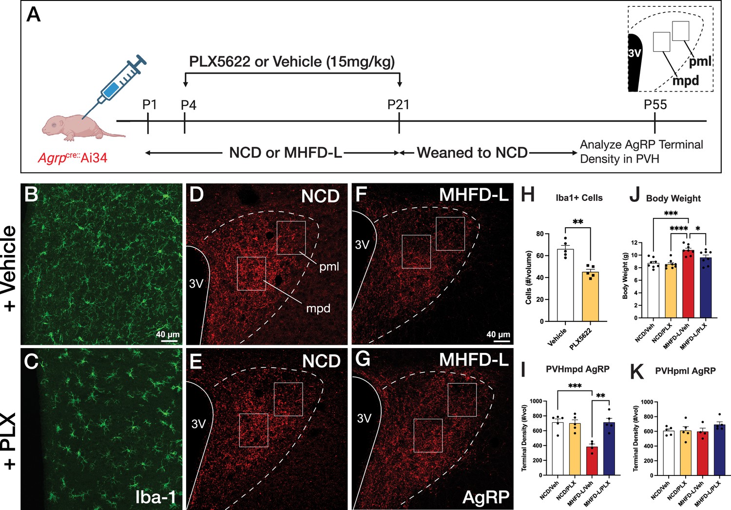
Microglial depletion during lactation period.
Schematic of MHFD-L exposure and PLX5622 treatment experimental design (A). Microglial cells (green) in the PVH of adult mice (P55) treated during lactation with vehicle (B) or PLX5622 (C). Images of labeled AgRP terminals (red) to illustrate the density of labeling in distinct compartments of the PVH (white boxes denote locations of ROIs) of normal chow diet (NCD) offspring (D ,E) and MHFD-L offspring (F, G). Graphical comparisons to illustrate the effects of postnatal PLX5622 treatments on microglia density in the PVH (H), body weight (J), and the density of AgRP terminals in the PVHmpd (I) and PVHpml (J). Bars represent the mean ± SEM and each point represents one animal. Unpaired t-test was used to compare cell number in 4 H; two-way ANOVA was used to test for differences in group means, followed by Tukey’s multiple comparisons posthoc test to identify specific group differences in 4I-K. Pp-values less than 0.05 were considered significant; *p<0.05, **p<0.005, ***p<0.0005. Abbreviations: AgRP, agouti-related peptide; CSF1R, Colony-stimulating factor 1 receptor; MHFD-L, maternal HFD during lactation; PVH, paraventricular nucleus of the hypothalamus; mpd, medial parvocellular compartment of the PVH; pml, posterior magnocellular compartment of the PVH.
Figure 5
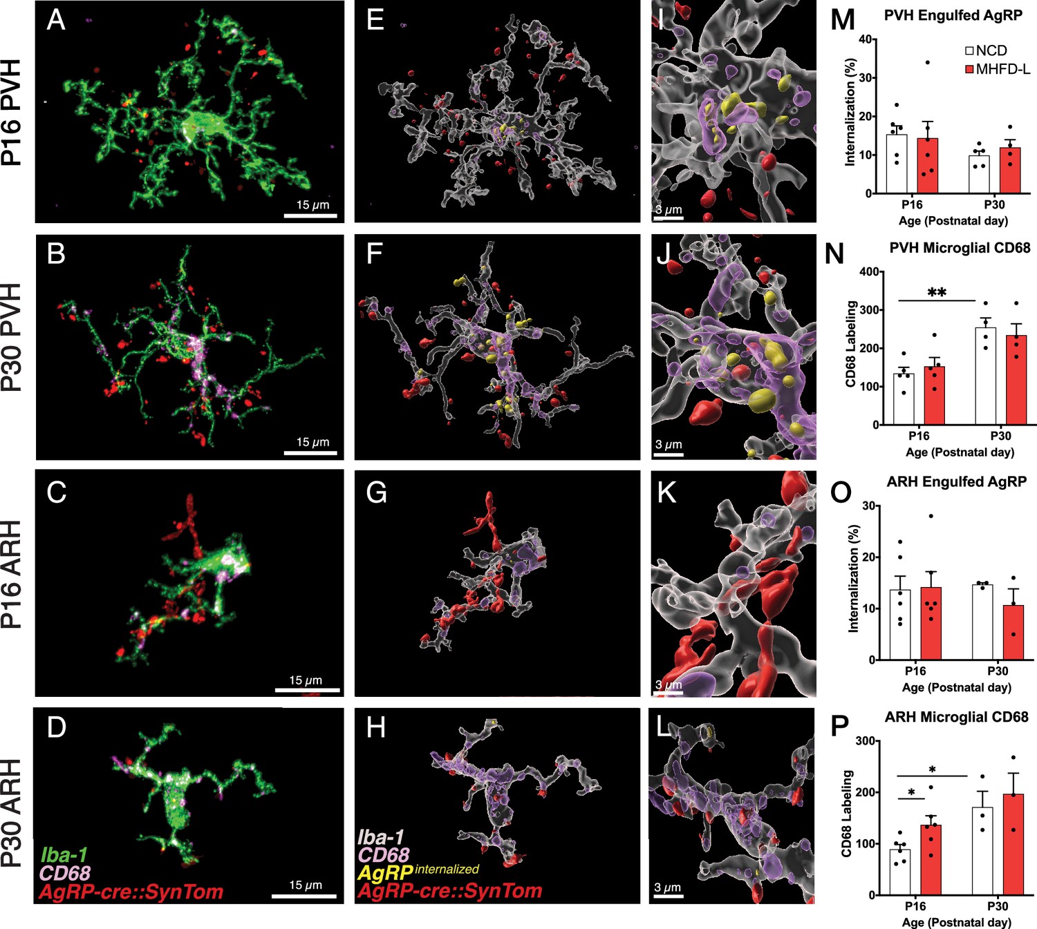
Microglial interaction with AgRP axon terminals in the PVH and arcuate nucleus of the hypothalamus (ARH).
(A–D) Representative images of microglial cells (green), labeled AgRP terminals (red), and CD68 (lysosomal associated membrane protein and phagocytic capacity marker, pink) that compare their cellular relationships in the PVH (A, B) and ARH (C, D) at P16 and P30. (E–H) Digital 3D reconstructions of cells shown in (A–D) after application of filaments tool to visualize internalized AgRP terminals. (I–L) Cells shown in E-H after application of digital zoom to more clearly illustrate engulfment of labeled AgRP terminals by microglia and location of CD-68 labeled profiles. (M–P) Graphical comparisons between groups to illustrate the effects of age and MHFD-L exposure on CD68 expression and AgRP terminal engulfment. Bars represent the mean ± SEM and each point represents one animal. *Pp<0.05, **Pp<0.005. Abbreviations: AgRP, agouti-related peptide; ARH, arcuate nucleus or the hypothalamus; CD68, Cluster of Differentiation 68; MHFD-L, maternal HFD during lactation; PVH, paraventricular nucleus of the hypothalamus.
Tables
Key resources table
| Reagent type (species) or resource | Designation | Source or reference | Identifiers | Additional information |
|---|---|---|---|---|
| Genetic reagent (M. musculus) | Agrptm1(cre)Lowl/J | Jackson Laboratory | Stock #: 012899 RRID:IMSR_JAX:012899 | MGI ID: J:140858 |
| Genetic reagent (M. musculus) | Ai34(RCL-Syp/tdT)-D (B6;129S-Gt(ROSA)26Sortm34.1(CAG-Syp/tdTomato)/Hze/J) | Jackson Laboratory | Stock #: 012570 RRID:IMSR_JAX:012570 | MGI ID: J:170755 |
| Antibody | Rabbit polyclonal anti-Iba1 | FUJIFILM Wako | Cat. #: 019–19741 RRID:AB_839504 | IHC (1:2000) |
| Antibody | Rat monoclonal anti-CD68 [FA-11] | Abcam | Cat. #: ab53444 RRID:AB_869007 | IHC (1:500) |
| Antibody | Donkey polyclonal anti-rabbit Alexa Fluor 488 | ThermoFisher Scientific | Cat. #: A32790 RRID:AB_2762833 | IHC (1:500) |
| Antibody | Donkey polyclonal anti-rat Alexa Fluor 647 | ThermoFisher Scientific | Cat. #: A48272 RRID:AB_2893138 | IHC (1:500) |
| Chemical compound | PLX5622 hemifumarate, CSF1R inhibitor | MedChemExpress | Cat. #: HY114153A | |
| Software, Algorithm | Imaris | Bitplane | V9.5 | |
| Software, Algorithm | GraphPad Prism | Prism | Prism 10 |
Additional files
Download links
A two-part list of links to download the article, or parts of the article, in various formats.
Downloads (link to download the article as PDF)
Open citations (links to open the citations from this article in various online reference manager services)
Cite this article (links to download the citations from this article in formats compatible with various reference manager tools)
Microglia are required for developmental specification of AgRP innervation in the hypothalamus of offspring exposed to maternal high-fat diet during lactation
eLife 13:RP101391.
https://doi.org/10.7554/eLife.101391.3


