The first complete 3D reconstruction and morphofunctional mapping of an insect eye
- Department of Entomology, Faculty of Biology, Lomonosov Moscow State University, Russian Federation
- Center for Computational Neuroscience, Flatiron Institute, United States
- Janelia Research Campus, Howard Hughes Medical Institute, United States
- Yale School of Medicine, United States
- Department of Cellular and Molecular Physiology, Yale School of Medicine, United States
- Neuroscience Institute, NYU Langone Medical Center, United States
Figures
Figure 1
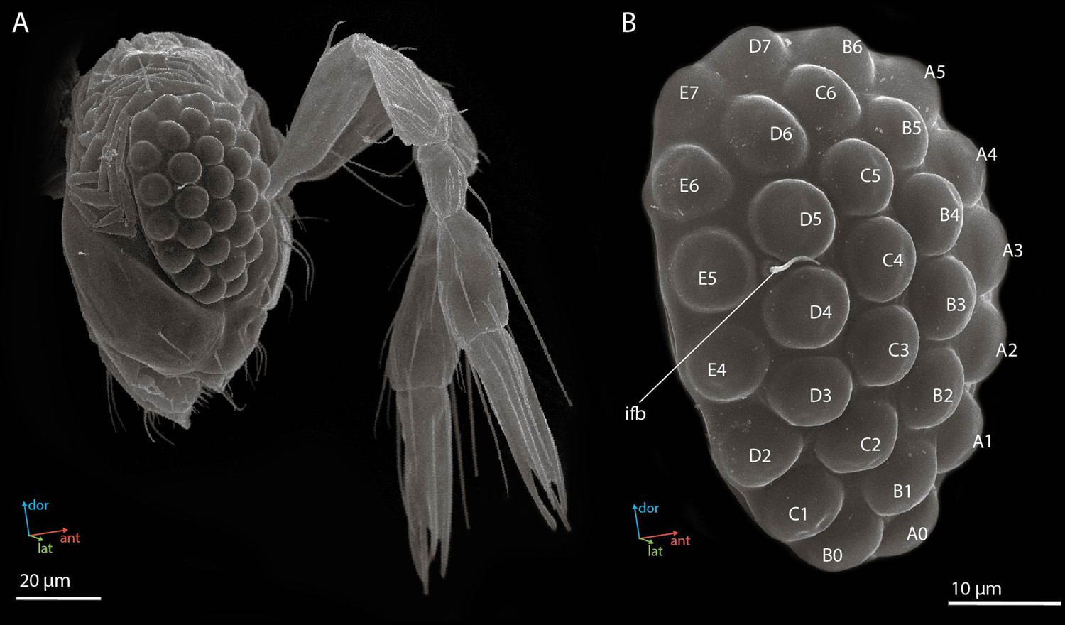
Scanning electron microscopy (SEM) images of the head (A) and the compound eye (B) of a female Megaphragma viggianii (side view).
Ifb, interfacet bristle. The compound eye comprises 29 ommatidia named here as in Chua et al., 2023.
Figure 2
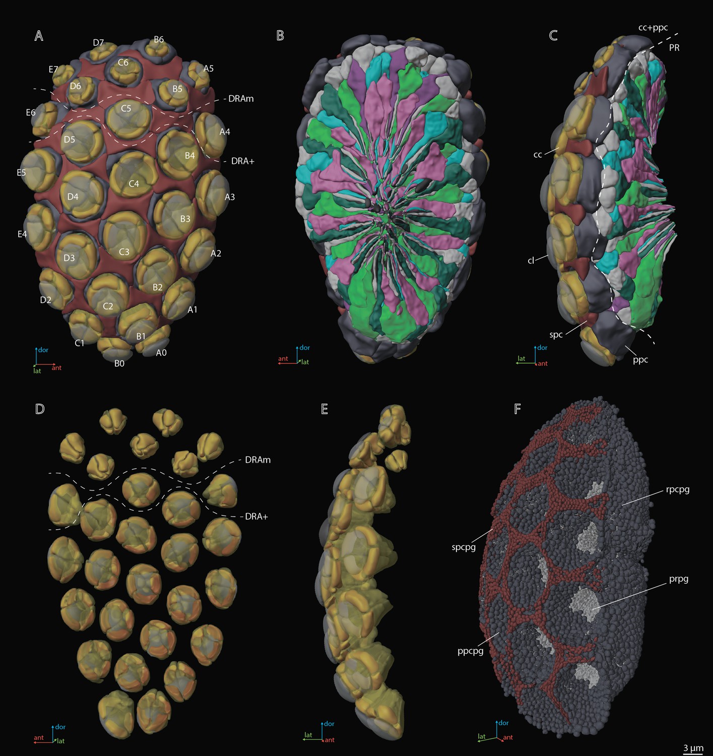
A three-dimensional (3D) reconstruction of the compound eye of M. viggianii.
(A) A front view from the cornea side; (B) a rear view from the retinal side; (C) a side view; (D) a rear view of the ommatidia DA; (E) a rear view of the ommatidia DA; (F) a semi-side view of pigment granules of all cells. cc, crystalline cones; cl, corneal lense; DRAm, dorsal rim area ommatidia (morphological specialization); DRA+, transitional zone ommatidia; ppc, primary pigment cells; ppcpg, pigment granules of PPC; prpg, retinal (photoreceptor) pigment granules; rpcpg, rim cells pigment granules; spc, secondary pigment cells; spcpg, secondary pigment cell pigment granules. Ommatidia are named as in Chua et al., 2023.
Figure 3 with 5 supplements
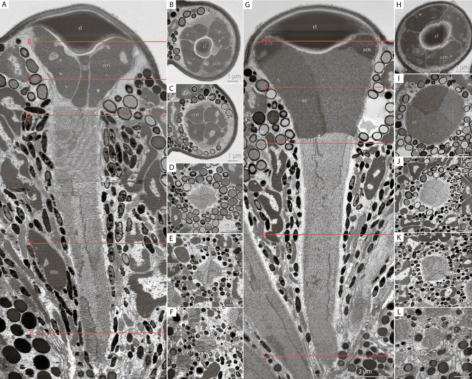
Cross-sections of M. viggianii ommatidia sampled from a volume electron microscopy (vEM) (FIB-SEM) dataset.
(A–F) Dorsal rim area (DRA) ommatidia (B5); (G–L) non-DRA ommatidia (C3). (A, G) A longitudinal section through one ommatidium; (B, H) a cross-section through the proximal part of a corneal lens; (C, I) a cross-section through the center of a cone; (D, J) a cross-section through a distal rhabdom, directly under the cone; (E, K) a cross-section through the center of a rhabdom; (F, L) a cross-section through a distal rhabdom. cc, crystalline cone; ccn, nuclei of crystalline cone cells; cl, corneal lens; ppc, primary pigment cells; R1–R8, retinal cells; spc, secondary pigment cells; asterisk (*) marks cone cell projections.
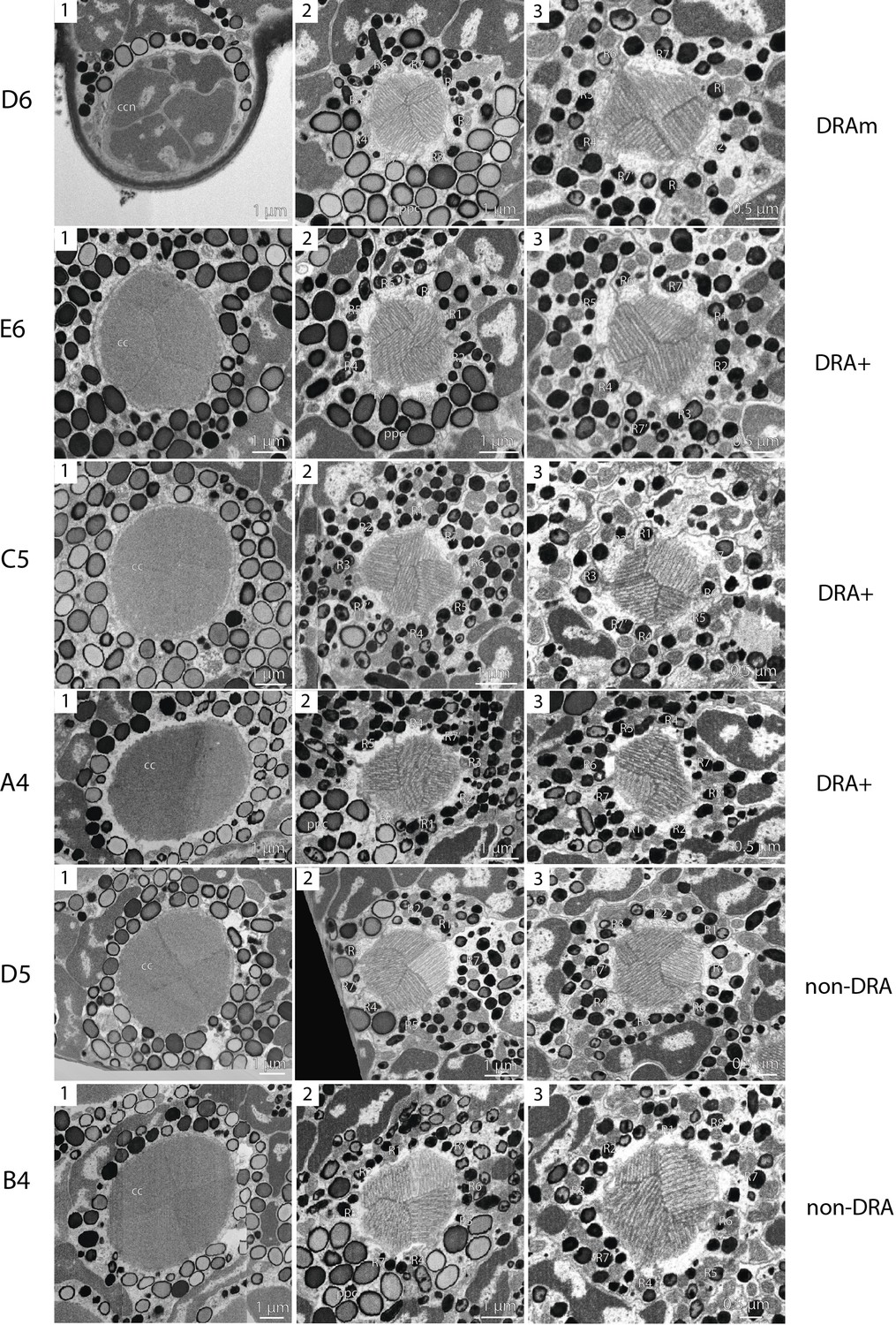
Figure 3—figure supplement 1
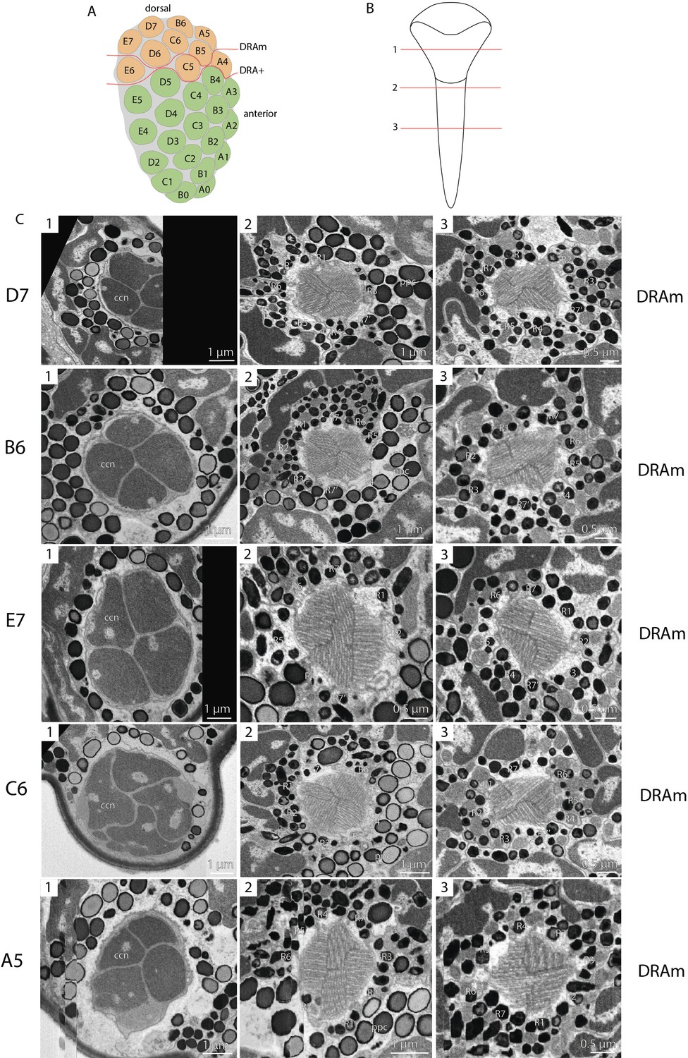
Scheme of the compound eye of Megaphragma viggianii (A); Scheme of cross-sections of ommatidia (B); EM sections through an ommatidium of M. viggianii (C).
(A) DRAm, dorsal rim area ommatidia (morphological specialization); DRA+, transitional zone ommatidia. The ommatidia are marked as follows: DRA orange; non-DRA green. Ommatidia are named as in Chua et al., 2023. Scheme of cross-sections of ommatidia (B): 1, through the center of the cone; 2, through the distal rhabdom, directly under the cone; 3, through the center of the rhabdom. EM sections through an ommatidium of M. viggianii (C), as in scheme (B). cc, crystalline cones; ccn, nuclei of crystalline cone cells; ppc, primary pigment cells; R1–R8, retinal cells; spc, secondary pigment cells. In ommatidia D7, E7, D6, E5, C4, D4, and E4, the major part of the dioptric apparatus was damaged due to milling (black area in slides).
Figure 3—figure supplement 2
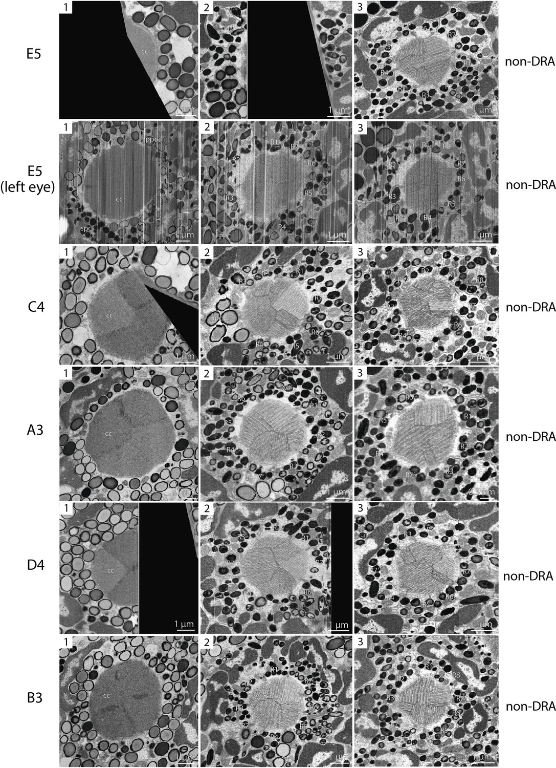
EM sections through an ommatidium of M. viggianii, continuation of Figure 3—figure supplement 1 (C).
Figure 3—figure supplement 3
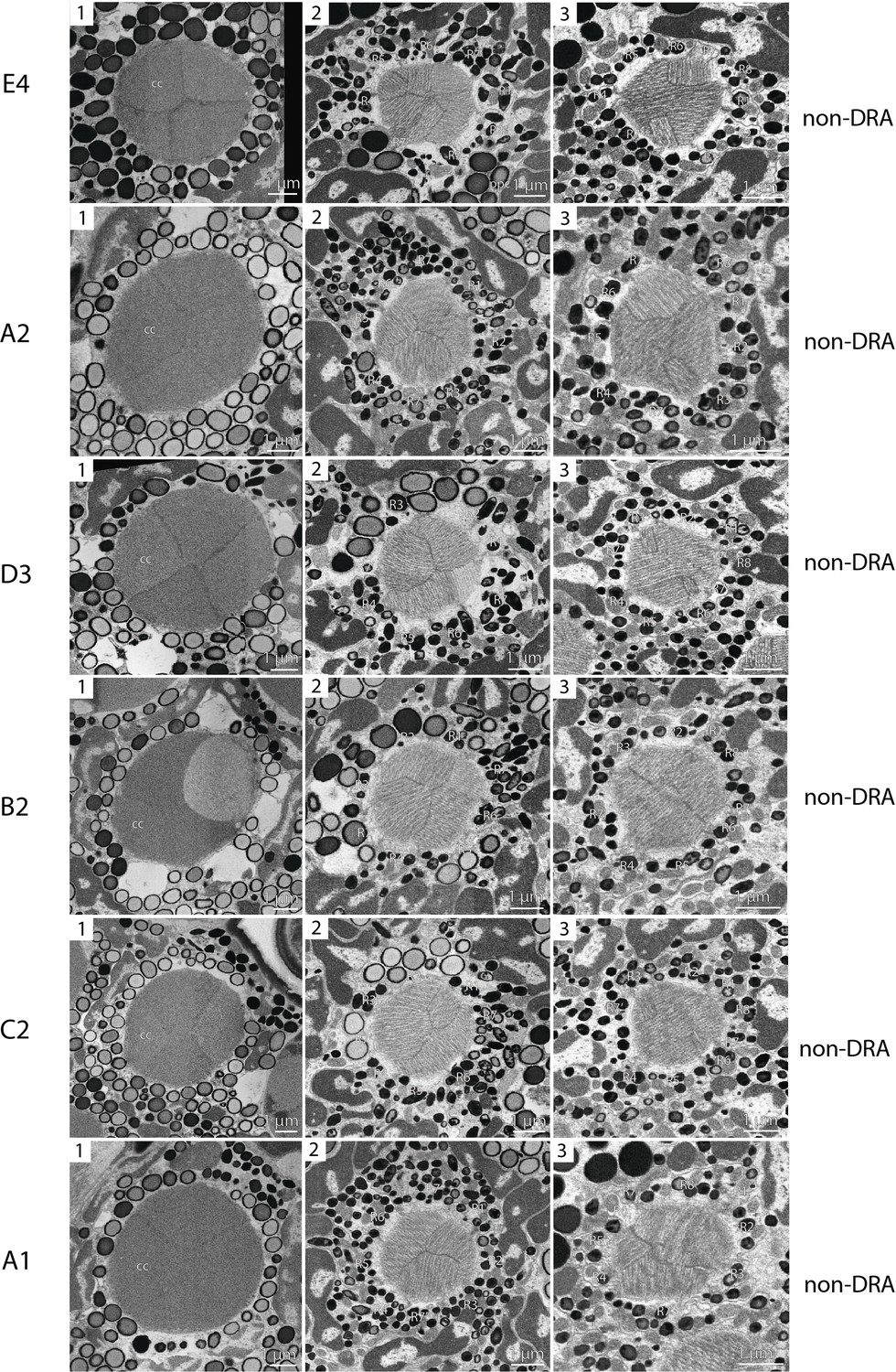
EM sections through an ommatidium of M. viggianii, continuation of Figure 3—figure supplement 1 (C).
Figure 3—figure supplement 4
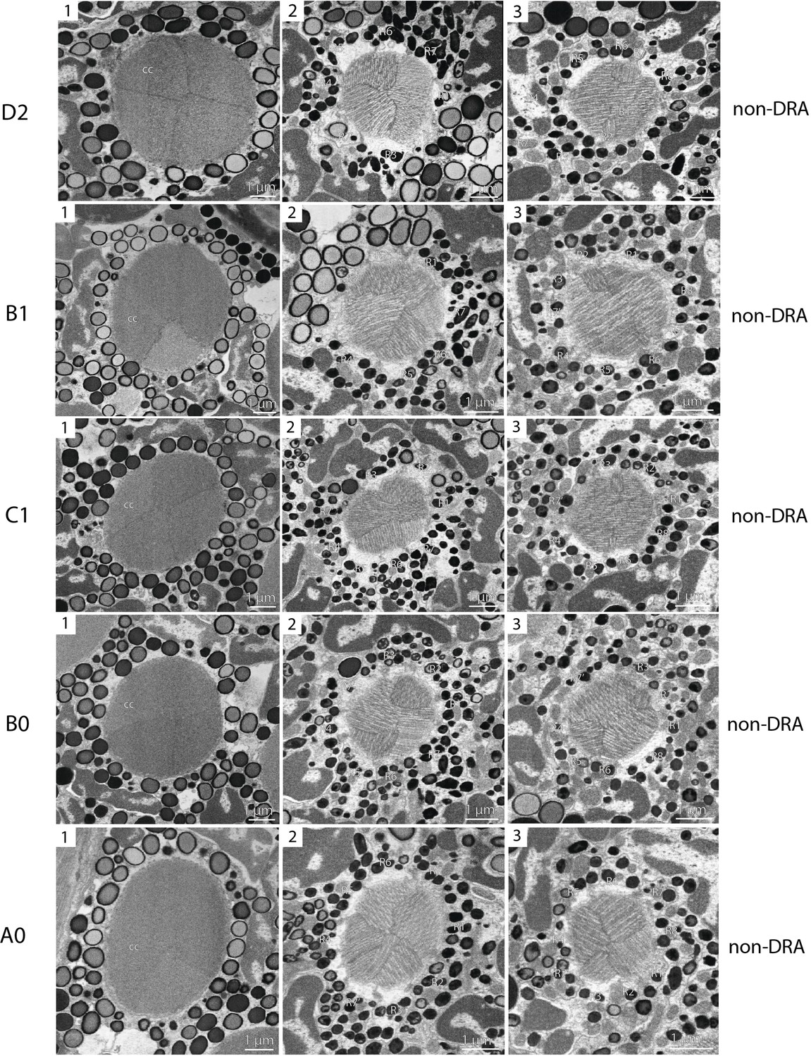
EM sections through an ommatidium of M. viggianii, continuation of Figure 3—figure supplement 1 (C).
Figure 3—figure supplement 5
EM sections through an ommatidium of M. viggianii, continuation of Figure 3—figure supplement 1 (C).
Figure 4
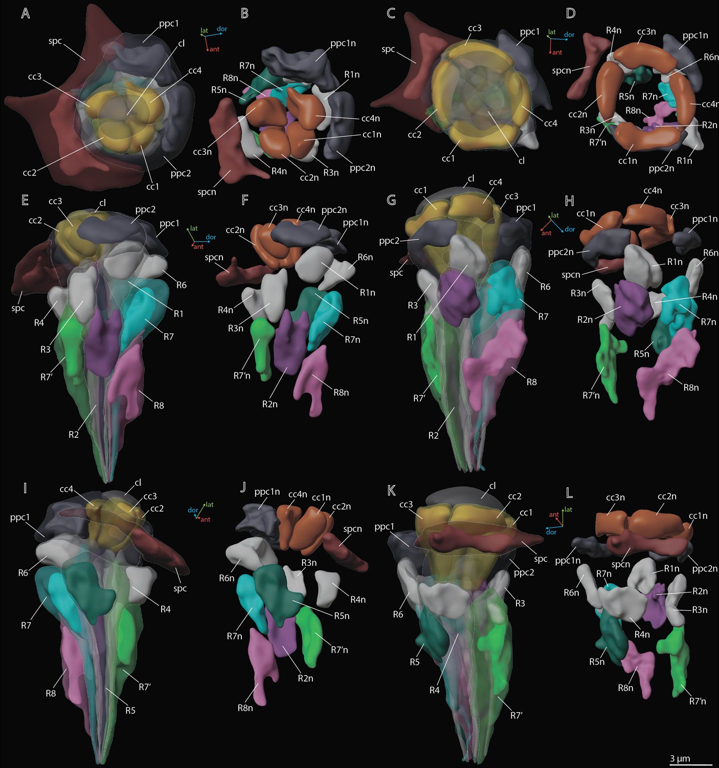
3D reconstruction of nuclei in ommatidium cells of M. viggianii.
(A, B, E, F, I, J) Dorsal rim area (DRA) ommatidia (B6; C, D, G, H, K, L) non-DRA ommatidia (C4). cc1–4, crystalline cone cells; cc1n–4 n, nuclei of crystalline cone cells; cl, corneal lens; ppc1, 2, primary pigment cells; ppc1n, ppc2n, nuclei of PPC; R1–R8, retinal cells; R1n–8 n, nuclei of retinal cells; spc, secondary pigment cells; spcn, nuclei of secondary pigment cells. Colors of nuclei same as colors of their cells.
Figure 5
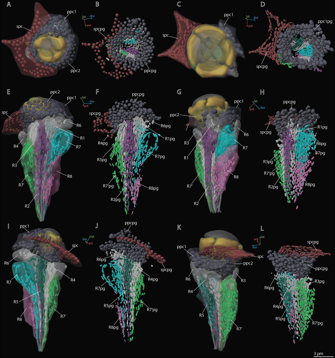
3D reconstruction of pigment granules in the ommatidium cells of M. viggianii.
(A, B, E, F, I, J) Dorsal rim area (DRA) ommatidia (B6); (C, D, G, H, K, L) non-DRA ommatidia (C4). ppc1, 2, primary pigment cells; ppc1pg, ppc2pg, pigment granules of PPC; R1–R8, retinal cells; R1pg–R8pg, pigment granules of retinal cells; spc, secondary pigment cells; spcpg, pigment granules of secondary pigment cells. Colors of pigment granules are the same as the colors of their cells.
Figure 6
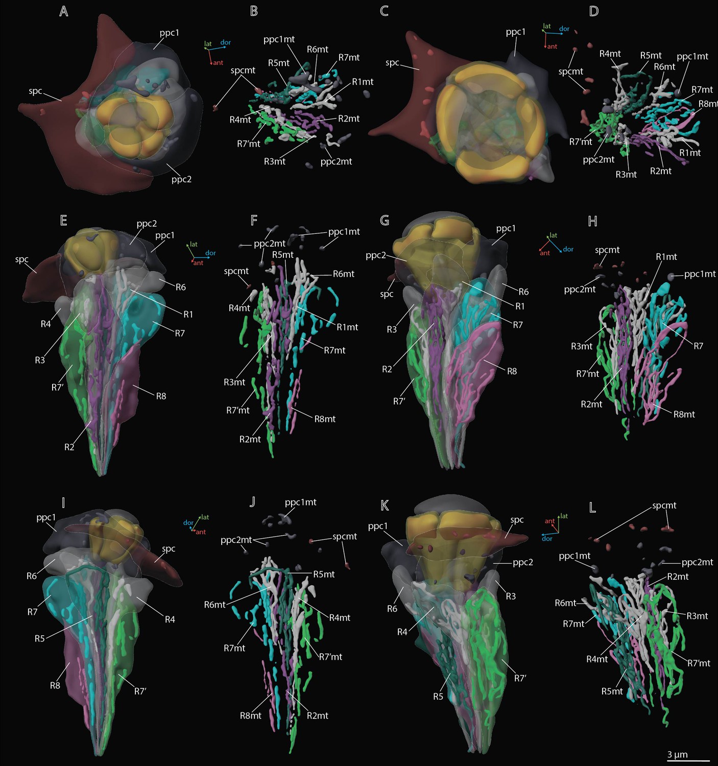
3D reconstruction of mitochondria in the ommatidium cells of M. viggianii.
(A, B, E, F, I, J) Dorsal rim area (DRA) ommatidia (B6); (C, D, G, H, K, L) non-DRA ommatidia (C4). ppc1, 2, primary pigment cells; ppc1mt, ppc 2mt, mitochondria of PPC; R1–R8, retinal cells; R1mt–R8mt, mitochondria of retinal cells; spc, secondary pigment cells; spcpg, mitochondria of secondary pigment cells. Colors of mitochondria are the same as the colors of their cells.
Figure 7
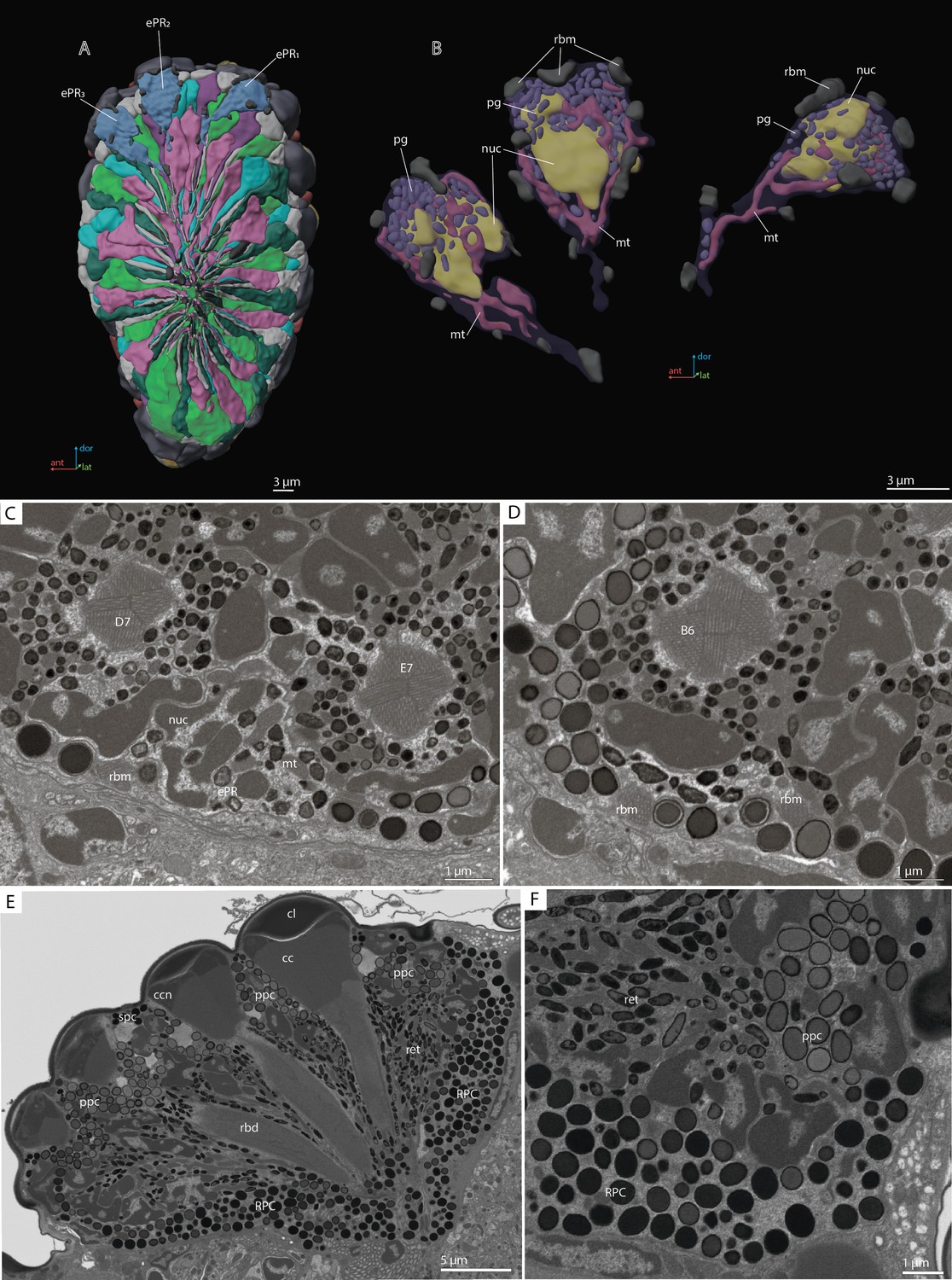
‘Ectopic’ photoreceptors (ePRs) and rind photoreceptor shield in the compound eye of M. viggianii.
(A) A 3D reconstruction of the eye: posterior view from the retinal area with labeled ePRs; (B) a 3D reconstruction of ePRs; (C, D) an EM section through dorsal border of the eye; (F) an EM section through the eye showing rim pigment cells (RPC). cc, crystalline cones; cl, corneal lens; ePR1-3, ‘ectopic’ photoreceptors; mt, mitochondria; nuc, nuclei; pg, pigment granules; ppc, primary pigment cells; rbd, rhabdom; rbm, rhabdomeres of ‘ectopic’ photoreceptors; spc, secondary pigment cells. B6, D7, E7, DRA ommatidia abutting ‘ectopic’ photoreceptors.
Figure 8
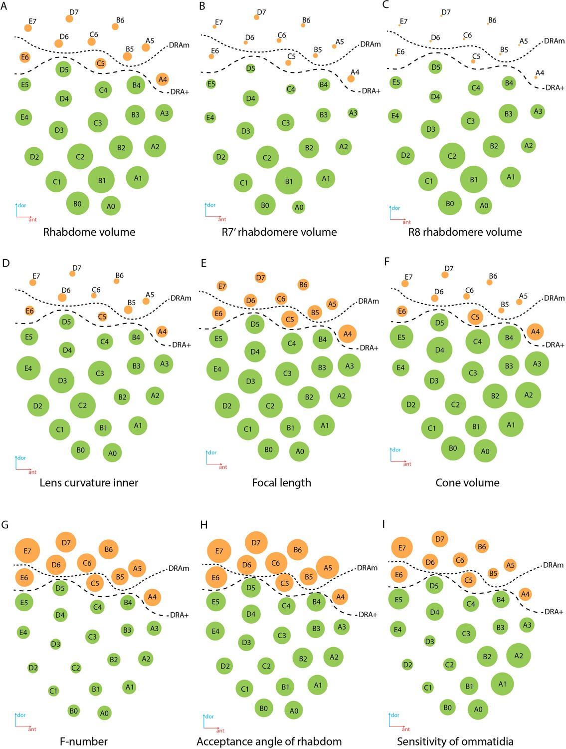
Regional specialization of the compound eye in M. viggianii.
Bubble size indicates the value of each parameter. (A) Rhabdom volume; (B) volume of rhabdomere R7’; (C) volume of rhabdomere R8; (D) inner curvature of the lens; (E) focal length; (F) cone volume; (G) f-number; (H) acceptance angle of the rhabdom; (I) sensitivity of the ommatidium. DRAm, dorsal rim area ommatidia (morphological specialization); DRA+, transitional zone ommatidia. Ommatidia are named as in Chua et al., 2023.
Videos
Video 1
3D reconstruction of the ultrastructure of the compound eye of M. viggianii.
cc, crystalline cones; cl, corneal lense; ccn, nuclei of crystalline cone cells; ePR, ‘ectopic’ photoreceptors; ppc, primary pigment cells; rbd, rhabdoms; spc, secondary pigment cells; R1–R8, retinal cells.
Video 2
3D reconstruction of the ultrastructure of the ePR in the compound eye of M. viggianii.
cc, crystalline cones; cl, corneal lense; ccn, nuclei of crystalline cone cells; ePR, ‘ectopic’ photoreceptors; mt, mitochondria; nuc, nuclei; pg, pigment granules; ppc, primary pigment cells; R1–R8, retinal cells; rbm, rhabdomeres; spc, secondary pigment cells.
Video 3
3D reconstruction of the ultrastructure of the DRA and Reg ommatidia in M. viggianii.
DRAm, dorsal rim area ommatidia (morphological specialization); DRA+, transitional zone ommatidia; cc, crystalline cones; cl, corneal lense; ccn, nuclei of crystalline cone cells; ppc, primary pigment cells; R1–R8, retinal cells; rbm, rhabdomeres; spc, secondary pigment cells. Colors of nuclei, mitochondria, and pigment granules are the same as the colors of their cells.
Tables
Table 1
Linear measurements (µm) of M. viggianii eye components.
Diameter*,†, diameter of rhabdom measured in orthogonal planes, according to its not round shape. Hereinafter mean ± s.d. DRA, dorsal rim ommatidia in general (DRAm and DRA+); DRAm, dorsal rim area ommatidia (morphological specialization); DRA+, transitional zone ommatidia; non-DRA, regular (non-DRA) ommatidia. Raw data, Supplementary file 1a.
| Ommatidium length | Lense | Cone | Rhabdom | |||||||||
|---|---|---|---|---|---|---|---|---|---|---|---|---|
| Diameter | Thickness | Curvature | Length | Width | Diameter (distal) | Diameter central* | Diameter central† | Diameter mean | Length | |||
| inner | outer | |||||||||||
| DRA | 19.2±0.37 | 6.9±0.89 | 2.5±0.56 | 1.2±0.32 | 3.3±0.64 | 3.0±0.38 | 4.9±0.66 | 2.0±0.16 | 2.1±0.32 | 2.2±0.23 | 2.1±0.18 | 13.4±0.64 |
| DRAm | 19.0±0.20 | 6.5±0.51 | 2.2±0.28 | 1.1±0.21 | 3.1±0.51 | 2.8±0.28 | 4.5±0.21 | 1.9±0.11 | 1.9±0.16 | 2.2±0.24 | 2.1±0.11 | 13.7±0.29 |
| DRA+ | 19.7±0.15 | 7.9±0.85 | 3.2±0.26 | 1.6±0.19 | 3.9±0.68 | 3.4±0.14 | 5.8±0.21 | 2.2±0.068 | 2.5±0.21 | 2.2±0.25 | 2.4±0.11 | 12.6±0.63 |
| Non-DRA | 22.3±2.5 | 8.0±0.77 | 3.3±0.40 | 3.0±0.52 | 4.7±0.39 | 4.6±0.60 | 6.7±0.25 | 2.7±0.26 | 2.9±0.25 | 2.8±0.27 | 2.9±0.21 | 14.2±1.8 |
Table 2
Mean volumes (µm3) for cellular and subcellular elements of ommatidia in M. viggianii.
The volumes were obtained from 3D models. DRA, dorsal rim ommatidia in general (DRAm and DRA+); DRAm, dorsal rim area ommatidia (morphological specialization); DRA+, transitional zone ommatidia; non-DRA, regular (non-DRA) ommatidia; R1-R8, retinal cells; PPC, primary pigment cells; SPC, secondary pigment cells. Raw data, Supplementary file 1b.
| Retinal cell | Cone | PPC | SPC | Lense | ||||||||||
|---|---|---|---|---|---|---|---|---|---|---|---|---|---|---|
| R1 | R2 | R3 | R7' | R4 | R5 | R6 | R7 | R8 | ||||||
| Soma | DRA | 17.5±2.0 | 24.4±3.8 | 17.9±2.3 | 33.0±2.5 | 18.0±2.0 | 25.2±4.1 | 17.9±1.6 | 32.1±4.8 | 15.8±1.9 | 13.1±6.0 | 47.1±14.4 | 22.0±4.7 | 16.5±14.0 |
| DRAm | 16.4±1.2 | 22.3±2.3 | 17.7±2.3 | 32.2±2.0 | 17.2±1.4 | 23.4±3.1 | 17.1±0.8 | 30.0±3.4 | 14.8±0.8 | 9.5±1.2 | 40.6±9.5 | 20.2±4.3 | 8.6±3.3 | |
| DRA+ | 19.8±2.7 | 29.2±3.7 | 18.3±3.1 | 34.8±3.7 | 20.0±3.0 | 29.4±4.3 | 19.9±1.9 | 37.2±6.7 | 18.2±2.5 | 21.3±4.5 | 61.4±13.0 | 26.2±2.6 | 34.8±17.2 | |
| Non-DRA | 21.0±3.3 | 29.5±3.1 | 22.0±2.6 | 53.3±11.1 | 22.4±3.5 | 31.1±3.2 | 20.5±4.5 | 30.6±8.5 | 28.2±4.3 | 32.9±5.9 | 64.5±11.4 | 24.5±3.2 | 41.4±9.0 | |
| Nuclei | DRA | 6.5±0.46 | 7.2±0.89 | 6.7±0.64 | 8.2±0.83 | 6.7±0.52 | 7.4±0.73 | 6.8±0.36 | 8.4±1.03 | 7.0±0.63 | 6.6±0.59 | 7.5±0.86 | 6.6±0.78 | n/a |
| DRAm | 6.4±0.42 | 6.8±0.72 | 6.8±0.62 | 8.4±0.61 | 6.7±0.53 | 7.3±0.66 | 6.8±0.38 | 8.3±0.98 | 6.8±0.53 | 6.5±0.65 | 7.4±0.93 | 6.8±0.76 | n/a | |
| DRA+ | 6.8±0.73 | 8.0±1.2 | 6.4±0.75 | 7.6±1.1 | 6.8±0.60 | 7.7±0.72 | 6.9±0.47 | 8.6±0.87 | 7.4±0.73 | 6.9±0.38 | 7.5±0.75 | 5.9±0.42 | n/a | |
| Non-DRA | 7.0±0.56 | 7.6±0.91 | 7.2±0.73 | 9.2±1.1 | 7.2±0.85 | 7.9±0.71 | 6.9±0.98 | 7.9±1.2 | 8.3±0.80 | 7.5±0.93 | 7.6±0.98 | 6.4±1.0 | n/a | |
| Rhabdomere | DRA | 1.8±0.55 | 3.1±0.74 | 2.3±0.58 | 3.9±0.81 | 1.7±0.27 | 3.2±0.69 | 1.8±0.83 | 4.2±1.1 | 0.86±0.25 | n/a | n/a | n/a | n/a |
| DRAm | 1.5±0.28 | 2.7±0.46 | 2.0±0.44 | 3.5±0.4 | 1.7±0.26 | 2.9±0.34 | 1.4±0.59 | 3.6±0.47 | 0.73±0.05 | n/a | n/a | n/a | n/a | |
| DRA+ | 2.4±0.19 | 4.0±0.46 | 2.9±0.58 | 4.8±0.61 | 1.9±0.21 | 4.1±0.53 | 2.6±0.39 | 5.7±1.6 | 1.2±0.42 | n/a | n/a | n/a | n/a | |
| Non-DRA | 4.1±1.6 | 5.6±1.3 | 3.2±0.9 | 12.7±3.9 | 3.6±1.3 | 5.9±1.0 | 3.2±0.74 | 5.5±2.1 | 6.7±1.6 | n/a | n/a | n/a | n/a | |
Table 3
Volumes (µm3) of pigment granules and mitochondria in ommatidia of M. viggianii and Trichogramma evanescens.
The data for T. evanescens are from Fischer et al., 2019. R1–R8, retinal cells; mt, mitochondria; pg, pigment granules; PPC, primary pigment cells; SPC, secondary pigment cells. B6, C4, A3, A0, ommatidia on which cells pigment granules and mitochondria were reconstructed. Raw data, see Supplementary file 1c and e.
| Organelle type | R1 | R2 | R3 | R7' | R4 | R5 | R6 | R7 | R8 | PPC | SPC | ||
|---|---|---|---|---|---|---|---|---|---|---|---|---|---|
| M. viggianii | B6, D7, C4, A3, A0 | PG | 2.5±0.71 | 3.2±0.86 | 2.1±0.51 | 6.5±2.1 | 2.4±0.31 | 2.7±0.76 | 2.2±0.36 | 5.5±2.3 | 2.0±1.0 | 20.6±10.5 | 7.9±3.0 |
| Mt | 1.2±0.40 | 1.8±0.56 | 1.0±0.32 | 2.8±0.12 | 1.2±0.39 | 1.8±0.54 | 1.0±0.42 | 2.0±0.59 | 1.5±0.96 | 0.53±0.35 | 0.31±0.070 | ||
| T. evanescens | Three central ommatidia | PG | 2.0±0.12 | 2.1±0.16 | 1.9±0.07 | 2.5±0.19 | 2.0±0.14 | 2.1±0.07 | 1.9±0.15 | 3.0±0.23 | 0.66±0.12 | 8.5±1.4 | 4.0±0.53 |
| Mt | 1.8±0.08 | 2.8±0.19 | 1.9±0.05 | 1.4±0.23 | 2.1±0.08 | 2.8±0.14 | 1.9±0.14 | 1.8±0.29 | 0.59±0.05 | 0.46±0.14 | 0.32±0.09 | ||
| M. viggianii | % | PG | 12.1±1.9 | 11.4±1.8 | 11.2±2.6 | 13.2±1.7 | 10.5±1.9 | 10.5±1.5 | 10.3±1.5 | 13.2±1.7 | 8.9±3.4 | 32.1±11.5 | 26.1±10.9 |
| Mt | 5.7±1.0 | 7.0±1.8 | 4.9±1.3 | 6.6±1.8 | 5.5±1.5 | 6.8±1.0 | 5.0±1.0 | 5.2±0.51 | 5.0±1.9 | 0.60±0.0042 | 1.1±0.42 | ||
| T. evanescens | PG | 11.3±1.7 | 7.9±0.63 | 9.6±0.84 | 13.3±2.1 | 11.4±1.7 | 7.9±0.81 | 11.2±0.45 | 11.6±0.43 | 7.8±0.76 | 21.0±0.94 | 19.1±2.4 | |
| Mt | 9.8±1.1 | 10.3±0.72 | 9.5±0.86 | 7.6±0.51 | 11.6±0.82 | 10.5±0.27 | 11.0±0.24 | 6.9±0.75 | 6.9±0.45 | 1.1±0.17 | 1.5±0.31 |
Table 4
Volumes (µm3) and number for ‘ectopic’ photoreceptors in M. viggianii.
EPR1–3, ‘ectopic’ photoreceptors.
| Soma | Nuclei | Rhabdomeres (total volume) | Pigment granules (total volume) | Number of pigment granules per cell | Mitochondria total volume | Number of mitochondria per cell | |
|---|---|---|---|---|---|---|---|
| EPR1 | 26.5 | 8.8 | 2.5 | 2.8 | 117 | 1.8 | 5 |
| EPR2 | 25.1 | 7,5 | 2.4 | 3.2 | 130 | 2.2 | 5 |
| EPR3 | 23.6 | 7.6 | 2.2 | 3 | 105 | 1.9 | 10 |
| Mean | 25.1±1.4 | 7.9±0.73 | 2.4±0.15 | 2.9±0.22 | 117±12 | 1.9±0.19 | 7±3 |
Table 5
Features of DRA and non-DRA ommatidia obtained by reconstruction of compound eyes of M. viggianii.
DRA, dorsal rim ommatidia in general (DRAm and DRA+); DA, dioptric apparatus; non-DRA, regular (non-DRA) ommatidia; PC, pigment cells; PPC, primary pigment cells; R1–R8, retinal cells. t-test *0.001≤p<0.01, **0.0001≤p<0.001, ***p<0.0001.
| Ommatidial area | Features | DRA | Non-DRA | Difference | |
|---|---|---|---|---|---|
| Ommatidial length, µm | 19.2±0.37 | 22.3±2.5 | ** | ||
| DA | Cone | Length, µm | 3.0±0.37 | 4.6±0.59 | *** |
| Width, µm | 4.5±0.21 | 6.6±0.25 | *** | ||
| Volume, µm3 | 13.0±6.0 | 32.9±5.9 | *** | ||
| Lens | Lens diameter, µm | 6.9±0.89 | 8.0±0.77 | * | |
| Volume, µm3 | 16.5±14.0 | 41.4±9.0 | ** | ||
| Lens thickness, µm | 2.5±0.56 | 3.3±0.39 | * | ||
| Inner curvature, µm | 1.2±0.32 | 3.0±0.51 | *** | ||
| Outer curvature, µm | 3.3±0.63 | 4.7±0.39 | *** | ||
| Cone cell (CC) nuclei | CC nuclei | Fill most of the volume of the cell | Form a ring in the upper third of the cone | n/a | |
| % of cone volume | 58±0.17 | 23±0.02 | *** | ||
| Volume, µm3 | 6.6±0.68 | 7.5±1.1 | *** | ||
| PC | PPC volume | 47.1±14.3 | 64.5±11.4 | *** | |
| Retina | Rhabdom | Shape | Rectangular shape of rhabdom from center to lower part | Spheric shape of the rhabdom along whole length | n/a |
| Diameter, µm | 2.0±0.16 | 2.7±0.75 | *** | ||
| Microvilli orientation | Orthogonal orientation of R7, R7' microvilli along the rhabdom length | Non-orthogonal orientation of R7, R7' | n/a | ||
| Volume, µm3 | Duet (R2, R5) | 24.8±3.9 | 30.3±3.2 | *** | |
| Quartet (R1, R3, R4, R6) | 17.8±1.9 | 21.4±3.5 | *** | ||
| R7' | 33.0±2.5 | 53.3±11.1 | *** | ||
| R7, R7' | 32.6±3.7 | 41.9±15.1 | *** | ||
| R8 | 15.8±1.9 | 28.2±4.2 | *** | ||
Table 6
Comparison of volumes (µm3) of ommatidial components for Trichogramma evanescens and M. viggianii.
Data on T. evanescens are from Fischer et al., 2019. CC, crystalline cones R1–R8, retinal cells; PPC, primary pigment cells; SPC, secondary pigment cells.
| CC | PPC | SPC | R1 | R2 | R3 | R4 | R5 | R6 | R7' | R7 | R8 | |
|---|---|---|---|---|---|---|---|---|---|---|---|---|
| Soma | ||||||||||||
| T. evanescens | 12.3±1.2 | 40.5±6.9 | 21.0±2.5 | 18.5±2.3 | 27.6±0.35 | 20.3±1.3 | 18.2±1.4 | 26.9±1.8 | 17.5±0.92 | 19.7±4.3 | 26.5±2.8 | 8.48±1.1 |
| M. viggianii (B3, C3, C4) | 32.9±7.5 | 61.8±9.2 | 23.2±3.8 | 20.4±0.81 | 30.3±3.0 | 19.4±1.8 | 21.2±1.6 | 30.2±2.0 | 18.1±1.7 | 48.4±8.4 | 33.8±10.9 | 26.9±1.7 |
| M. viggianii (all non-DRA) | 32.9±5.9 | 64.4±11.4 | 24.5±3.1 | 20.9±3.2 | 29.4±3.1 | 22.0±2.6 | 22.3±3.5 | 31.1±3.2 | 20.4±4.5 | 53.3±11.1 | 30.6±8.5 | 28.1±4.3 |
| Nuclei | ||||||||||||
| T. evanescens | 3.1±0.18 | 3.6±0.29 | 3.2±0.40 | 2.4±0.09 | 2.4±0.15 | 2.4±0.17 | 2.1±0.11 | 2.2±0.24 | 2.3±0.1 | 2.4±0.3 | 2.9±0.48 | 1.9±0.15 |
| M. viggianii (B3, C3, C4) | 7.0±1.0 | 7.2±1.1 | 5.7±0.28 | 7.0±0.11 | 7.9±0.18 | 6.7±0.63 | 6.9±0.65 | 8.0±0.48 | 5.9±0.36 | 9.1±1.1 | 8.1±1.3 | 8.3±0.73 |
| M. viggianii (all non-DRA) | 7.5±1.1 | 7.6±0.98 | 6.4±1.03 | 7.0±0.56 | 7.6±0.90 | 7.2±0.72 | 7.2±0.84 | 7.9±0.71 | 6.5±1.7 | 9.2±1.0 | 7.9±1.2 | 8.3±0.80 |
| Rhabdomere | ||||||||||||
| T. evanescens | n/a | n/a | n/a | 2.4±0.2 | 3.5±0.19 | 2.2±0.02 | 2.3±0.08 | 3.5±0.15 | 2.4±0.25 | 2.1±0.27 | 3.8±0.28 | 0.79±0.07 |
| M. viggianii (B3, C3, C4) | n/a | n/a | n/a | 3.7±0.92 | 6.0±0.79 | 2.3±1.0 | 3.2±0.46 | 5.9±0.54 | 2.8±0.071 | 11.1±3.0 | 6.5±2.3 | 6.6±0.80 |
| M. viggianii (all non-DRA) | n/a | n/a | n/a | 4.1±1.6 | 5.6±1.3 | 3.2±0.94 | 3.6±1.3 | 5.9±0.97 | 3.2±0.74 | 12.7±3.9 | 5.5±2.1 | 6.7±1.6 |
| Nuclei/soma, % | ||||||||||||
| T. evanescens | 25.0±2.7 | 9.1±1.2 | 15.2±2.5 | 13.0±1.1 | 8.8±0.55 | 11.6±0.67 | 12.4±1.6 | 12.0±0.41 | 8.1±0.44 | 13.3±1.02 | 11.0±1.02 | 23.0±1.3 |
| M. viggianii (B3, C3, C4) | 22.5±7.1 | 11.7±1.7 | 25.0±4.5 | 34.4±1.7 | 26.5±3.3 | 34.7±1.0 | 19.0±0.98 | 32.4±1.2 | 26.6±0.33 | 32.9±1.3 | 25.1±6.8 | 30.8±0.74 |
| M. viggianii (all non-DRA) | 35.3±19.9 | 15.1±11.6 | 28.6±7.9 | 34.5±3.4 | 27.1±2.9 | 34.4±3.1 | 20.1±4.2 | 34.2±3.5 | 27.0±3.1 | 33.5±6.2 | 26.7±4.6 | 34.8±7.8 |
Additional files
-
Supplementary file 1
Raw data for all measurements of ommatidia.
- https://cdn.elifesciences.org/articles/103247/elife-103247-supp1-v1.xlsx
-
MDAR checklist
- https://cdn.elifesciences.org/articles/103247/elife-103247-mdarchecklist1-v1.pdf
Download links
A two-part list of links to download the article, or parts of the article, in various formats.
Downloads (link to download the article as PDF)
Open citations (links to open the citations from this article in various online reference manager services)
Cite this article (links to download the citations from this article in formats compatible with various reference manager tools)
The first complete 3D reconstruction and morphofunctional mapping of an insect eye
eLife 14:RP103247.
https://doi.org/10.7554/eLife.103247.3


