Shc1 cooperates with Frs2 and Shp2 to recruit Grb2 in FGF-induced lens development
- Department of Ophthalmology, Columbia University, United States
- Department of Pathology and Cell Biology, Columbia University, United States
Figures
Figure 1 with 1 supplement
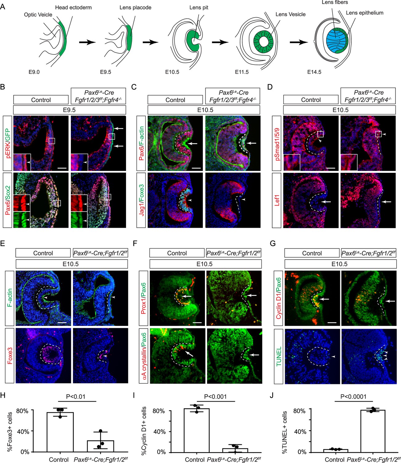
Fibroblast growth factor (FGF) signaling regulates lens development in a dose-dependent manner.
(A) Schematic diagram of murine lens development. The head ectoderm is induced by the underlying optic vesicle to become the lens placode, which subsequently folds inwards to become the lens pit. The closure of the lens vesicle sets the stage for the differentiation of the lens epithelium into the lens fibers. (B) Depletion of all four Fgfr1/2/3/4, driven by Pax6Le-Cre and traced with GFP (arrows), led to a thinner lens placode, evident from the absence of pERK signals and the failure to upregulate Sox2 like Pax6 (inserts, arrowheads). (C) Fgfr1/2/3/4 mutants displayed disrupted apical constriction (F-actin accumulation, arrows) and lacked lens-specific expression of Foxe3 and Jag1 (arrowheads). Dotted lines outline the lens pit. (D) Despite Fgfr1/2/3/4 mutations, BMP (pSmad1/5/9 staining, arrowheads) and Wnt signaling (Lef1 expression, arrows) remained unaffected. (E) The absence of Fgfr1/2 alone did not impede the apical buildup of F-actin nor the expression of Foxe3, indicating partial retention of lens development processes. (F) Crucial lens markers, Prox1 and αA-crystallin, were absent in Fgfr1/2 mutants, pointing to a significant developmental defect after the lens induction stage. Biological replicates, n=3. (G) Fgfr1/2 mutants exhibited loss of cell proliferation marker Cyclin D1 (arrows) and widespread apoptosis (TUNEL staining, arrowheads). (H) Quantification of Foxe3+ cells in Fgfr1/2 mutants. Student’s t-test, n=3, p<0.01. (I) Quantification of Cyclin D1+ cells in Fgfr1/2 mutants. Student’s t-test, n=3, p<0.001. (J) Quantification of TUNEL+ cells in Fgfr1/2 mutants. Student’s t-test, n=3, p<0.0001. Scale bars:25 µm.
Figure 1—figure supplement 1
Lens development in Fgf receptor mutants.
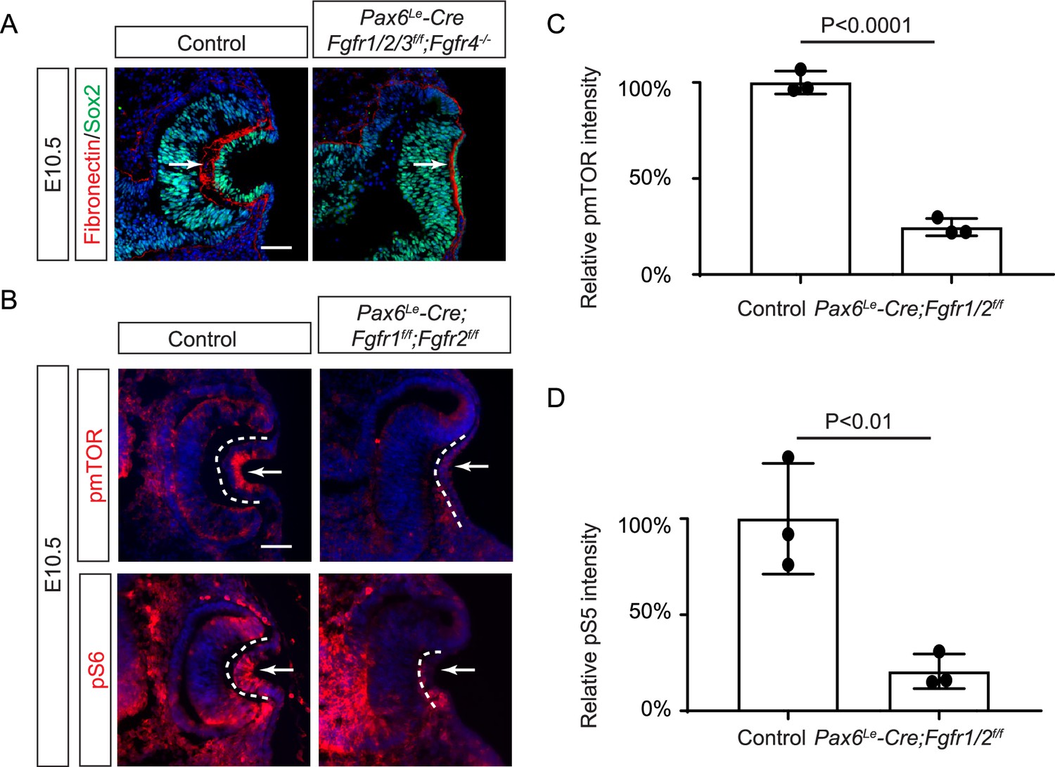
(A) Depletion of all four Fgfr1/2/3/4 did not disrupt the Fibronectin expression at the basal side of the lens placode (arrows). (B) pmTOR and pS6 staining were reduced in Pax6Le-Cre;Fgfr1f/f;Fgfr2f/LR lens. (C) Quantification of pmTOR fluorescent intensity in Fgfr1/2 mutants. Student’s t-test, n=3, p<0.0001. (D) Quantification of pmS6 fluorescent intensity in Fgfr1/2 mutants. Student’s t-test, n=3, p<0.01. Scale bars:25 µm.
Figure 2
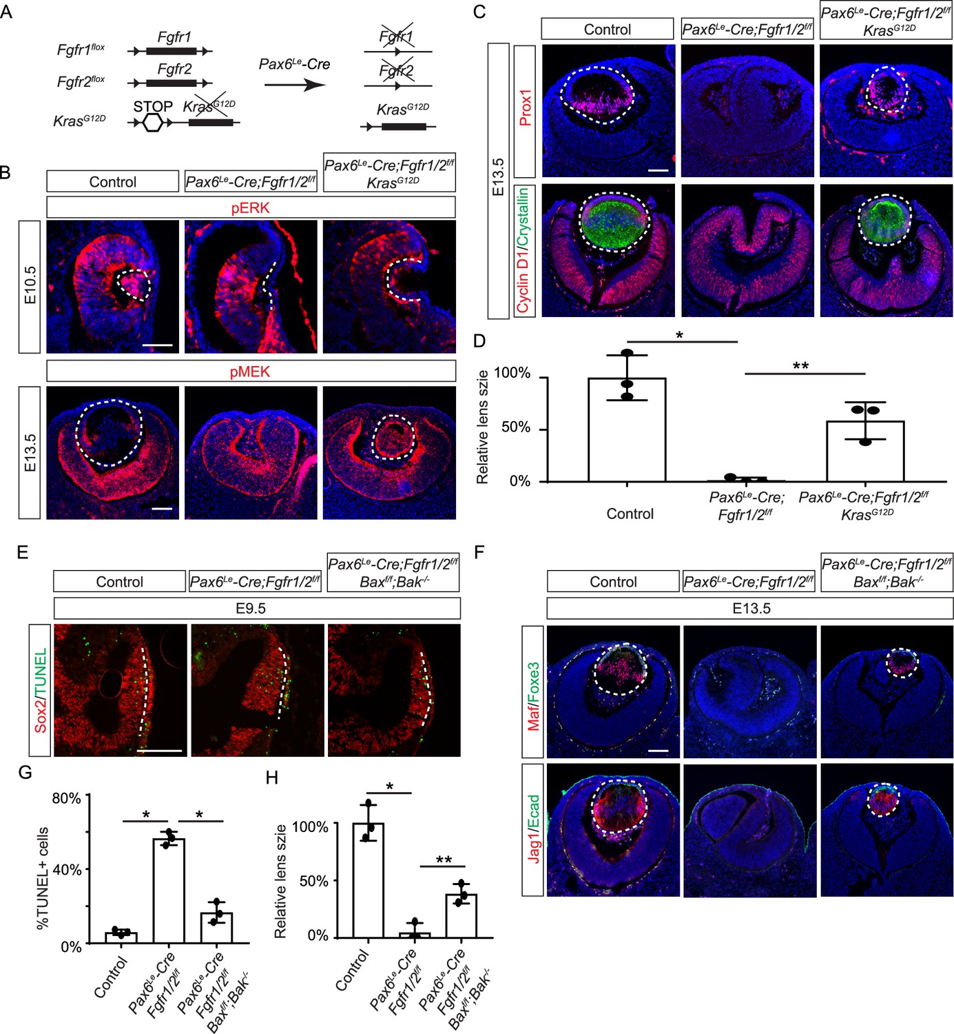
Restoration of lens development in fibroblast growth factor (FGF) signaling mutants via Kras activation and apoptosis inhibition.
(A) The Pax6Le-Cre driver facilitated the excision of the floxed alleles of Fgf1/2 along with the LSL-STOP cassette at the Kras locus, leading to the expression of the constitutively active KrasG12D allele within the Fgf1/2 mutant background. (B) The activation of Kras signaling in the FGF signaling mutant lenses reinstated pERK activity at E10.5 and pMEK expression at E13.5, indicating restoration of MAPK signaling. (C) The lens-specific expression of Prox1 and αA-crystallin were also recovered, indicating successful lens development rescue. (D) Quantification of the lens size. One way ANOVA, n=3, *p<0.001, **p<0.02. (E) The deletion of pro-apoptotic genes Bak and Bax in Fgf1/2 mutants suppressed apoptosis as shown by TUNEL staining. (F) Inhibiting apoptosis in Fgfr1/2 mutants facilitated lens formation, as indicated by the expression of lens differentiation markers Prox1, Maf, and Jag1. (G) Quantification cell apoptosis at E9.5. One-way ANOVA, n=3, *p<0.001. (H) Quantification of the lens size at E13.5. One-way ANOVA, n=3, *p<0.0001, **p<0.05. Scale bars:50 µm.
Figure 3
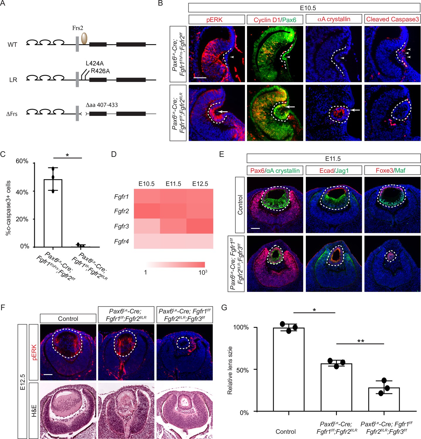
The Frs2 binding site on fibroblast growth factor receptor 2 (FGFR) is only required for lens vesicle differentiation.
(A) Overview of Fgfr mutant alleles. Fgfr1ΔFrs lacks the Frs2 binding domain (amino acid 407–433), and Fgfr2LR has point mutations disrupting Frs2 binding (L424A and L426A). (B) In Fgf1/2 compound mutants, loss of pERK, CyclinD1, αA-crystallin, and increased cleaved caspase3 were observed with the Fgfr1ΔFrs allele but not the Fgfr2LR allele. (C) Quantification of cleaved caspase3 staining. Student’s t-test, n=3, *p<0.001. (D) Heatmap depicts Fgfr expression levels during lens development. (E) Fgfr2LR mutants in the Fgfr1/2/3 genetic background showed impaired lens vesicle differentiation, with posterior lens epithelial cells failing to elongate and activate lens fiber cell markers Jag1 and Maf. (F) Fgfr1/2/3 triple mutants with Fgfr2LR lost pERK staining and displayed a shallow lens vesicle at E12.5. (G) Quantification of the lens size. One-way ANOVA n=3, *p<0.001, **p<0.05. Scale bars:50 µm.
Figure 4

Grb2 is essential for lens vesicle survival, proliferation, and differentiation.
(A) The targeted removal of Grb2 in the lens led to a loss of pERK signaling, reduced CyclinD3 expression, increased apoptosis (TUNEL staining), and disrupted expression of critical lens development genes Maf, Foxe3, Jag1, and γ-crystallin expression at E12.5. (B) Grb2 mutants displayed absent CyclinD1, Prox1, and p57 expression at E11.5 and remained an undifferentiated hollow vesicle at E13.5, failing to undergo normal lens fiber elongation. (C) Quantification of the lens size. Student’s t-test, n=3, *p<0.0001. Scale bars:50 µm.
Figure 5
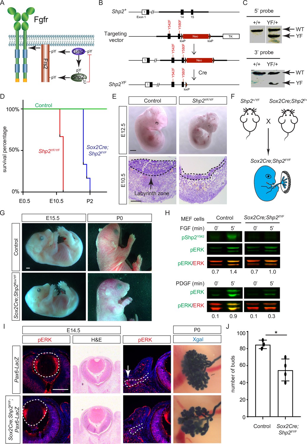
Shp2 C-terminal tyrosine phosphorylation is required for embryonic survival but dispensable for lens development.
(A) Schematic of the core FGF signaling pathway. FGFR activation leads to phosphorylation of the adaptor Frs2 on N- and C-terminal tyrosines, recruiting Grb2 and Shp2, respectively. Shp2 can also bind Grb2 via its own C-terminal phosphotyrosines, and dephosphorylates Grb2 via its catalytic cysteine residue. (B) Generation of the Shp2YF allele by homologous recombination to introduce loxP-flanked Neo and point mutations (Y542F and Y580F) disrupting the Shp2 C-terminal phosphotyrosine sites. The Neo cassette was subsequently excised by Cre-mediated recombination. (C) Validation of the Shp2YF allele targeting was confirmed through Southern blot analysis with both 5’ and 3’ probes. (D) Kaplan-Meier survival curves demonstrate early lethality of Shp2YF embryos (E12.5) and perinatal lethality of Sox2Cre;Shp2f/YF mutants. n=5 for Shp2YF/YF and n=6 for Sox2Cre;Shp2f/YF mutants. (E) Shp2YF/YF embryos displayed reduced body size at E12.5 and thinner labyrinth zones in their placenta at E10.5. (F) Sox2Cre-mediated targeting restricts Shp2 deficiency to the embryonic proper, circumventing placental abnormalities. (G) Sox2Cre;Shp2f/YF mutants appeared grossly normal at E15.5 but failed to survive after birth. (H) While Y542 phosphorylation in Shp2 was lost as expected, Sox2Cre;Shp2f/YF MEFs exhibited a more pronounced reduction in pERK response to PDGF stimulation compared to FGF stimulation. (I) Sox2Cre;Shp2f/YF mutant lens displayed normal pERK staining and morphology, but reduced pERK in lacrimal glands at E14.5 and decreased bud numbers at P0. (J) Quantification of the number of lacrimal gland buds. Student’s t-test, n=3, *p<0.02. Scale bars: 100 µm.
-
Figure 5—source data 1
Original files for southern blot analysis displayed in Figure 5C.
- https://cdn.elifesciences.org/articles/103615/elife-103615-fig5-data1-v1.zip
-
Figure 5—source data 2
Original membranes corresponding to Figure 5, panel C, with the relevant lanes are outlined in yellow.
- https://cdn.elifesciences.org/articles/103615/elife-103615-fig5-data2-v1.zip
-
Figure 5—source data 3
Original files for western blot analysis displayed in Figure 5H.
- https://cdn.elifesciences.org/articles/103615/elife-103615-fig5-data3-v1.zip
-
Figure 5—source data 4
Original membranes corresponding to Figure 5, panel H, with the relevant lanes are outlined in yellow.
- https://cdn.elifesciences.org/articles/103615/elife-103615-fig5-data4-v1.zip
Figure 6 with 1 supplement
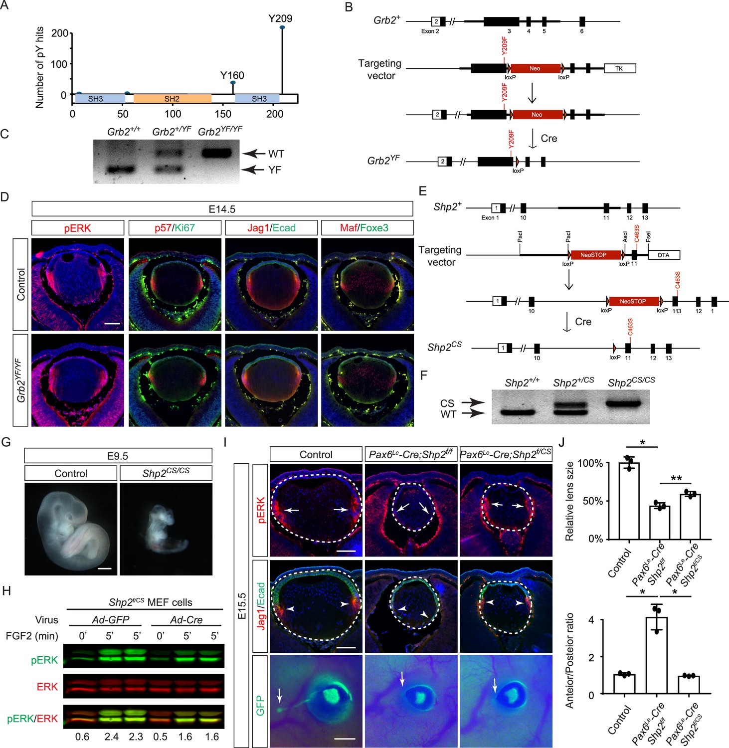
Shp2 phosphatase activity is partially required for fibroblast growth factor (FGF) signaling independently of Grb2 dephosphorylation.
(A) PhosphositePlus database indicates that Grb2 predominantly undergoes phosphorylation at Y209 and less frequently at Y160. (B) The Grb2YF allele was constructed by homologous recombination to integrate a loxP-flanked Neo cassette and a Y209F point mutation into the Grb2 locus. (C) PCR genotyping confirmed the presence of the Grb2YF allele. (D) Grb2 mutant lens typical expression patterns of pERK, cell cycle markers p57 and Ki67 and differentiation markers Foxe3, Maf, and Jag1. (E) The Shp2CS allele was generated by inserting the C459S mutation into the Shp2 locus by homologous recombination, followed by Cre-mediated removal of the loxP-flanked Neo cassette. (F) Shp2CS allele validated by PCR genotyping. (G) Shp2CS/CS mutants exhibited growth retardation and died at E9.5. (H) Shp2f/CS MEF cells retained a significant capacity to activate pERK upon FGF stimulation after Cre virus infection. (I) Pax6Le-Cre;Shp2f/f mutants exhibited loss of pERK and Jag1 staining at the lens transition zone (arrowheads), which also shifted posteriorly. Pax6Le-Cre;Shp2f/CS mutant lens, in contrast, maintained staining at the equatorial region. Notably, both mutant types lacked lacrimal gland buds (arrows). (J) Quantification of the lens size. One-way ANOVA, n=3, *p<0.0001, **p<0.001. (K) Quantification of the lens perimeter spanning the anterior epithelium versus that of the posterior lens fiber. One-way ANOVA, n=3, *p<0.001. Scale bars:50 µm.
-
Figure 6—source data 1
Original files for PCR analysis displayed in Figure 6C.
- https://cdn.elifesciences.org/articles/103615/elife-103615-fig6-data1-v1.zip
-
Figure 6—source data 2
Original image corresponding to Figure 6, panel C, with the relevant lanes are outlined in yellow.
- https://cdn.elifesciences.org/articles/103615/elife-103615-fig6-data2-v1.zip
-
Figure 6—source data 3
Original files for PCR analysis displayed in Figure 6F.
- https://cdn.elifesciences.org/articles/103615/elife-103615-fig6-data3-v1.zip
-
Figure 6—source data 4
Original image corresponding to Figure 6, panel F, with the relevant lanes are outlined in yellow.
- https://cdn.elifesciences.org/articles/103615/elife-103615-fig6-data4-v1.zip
-
Figure 6—source data 5
Original files for western blot analysis displayed in Figure 6H.
- https://cdn.elifesciences.org/articles/103615/elife-103615-fig6-data5-v1.zip
-
Figure 6—source data 6
Original membranes corresponding to Figure 6, panel H, with the relevant lanes are outlined in yellow.
- https://cdn.elifesciences.org/articles/103615/elife-103615-fig6-data6-v1.zip
Figure 6—figure supplement 1
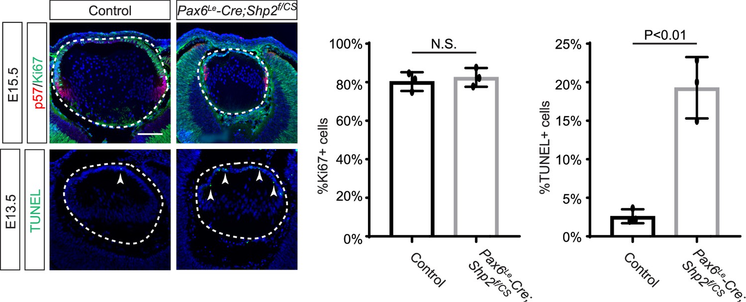
Cell proliferation and apoptosis inShp2CS mutants.
Pax6Le-Cre;Shp2f/CS lens exhibits normal expression of proliferation marker Ki67, but there is a significant increase in TUNLE+ cells (arrowheads). Student’s t-test, N.S. (not significant) for %Ki67+ cells and p<0.01 for %TUNEL+ cells in the lens epithelium. Scale bar:50 µm.
Figure 7

Shc1 complements Frs2 and Shp2 in mediating fibroblast growth factor (FGF) signaling in lens development.
(A) Fgfr1f/f;Fgfr2f/LR infected with Cre virus showed stronger pERK and pShc activation than Fgfr1f/ΔFrs;Fgfr2f/f mouse embryonic fibroblast (MEF) cells, despite both losing Frs2, Shp2, Gab1, and Crk phosphorylation. (B) Quantification of pShc levels. One-way ANOVA, n=3, *p<0.02. N.S. Not significant. (C) pShc staining was lost in Pax6Le-Cre;Fgfr1f/ΔFrs;Fgfr2f/f mutant lens (arrowhead) but preserved in Pax6Le-Cre;Fgfr1f/f;Fgfr2f/LR lens. (D) Shc1-deficient lenses showed a slight decrease in both pERK staining intensity and overall lens size. (E) E12.5 Frs2/Shc1 mutant lenses were smaller than controls, while lenses from Frs2/Shp2 mutants showed a pronounced hollow vesicle structure, a condition that worsened in Frs2/Shp2/Shc1 triple mutants. (F) Quantification of the lens size. One-way ANOVA, n=4, *p<0.001, **p<0.05. (G) Model of FGF signaling network. Frs2 recruits Grb2 directly and indirectly through Shp2, while Shc1 provides an alternate Grb2 recruitment route independent of Frs2. Scale bars:50 µm.
-
Figure 7—source data 1
Original files for western blot analysis displayed in Figure 7A.
- https://cdn.elifesciences.org/articles/103615/elife-103615-fig7-data1-v1.zip
-
Figure 7—source data 2
Original membranes corresponding to Figure 7, panel A, with the relevant lanes are outlined in yellow.
- https://cdn.elifesciences.org/articles/103615/elife-103615-fig7-data2-v1.zip
Tables
Key resources table
| Reagent type (species) or resource | Designation | Source or reference | Identifiers | Additional information |
|---|---|---|---|---|
| Strain (Adenovirus) | Ade-GFP | Iowa viral vector core | Cat#VVC-U of Iowa-4 | |
| Strain (Adenovirus) | Ade-cre | Iowa viral vector core | Cat#VVC-U of Iowa-1174 | |
| Genetic reagent (M. musculus) | Fgfr1DFrs | Hoch and Soriano, 2006 | RRID:MGI:3620075 | Dr. Raj Ladher (RIKEN Kobe Institute-Center for Developmental Biology, Kobe, Japan) |
| Genetic reagent (M. musculus) | Fgfr2LR | Eswarakumar et al., 2006 | RRID:MGI:3699819 | Dr. Jacob V.P. Eswarakumara (Yale University School of Medicine, New Haven, CT) |
| Genetic reagent (M. musculus) | Fgfr3flox | Su et al., 2010 | RRID:MGI:4459831 | Dr. Xin Sun (University of California at San Diego, San Diego, CA) |
| Genetic reagent (M. musculus) | Fgfr4-/- | Weinstein et al., 1998 | RRID:MGI:3653043 | Dr. Chu-Xia Deng (National Institute of Health, Bethesda, MD) |
| Genetic reagent (M. musculus) | Frs2flox | Lin et al., 2007 | RRID:MGI:3768915 | Fen Wang (Texas A&M, Houston, TX) |
| Genetic reagent (M. musculus) | Grb2flox | Ackermann et al., 2011 | RRID:MGI:4949890 | Dr. Lars Nitschke (University of Erlangen-Nürnberg, Erlangen, Germany) |
| Genetic reagent (M. musculus) | P6 5.0 lacZ | Makarenkova et al., 2000 | Dr. Paul A. Overbeek (Baylor College of Medicine, Houston, TX) | |
| Genetic reagent (M. musculus) | Shc1flox | Hardy et al., 2007 | RRID:MGI:3716783 | Tony Pawson (University of Toronto, Ontario, Canada) |
| Genetic reagent (M. musculus) | Shp2flox | Zhang et al., 2004 | RRID:MGI:3522138 | Gen-sheng Feng (UCSD, Sad Diego, CA) |
| Genetic reagent (M. musculus) | KrasG12D | Tuveson et al., 2004 | RRID:MGI:3044567 | Mouse Models of Human Cancers Consortium (MMHCC) Repository at National Cancer Institute |
| Genetic reagent (M. musculus) | Pax6Le-Cre | Ashery-Padan et al., 2000 | RRID:MGI:3045795 | Richard Lang (Children's Hospital Research Foundation, Cincinnati, OH) |
| Genetic reagent (M. musculus) | Fgfr1flox | Jackson Lab | RRID:MGI:3713779 | |
| Genetic reagent (M. musculus) | Baxflox/flox;BakKO/KO | Jackson Lab | RRID:IMSR_JAX:006329 | |
| Genetic reagent (M. musculus) | Sox2Cre | Jackson Lab | Stock #: 008454 RRID:IMSR_JAX:008454 | |
| Genetic reagent (M. musculus) | Fgfr2flox | Yu et al., 2003 | RRID:MGI:3044690 | Dr. David Ornitz, Washington University Medical School, St Louis, MO |
| Genetic reagent (M. musculus) | Shp2YF | This study | Described in Methods and Materials. Mice are available upon request. | |
| Genetic reagent (M. musculus) | Shp2CS | This study | Described in Methods and Materials. Mice are available upon request. | |
| Genetic reagent (M. musculus) | Grb2YF | This study | Described in Methods and Materials. Mice are available upon request. | |
| Cell line | Mouse embryonic fibroblast cells | Made from E13.5 embryos | Primary cells | |
| Antibody | Rabbit monoclonal anti-phospho-ERK1/2 | Cell Signaling | Cat#4370 RRID:AB_2315112 | IHC (1:200) |
| Antibody | Rabbit polyclonal anti-phospho-mTOR | Cell Signaling | Cat#2971 RRID:AB_330970 | IHC (1:100) |
| Antibody | Rabbit monoclonal anti-phospho-pS6 | Cell Signalling | Cat#5364 RRID:AB_10694233 | IHC (1:300) |
| Antibody | Rabbit polyclonal anti-phospho-Shc | Cell Signalling | Cat#2434 RRID:AB_10841301 | IHC (1:100) WB (1:500) |
| Antibody | Rabbit monoclonal anti-phospho-MEK1/2 | Cell Signalling | Cat#2338 RRID:AB_490903 | IHC (1:200) |
| Antibody | Rabbit monoclonal anti-phospho-Smad1/5/9 | Cell Signalling | Cat#13820 RRID:AB_2493181 | IHC (1:500) |
| Antibody | Rabbit Polyclonal Anti- Phospho-FRS2 (Y436) | Cell Signalling | Cat#3861 RRID:AB_2231950 | IHC (1:500) |
| Antibody | Rabbit polyclonal Anti-phospho-Shp2 (Tyr542) | Cell Signalling | Cat#3751 RRID:AB_330825 | WB (1:1000) |
| Antibody | Mouse monoclonal Anti-pERK(E-4) | Santa Cruz Biotechnology | Cat#7383 RRID:AB_627545 | WB (1:2000) |
| Antibody | Rabbit monoclonalAnti-phospho-Gab1 (Y307) | Cell Signalling | Cat#3233 RRID:AB_2107683 | WB (1:500) |
| Antibody | Rabbit anti-phospho-crk II | Cell Signalling | Cat#3491 RRID:AB_2229920 | WB (1:1000) |
| Antibody | Rabbit monoclonal anti-LEF1 | Cell Signalling | Cat#2230 RRID:AB_823558 | IHC (1:300) |
| Antibody | Rabbit monoclonal anti-Cleaved caspase3 | Cell Signalling | Cat#9664 RRID:AB_2070042 | IHC (1:100) |
| Antibody | Mouse monoclonal anti-cyclin D1 | Cell Signalling | Cat#2926 RRID:AB_2070400 | IHC (1:100) |
| Antibody | Rabbit monoclonal anti-cyclin D1 | Cell Signalling | Cat#2978 RRID:AB_2259616 | IHC (1:100) |
| Antibody | Mouse monoclonal anti-cyclin D3 | Cell Signalling | Cat#2936 RRID:AB_2070801 | IHC (1:100) |
| Antibody | Rabbit monoclonal anti-N-cadherin | Cell Signalling | Cat#13116 RRID:AB_2687616 | IHC (1:300) |
| Antibody | Rabbit polyclonal anti-Fibronectin | Millipore Sigma | Cat#AB2033 | IHC (1:300) |
| Antibody | Mouse monoclonal anti-N-cadherin | BD | Cat#610920 RRID:AB_2077527 | IHC (1:200) |
| Antibody | Rabbit polyclonal anti-Maf | Santa Cruz Biotechnology | Cat#sc-7866 RRID:AB_638562 | IHC (1:200) |
| Antibody | Rabbit Polyclonal anti-Jag1 | Santa Cruz Biotechnology | Cat#sc-6011 RRID:AB_649689 | IHC (1:200) |
| Antibody | Mouse monoclonal anti-Foxe3 | Santa Cruz Biotechnology | Cat#sc-377465 | IHC (1:100) |
| Antibody | Mouse monoclonal anti-Ecadherin | BD | Cat#610181 RRID:AB_397580 | IHC (1:200) |
| Antibody | Mouse monoclonal anti-Ki67 | BD | Cat#550609 RRID:AB_393778 | IHC (1:200) |
| Antibody | Chicken Polyclonal anti-GFP | Aves Labs | Cat#GFP-1010 RRID:AB_2307313 | IHC (1:200) |
| Antibody | Rabbit monoclonal anti-p57 | Abcam | Cat# 75947 | IHC (1:200) |
| Antibody | Rabbit Polyclonal anti-Prox1 | Covance | Cat#PRB-238C RRID:AB_291595 | IHC (1:200) |
| Antibody | Rabbit Polyclonal anti-Pax6 | Covance | Cat#PRB-278P RRID:AB_291612 | IHC (1:300) |
| Antibody | Rat monoclonal anti-Sox2 | Thermo Fisher | Cat#14-9811-82 RRID:AB_11219471 | IHC (1:200) |
| Antibody | Rabbit Polyclonal anti-α-crystallin | Sam Zigler (National Eye Institute) | IHC (1:5000) | |
| Antibody | Rabbit Polyclonal anti-γ-crystallin | Sam Zigler (National Eye Institute) | IHC (1:5000) | |
| Peptide, recombinant protein | Recombinant murine FGF2 | ScienCell | Cat# 124–02 | |
| Peptide, recombinant protein | PDGF-AA | R&D | Cat#221-aa | |
| Chemical compound | Phallodin-488 | Life Technology | A12379 | IHC (1:200) |
| Commercial assay Kit | In situ cell death detection kit | Roche | Cat# 1168479510 |
Additional files
Download links
A two-part list of links to download the article, or parts of the article, in various formats.
Downloads (link to download the article as PDF)
Open citations (links to open the citations from this article in various online reference manager services)
Cite this article (links to download the citations from this article in formats compatible with various reference manager tools)
Shc1 cooperates with Frs2 and Shp2 to recruit Grb2 in FGF-induced lens development
eLife 13:RP103615.
https://doi.org/10.7554/eLife.103615.4


