Reward signal in a recurrent circuit drives appetitive long-term memory formation
- Tohoku University, Japan
- Max Planck Institute of Neurobiology, Germany
- Janelia Research Campus, Howard Hughes Medical Institute, United States
Figures
Figure 1 with 1 supplement
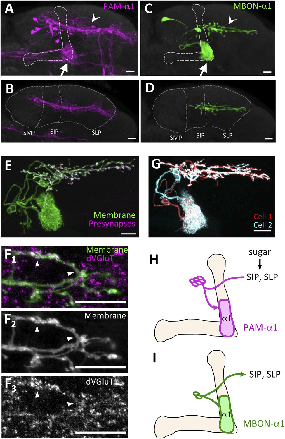
MBON-α1 receives inputs from MB-α1 and projects to the dendrites of PAM-α1.
(A–D) Anatomy of PAM-α1 (A, B) and MBON-α1 (C, D). Dotted line indicates α/β lobes in the mushroom body (MB) (A, C) or SMP, SIP, and SLP (superior medial, intermediate, and lateral protocerebra, respectively) (B, D). Arrows indicate α1 and arrowheads the SIP and SLP (A, C). PAM-α1 and MBON-α1 are visualized by pJFRC2-10xUAS-mCD8GFP in VK00005 and MB299B-GAL4 (A, B) or MB310C-GAL4 (C, D), respectively. (E) Presynaptic terminals of MBON-α1 are highly localized in SIP and SLP. MB310C-GAL4 is used to drive a general membrane marker (green) and a presynaptic marker (magenta) in MBON-α1. (F) Double labeling of the membrane of MBON-α1 (green) and anti-vesicular glutamate transporter (dVGluT, magenta) (F1), membrane staining (F2), and anti-dVGluT staining (F3). Arrowheads highlight the overlap. (G) Two individual MBON-α1 neurons visualized by multi-color flip-out with different colors (red and cyan). (H, I) Schematics of PAM-α1 and MBON-α1, respectively. The α/β lobe of the MB is outlined with light orange. Scale bars, 10 µm.
Figure 1—figure supplement 1
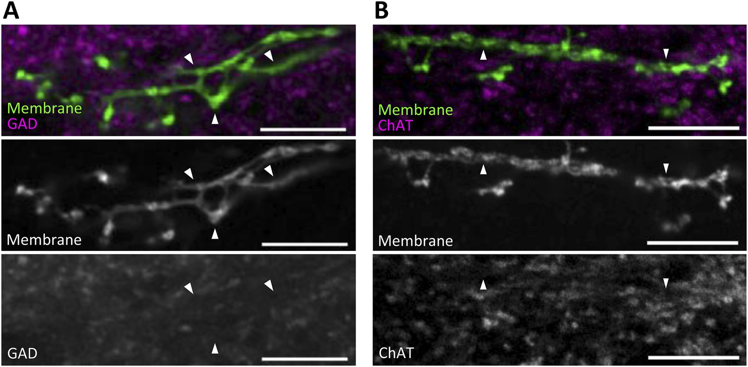
MBON-α1 is neither GABAergic nor cholinergic.
Double labeling of the membrane of MBON-α1 (green) and anti-glutamic acid decarboxylase (GAD1) (A, magenta) or choline acetyltransferase (ChAT) (B, magenta). Arrowheads highlight some of the processes, where no obvious overlaps are observed. Scale bars, 10 µm.
Figure 2 with 1 supplement
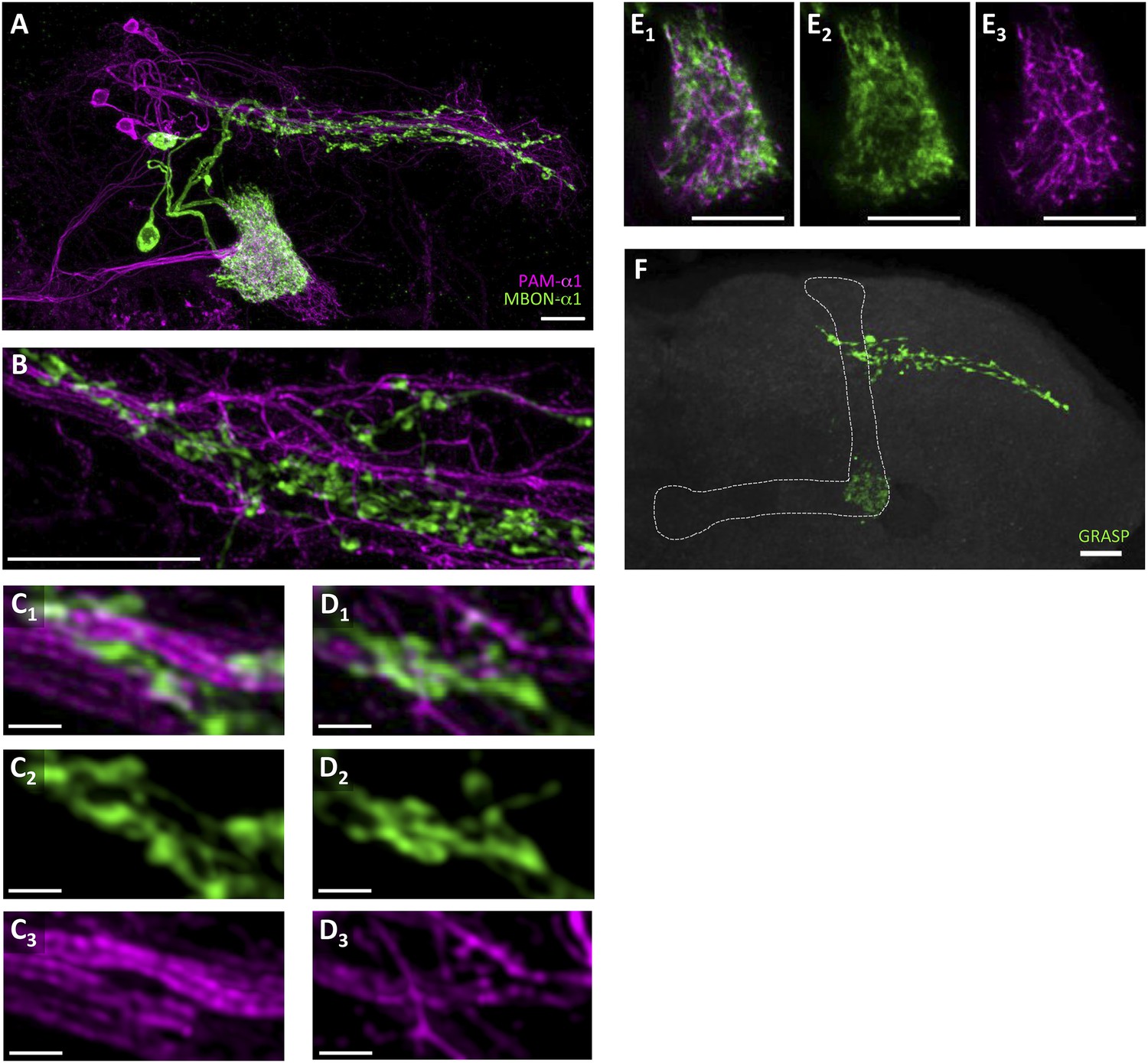
Processes of PAM-α1 and MBON-α1 intermingle each other.
(A) Double labeling of PAM-α1 and MBON-α1. PAM-α1 (magenta) and MBON-α1 (green) are visualized using R72D01-LexA and MB310C-GAL4, respectively. (B–D) Magnified substack images in SIP and SLP, obtained by a super-resolution detection system. (E) Magnified substack image in the α1 compartment in the MB. (F) GFP reconstitution across synaptic partner (GRASP) signals in SIP, SLP, and MB α1 support the contacts between PAM-α1 and MBON-α1. Scale bars, 10 µm (A, B, E, F), 1 µm (C, D).
Figure 2—figure supplement 1
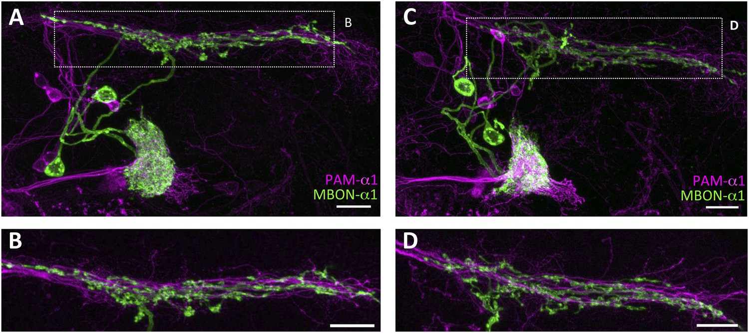
Stereotyped projections of PAM-α1 and MBON-α1.
(A, C) Two additional examples of the double labeling confirm stereotypy of the projections. (B, D) Magnified substacks for the insets in (A) and (C), respectively. Scale bars, 10 µm.
Figure 3 with 1 supplement
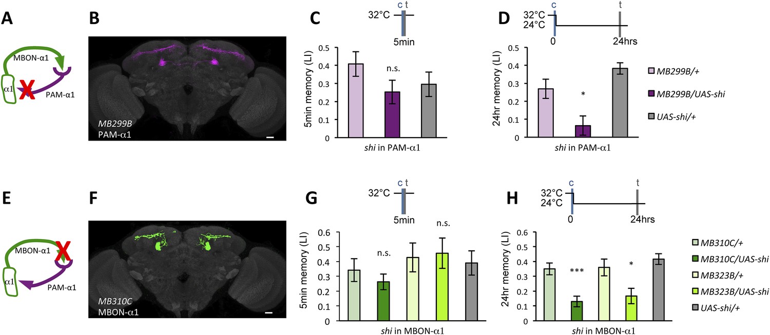
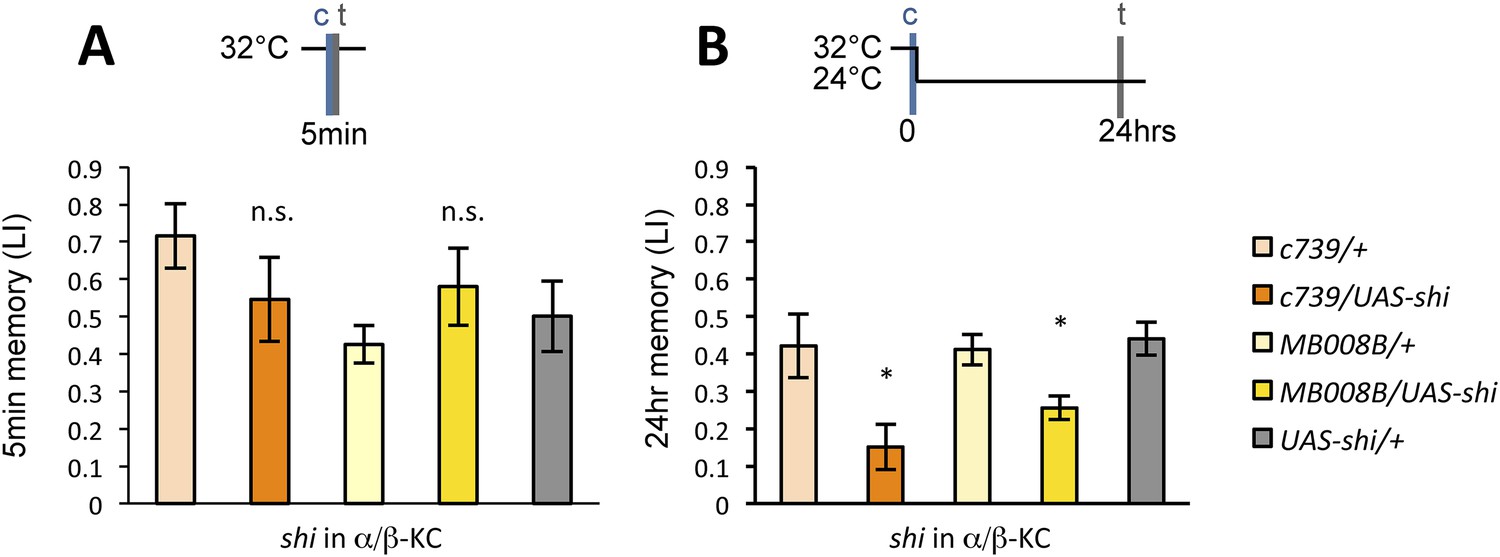
Feedback from MBON-α1 is required for appetitive LTM formation.
(A) Diagram of the experiment. (B) Expression pattern of MB299B-GAL4. (C) Blockade of PAM-α1 does not impair STM significantly. 5-min appetitive memory was measured (n = 10, 10, 12). c: conditioning, t: test. (D) Blockade of PAM-α1 during conditioning impairs LTM. 24-hr appetitive LTM was measured (n = 9, 12, 12). (E) Diagram of experiment. (F) Expression pattern of MB310C-GAL4. (G) Blockade of MBON-α1 does not impair STM significantly (n = 6, 7, 10, 6, 10). (H) Blockade of MBON-α1 during conditioning impairs LTM. MB323B-GAL4 is a second driver line that expresses in MBON-α1 (see figure supplement; n = 23, 24, 13, 14, 24). Bar graphs are mean ± s.e.m. *: p < 0.05, ***: p < 0.001, n.s.: p > 0.05. Scale bars, 20 µm.
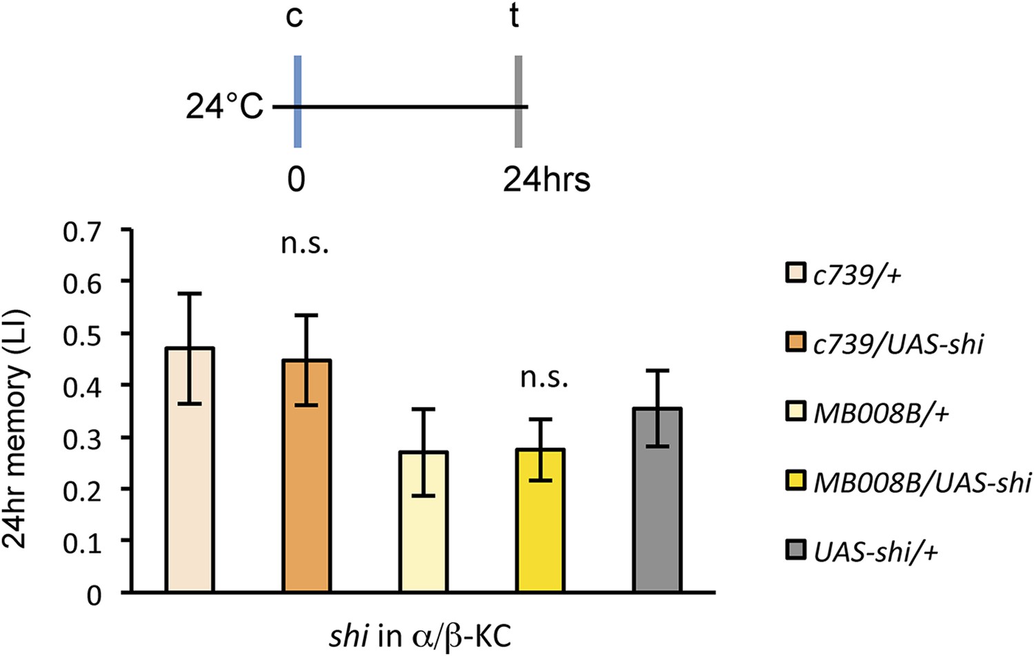
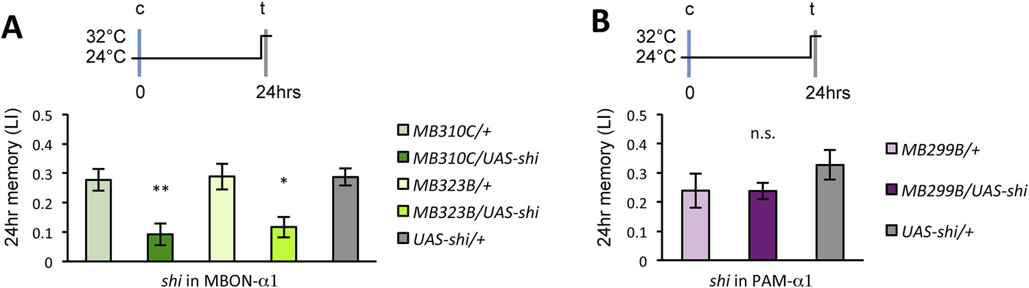
Figure 3—figure supplement 1
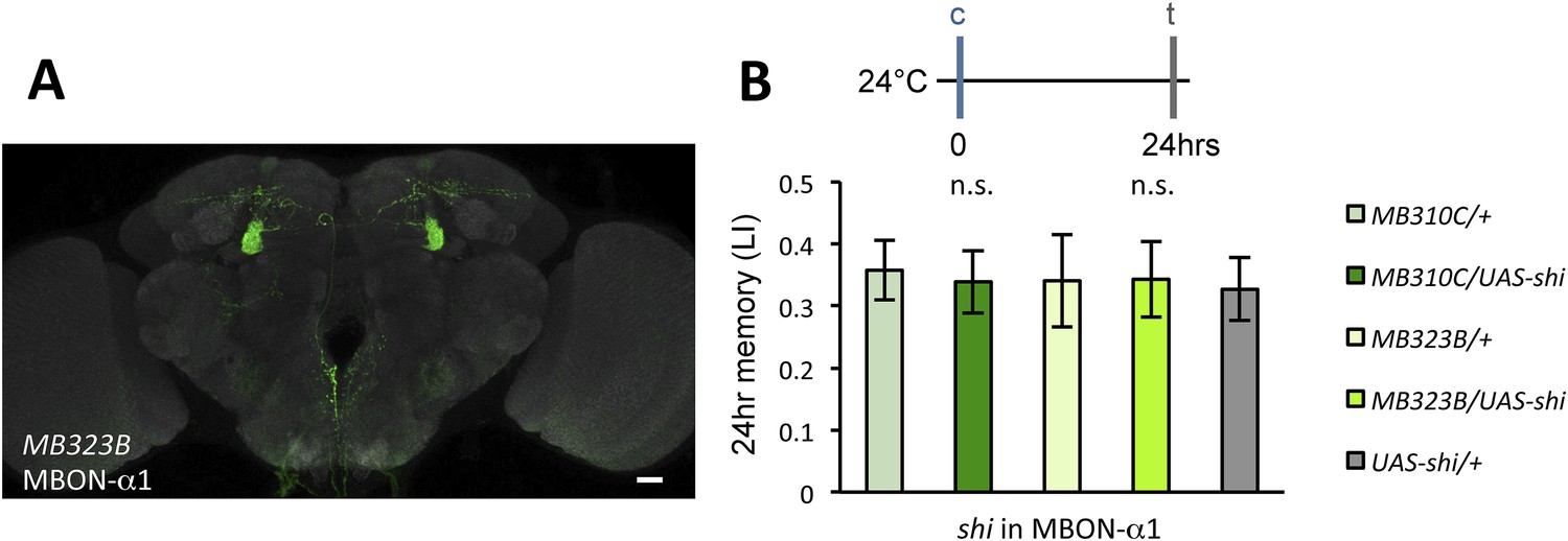
(A) Expression pattern of MB323B-GAL4.
Scale bar, 20 µm. (B) The LTM performance of MB310C/UAS-shi and MB323B/UAS-shi flies at the permissive temperature is not affected (n = 17, 16, 8, 9, 11). n.s.: p > 0.05.
Figure 4
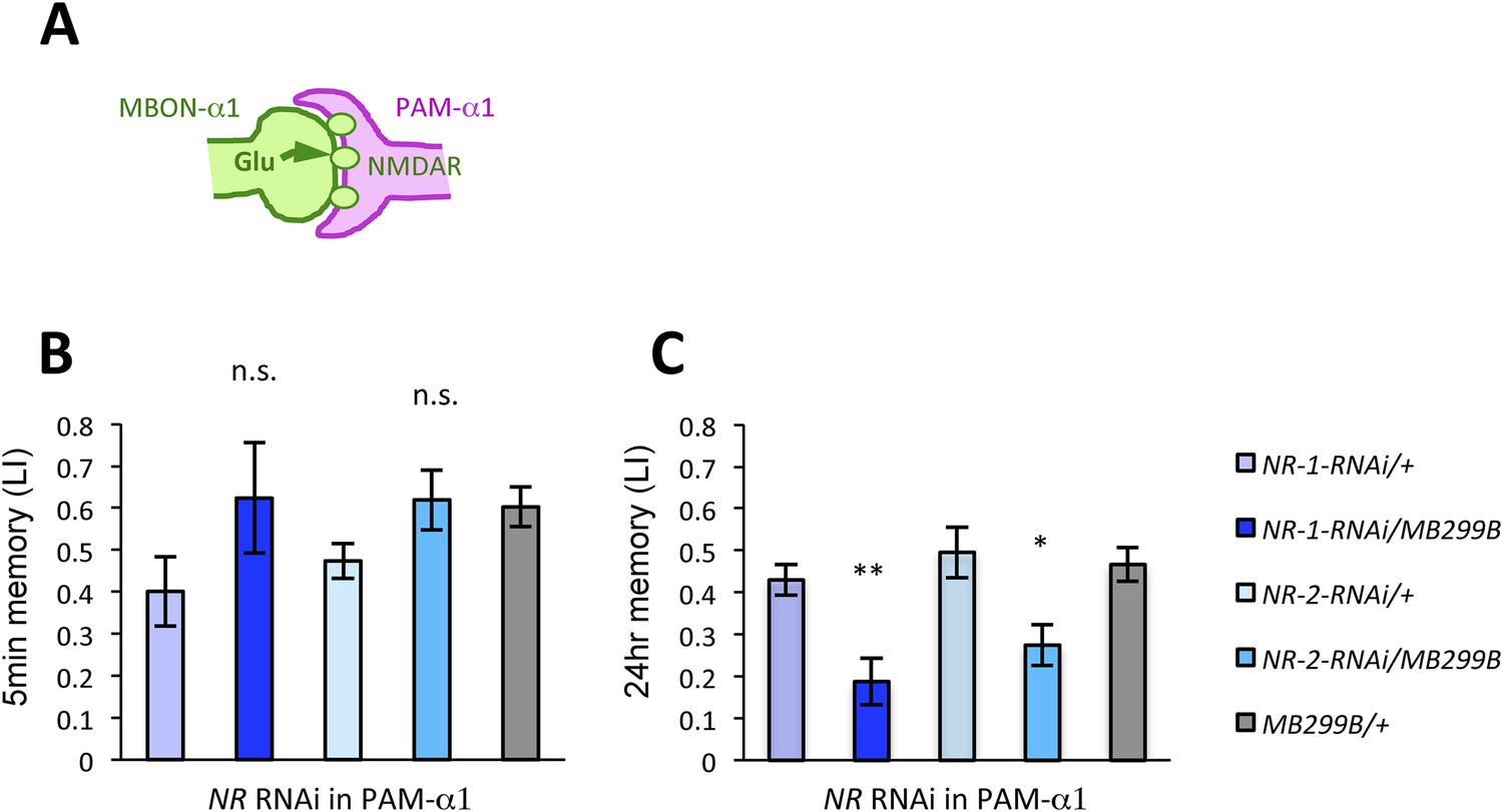
NMDA receptors in PAM-α1 are required for appetitive LTM.
(A) The subunits of NMDA receptor are down-regulated in PAM-α1. (B, C) Knocking down NMDA receptor subunits does not impair 5-min memory (B: n = 16, 8, 13, 12, 13) but impairs 24-hr memory significantly (C: n = 18, 8, 22, 20, 29). Bar graphs are mean ± s.e.m. *: p < 0.05, **: p < 0.01, n.s.: p > 0.05. Scale bars, 20 µm.
Figure 5

MBON-α1 signals reward for appetitive LTM.
(A) Activation of MBON-α1 was paired with odor presentations and 24-hr memory was measured. (B) Activation of MBON-α1 induces appetitive LTM formation (n = 16, 24, 24). Bar graphs are mean ± s.e.m. *: p < 0.05, n.s.: p > 0.05. Scale bars, 20 µm.
Figure 6 with 1 supplement
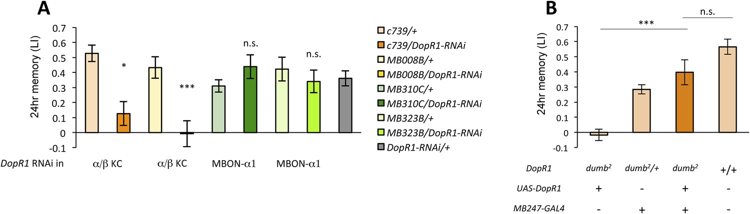
α/β KCs receive dopamine signals through DopR1 for LTM formation.
(A) Knocking down DopR1 in α/β Kenyon cells (KCs) but not in MBON-α1 impairs appetitive LTM (n = 17, 10, 11, 11, 18, 12, 7, 12, 27). (B) KC expression of DopR1 in the dumb2 mutant background fully rescues the LTM impairment (n = 12, 10, 7, 13). Bar graphs are mean ± s.e.m. *: p < 0.05, ***: p < 0.001.
Figure 6—figure supplement 1
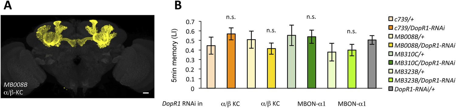
(A) Expression pattern of MB008B-GAL4.
Scale bar, 20 µm. (B) Knocking down DopR1 in α/β KCs or MBON-α1 does not impair appetitive STM, suggesting normal sugar or odor perception of these flies (n = 7, 7, 12, 10, 10, 10, 9, 9, 22). Bar graphs are mean ± s.e.m. n.s.: p > 0.05.
Figure 7 with 1 supplement
Output of α/β KCs has an essential role in the acquisition of appetitive LTM.
(A) Blockade of α/β KCs does not impair STM significantly (n = 8, 7, 7, 8, 9). (B) Blockade of α/β KCs during conditioning impairs LTM (n = 7, 7, 18, 19, 20). Bar graphs are mean ± s.e.m. *: p < 0.05, n.s.: p > 0.05.
Figure 7—figure supplement 1
Training and testing c739/UAS-shi and MB008B/UAS-shi flies at the permissive temperature do not impair appetitive LTM (n = 7, 6, 8, 9, 6).
Bar graphs are mean ± s.e.m. n.s.: p > 0.05.
Figure 8
Appetitive LTM is read out through MBON-α1.
(A) Blocking MBON-α1 during test impairs appetitive LTM retrieval (n = 20, 21, 11, 13, 20). (B) Blocking PAM-α1 during test does not significantly impair LTM retrieval (n = 7, 7, 10). Bar graphs are mean ± s.e.m. *: p < 0.05, **: p < 0.01, n.s.: p > 0.05.
Figure 9

Dopamine release from PAM-α1 is required for appetitive LTM consolidation.
(A, C, E) Blocking PAM-α1 (A), MBON-α1 (C), or α/β−KC (E) for 1 hr immediately after conditioning impairs appetitive LTM (n = 16, 15, 22 (A); n = 15, 16, 14, 20, 34 (C); n = 9, 8, 10, 7, 15 (E)). (B, D, F) Blocking PAM-α1 (B), MBON-α1 (D), or α/β−KC (F) for 1 hr, 22 hr after conditioning does not impair appetitive LTM (n = 15, 15, 14 (B); n = 16, 15, 13, 13, 23 (D); n = 12, 12, 15, 12, 15 (F)). Bar graphs are mean ± s.e.m. *: p < 0.05, ***: p < 0.001, n.s.: p > 0.05.
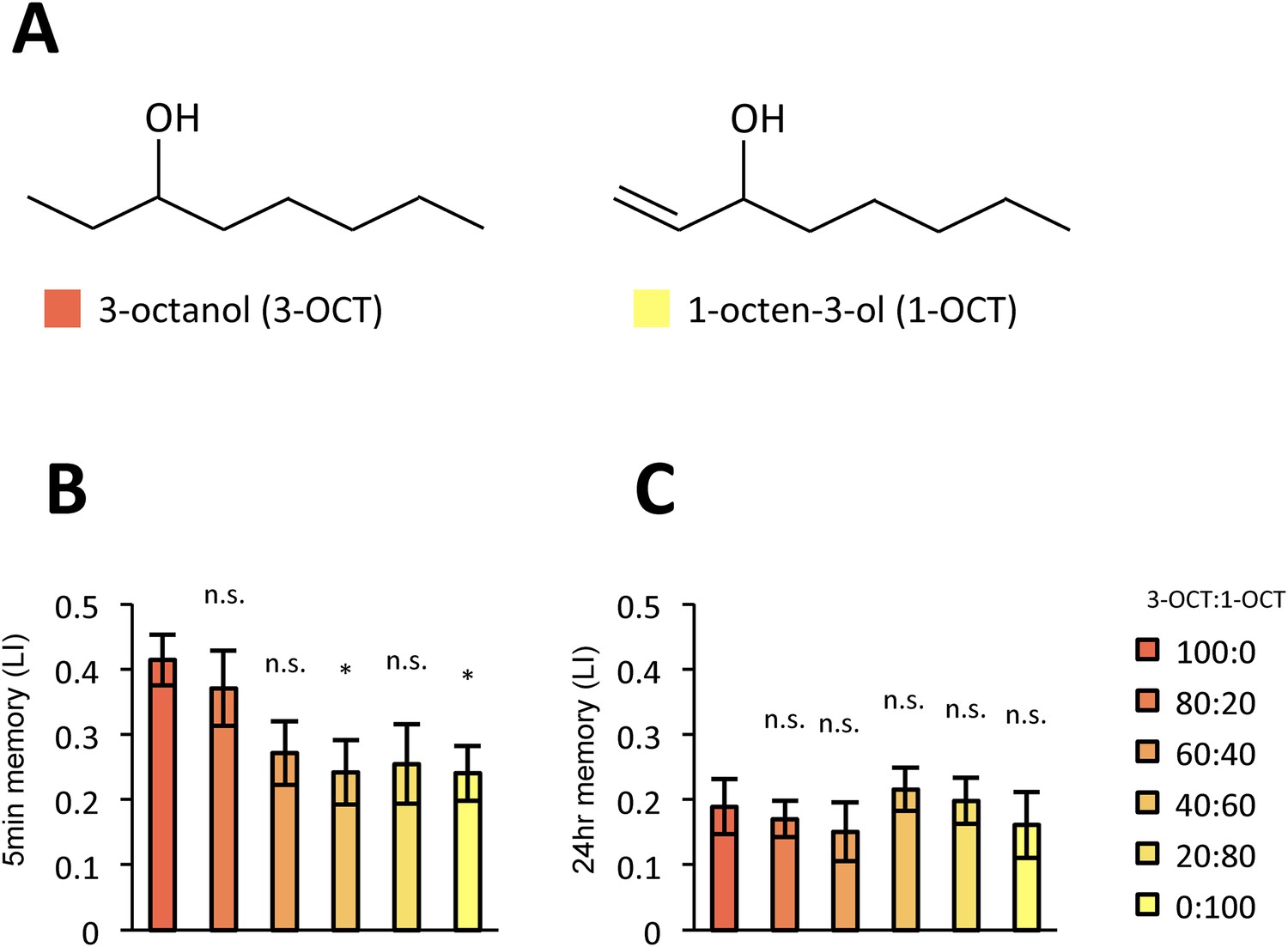
Figure 10 with 1 supplement

LTM specificity is compromised.
(A) Chemical structures of odorants. (B) Design of the experiment. For one group, presentation of 4-methylcyclohexanol was paired with sugar reward. The reciprocal group received sugar without odor. In the test situation, flies of each group were allowed to choose between the air and the mixture of the trained odor and the ‘contaminant’ (2-methylcyclohexanol). (C, D) The performance index declined more sharply with the increasing contamination ratio in STM (C; n = 16 for each) than in LTM (D; n = 24 for each). Each group was compared to the group that was tested without the contaminant (left most bar). Bar graphs are mean ± s.e.m. *: p < 0.05, **: p < 0.01, ***: p < 0.001, n.s.: p > 0.05.
Figure 10—figure supplement 1
LTM is less specific with another pair of odors.
(A) Chemical structures of odorants. (B, C) The performance declined more sharply in STM (B: n = 20 for each) than in LTM (C: n = 20 for each) also with this odor pair. Each group was compared to the group that was tested without the contaminant (left most bar). Bar graphs are mean ± s.e.m. *: p < 0.05, n.s.: p > 0.05.
Figure 11
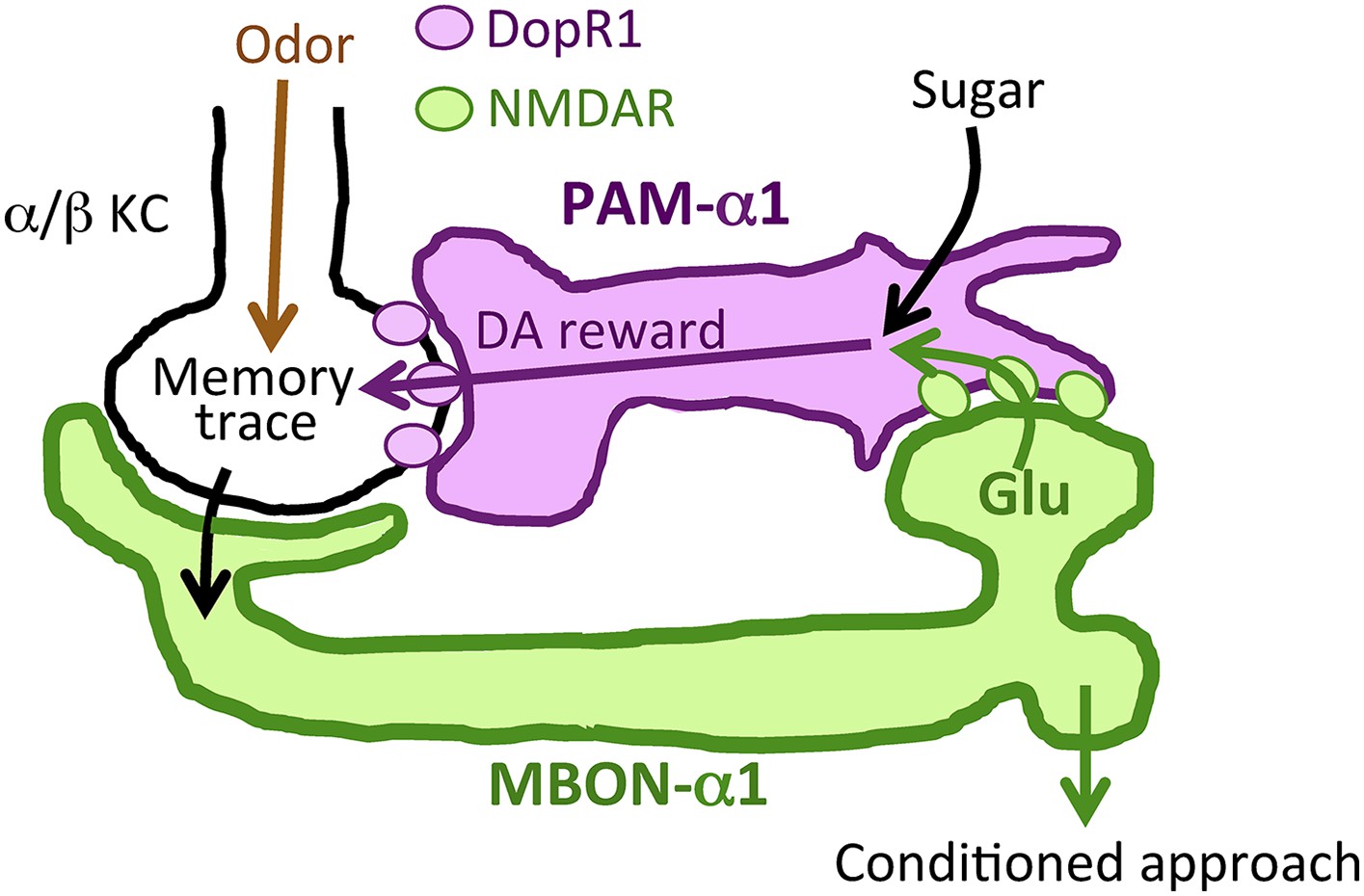
Recurrent reward circuit drives appetitive LTM formation.
Feedback circuit model of appetitive LTM formation. Neuronal signal of a sugar reward mediated by PAM-α1 converges with olfactory information in the α/β KCs. The coincidental signal is read out through MBON-α1 to give glutamatergic feedback onto PAM-α1 for the gain control of the dopamine release. Appetitive LTM is read out through MBON-α1 to activate other downstream targets.
Videos
Video 1
Volume-rendered image of processes of PAM-α1 (magenta) and MBON-α1 (green) in SIP and SLP.
https://doi.org/10.7554/eLife.10719.007Download links
A two-part list of links to download the article, or parts of the article, in various formats.
Downloads (link to download the article as PDF)
Open citations (links to open the citations from this article in various online reference manager services)
Cite this article (links to download the citations from this article in formats compatible with various reference manager tools)
Reward signal in a recurrent circuit drives appetitive long-term memory formation
eLife 4:e10719.
https://doi.org/10.7554/eLife.10719


