Direct neural pathways convey distinct visual information to Drosophila mushroom bodies
- Max-Planck Institut für Neurobiologie, Germany
- Tohoku University Graduate School of Life Sciences, Japan
- Janelia Research Campus, Howard Hughes Medical Institute, United States
- Cold Spring Harbor Laboratory, United States
Figures
Figure 1 with 1 supplement
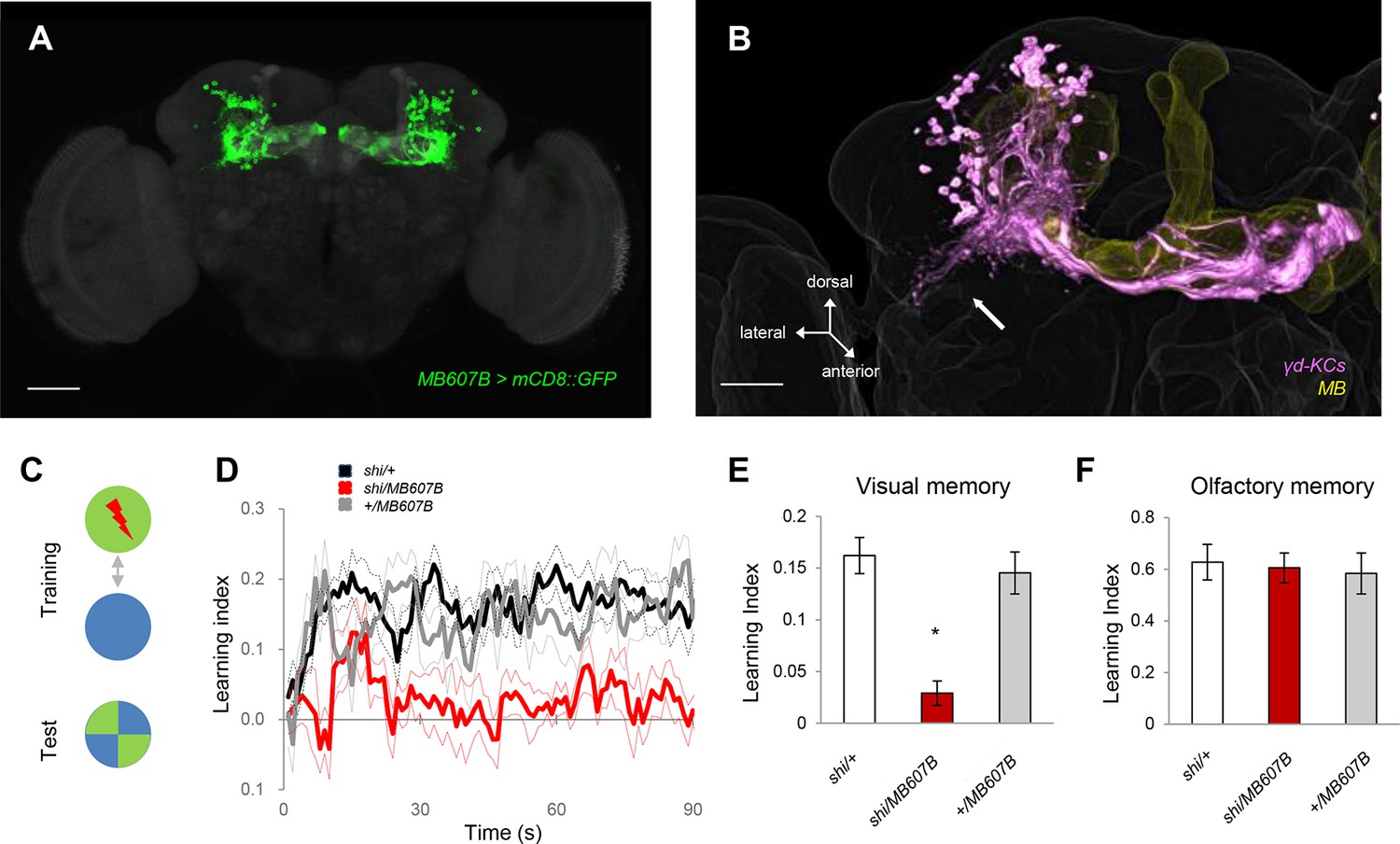
The γd KCs are required for visual memory.
(A) γd neurons labeled by MB607B-GAL4. (B) 3D reconstruction of γd neurons labeled by MB607B-GAL4 (purple) in the entire MB (yellow). Arrow indicates atypical dendritic protrusion of the γd neurons. Scale bars: 50 µm (A) and 20 µm (B). (C) Schematic diagram of color discrimination learning and test. (D) Average time courses of conditioned color avoidance in the test for flies with the blockade of the γd neurons with MB607B-GAL4 (red) and the parental controls (black and gray). (E) Pooled conditioned color avoidance. Blocking the γd neurons with MB607B-GAL4 impairs aversive color discrimination learning (one-way ANOVA, post-hoc pairwise comparison, p<0.05; n = 8–12). (F) The same Shits1 blockade of the γd neurons does not impair immediate aversive olfactory memory (one-way ANOVA, post-hoc pairwise comparison, p>0,05; n = 9–10). Throughout this study, bars and error bars display mean and SEM, respectively.
Figure 1—figure supplement 1
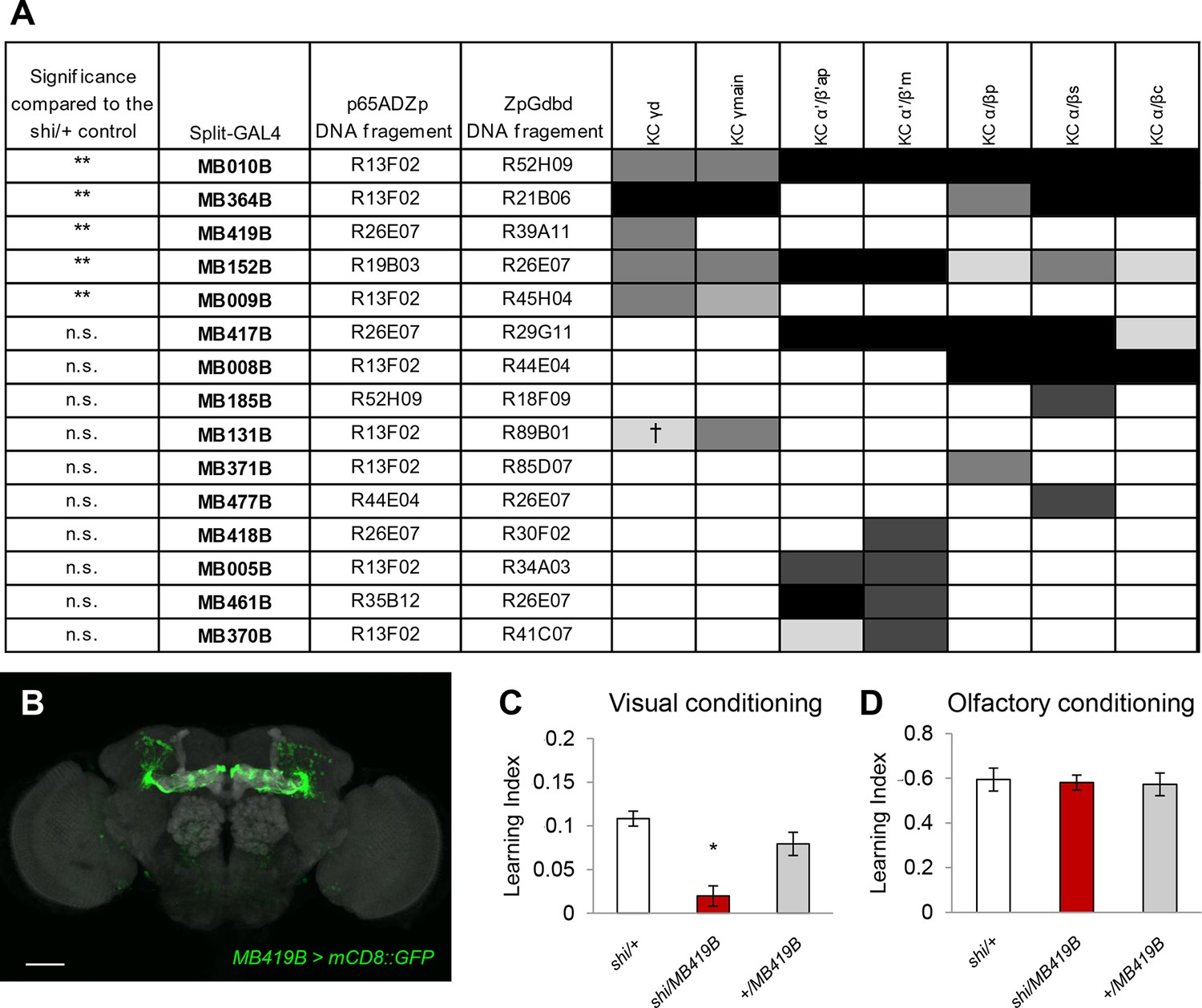
Behavioral screen identifies the requirement of the γd KCs in aversive visual conditioning but not olfactory conditioning.
(A) Subsets of KC types labelled in GAL4 lines and the resulting learning defects in the behavioral screen using UAS-shits1. Expression levels (represented in the gray scale) are determined as previously described (Paulk and Gronenberg, 2008); † stochastic expression. Stars in the left column indicate the statistical significance of memory impairment compared to the UAS-shi/+ control (Dunn’s multiple pairwise comparison); n = 8–119. (B) Expression pattern of MB419B-GAL4 which specifically labels γd KCs. Scale bar represents 50 µm. Grey background staining: Discs large. (C) Blocking output of the γd neurons with MB419B-GAL4 (B; one-way ANOVA, post-hoc pairwise comparison, p<0.05; n = 18–60) impairs aversive color discrimination learning. (D) The same blockade does not significantly affect aversive olfactory conditioning (one-way ANOVA, p>0.05; n = 8–10).
Figure 2

γd neurons are atypical KCs and respond to visual stimuli.
(A–B) Main output and input sites labeled by Syt::GFP (green) and DenMark::mCherry (red) are differentially localized to the dorsal γ lobe and the vAC (arrow). The MB lobe (A) and main calyx (B) are outlined. P: MB peduncle (C–D) The γd dendrites (green) enwrap presynaptic terminals (gray; arrows). A single optical slice of the inset in the projection in C is magnified in D-D’’. P: MB peduncle. Scale bars: 50 µm (A–B); 20 µm (C); 2 µm (D-D’’). (E) Responses to light and odor stimulation in γd KCs measured with whole-cell current-clamp recordings. Data from four representative neurons are shown (each column corresponds to the data from one cell). Voltage traces of individual trials (gray lines, 5–7 trials) are overlaid with the mean (colored line). Raster plots below the traces represent spikes. Stimulus presentation is indicated below each trace (duration = 1 s). For odors, three of five tested odors are displayed (OCT: 3-octanol; MCH: 4-methylcyclohexanol; HEP: 2-heptanone). (F) Responses in two representative α/β KCs. (G) Modality segregation by γd (n = 12 cells) and α/β KCs (n = 11 cells). Each of the pie charts represents 24 (γd) or 22 (α/β) light-cell pairs measured in 6 flies and 60 (γd) or 55 (α/β) odor-cell pairs measured in 3 flies. The distributions of all four response categories are significantly different between γd KCs and α/β KCs with respect to both visual (p<10–5, Fisher’s exact test) and odor responses (p<10–6) See Materials and methods for details.
Figure 3 with 3 supplements
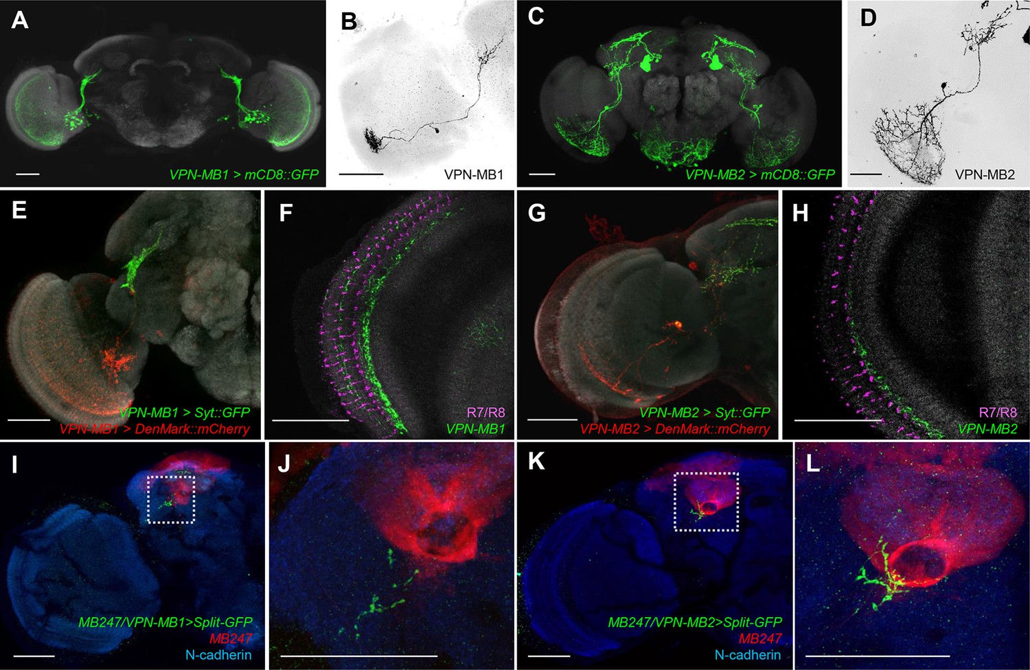
VPNs directly convey optic lobe inputs to the MB vAC.
(A) VPN-MB1 neurons labeled by MB425B-GAL4. (B) A single VPN-MB1 neuron, generated by heat shock flip out, connects the medulla and the central brain. (C) VPN-MB2 neurons labeled by MB334C-GAL4. (D) VPN-MB2 neurons connect the medulla and the central brain. (E) VPN-MB1 has dendrites (DenMark::mCherry, red) in the ventral medulla and presynaptic terminals in the central brain (Syt::GFP, green). (F) The dendrites of VPN-MB1 (green) arborize in the M8 layer. (G) VPN-MB2 has dendrites (DenMark::mCherry, red) in the ventral medulla and presynaptic terminals in the central brain (Syt::GFP, green). (H) The dendrites of VPN-MB2 (green) arborize in the M7 layer. (I–J) Reconstituted GFP signals visualize contacts between KCs and VPN-MB1 in the vAC. (K–L) Reconstituted GFP signals visualize contacts between KCs and VPN-MB2 in the vAC. J and L are magnifications of the insets in I and K. Scale bars represent 50 µm.
Figure 3—figure supplement 1
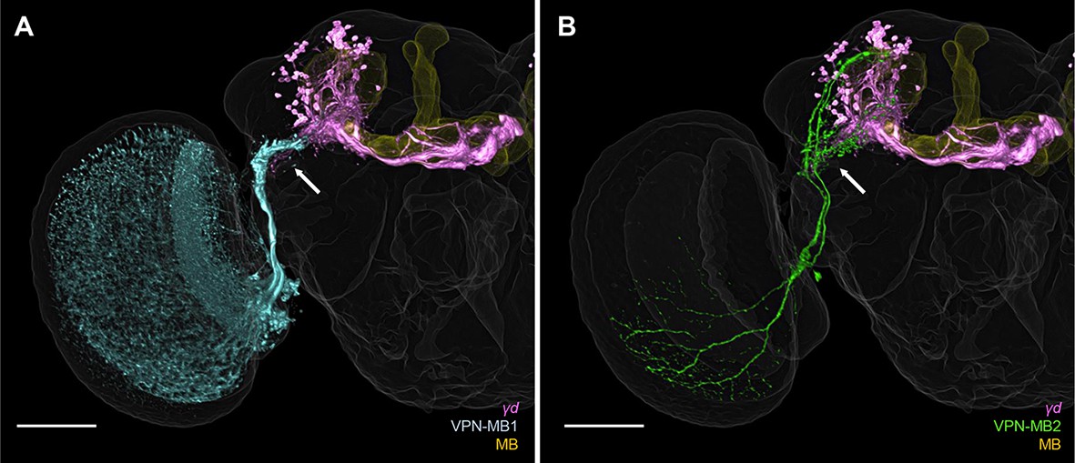
3D reconstruction of VPNs and γd neurons (purple: MB419B-GAL4) registered in a standard brain reveals overlapping processes in the vAC (arrow).
Yellow: entire MB. (A) Blue: MB425B-GAL4. (B) Green: MB334C-GAL4. Scale bar: 50 µm.
Figure 3—figure supplement 2
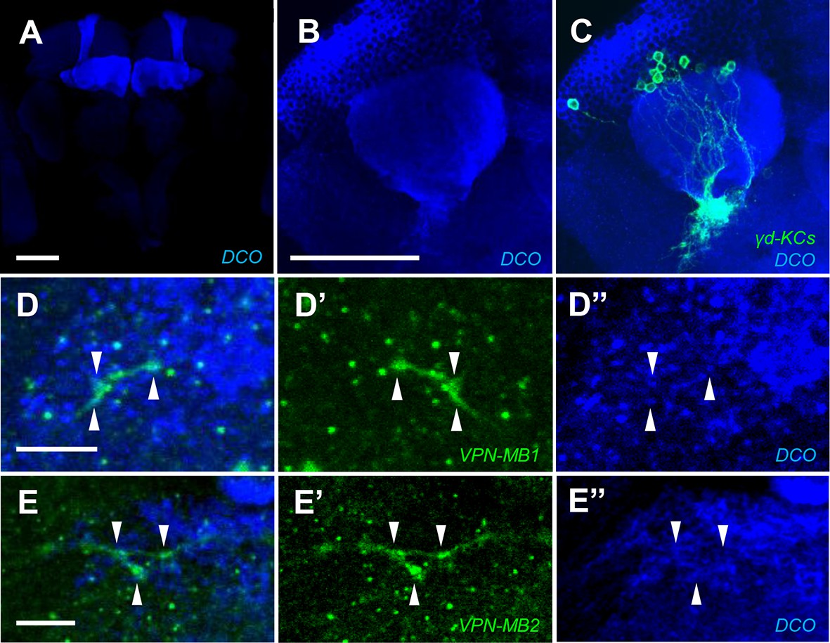
VPN axons overlap with KC processes in the vAC labeled with DC0.
(A–C) The DC0 antibody highlights KCs including the γd neurons labeled by MB419B-GAL4. (D and E) The γd dendrites overlap with the processes of VPN-MB1 (MB425B-GAL4) (D) and VPN-MB2 (MB334C-GAL4) (E) Scale bars represent 50 µm (A and B) and 5 µm (D and E).
Figure 3—figure supplement 3
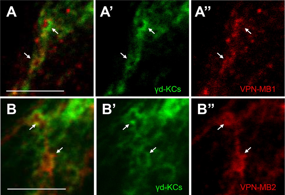
VPNs connect to the γd vAC.
(A–A”) Double labeling of VPN-MB1 (MB425B-GAL4, red) and KCs (R13F02-LexA for labeling most KCs, green) reveals overlap in the vAC (arrows). (B–B”) Double labeling of VPN-MB2 (MB334C-GAL4, red) and KCs (R13F02-LexA, green) reveals overlap in the vAC (arrows). Scale bar: 5 µm.
Figure 4 with 2 supplements
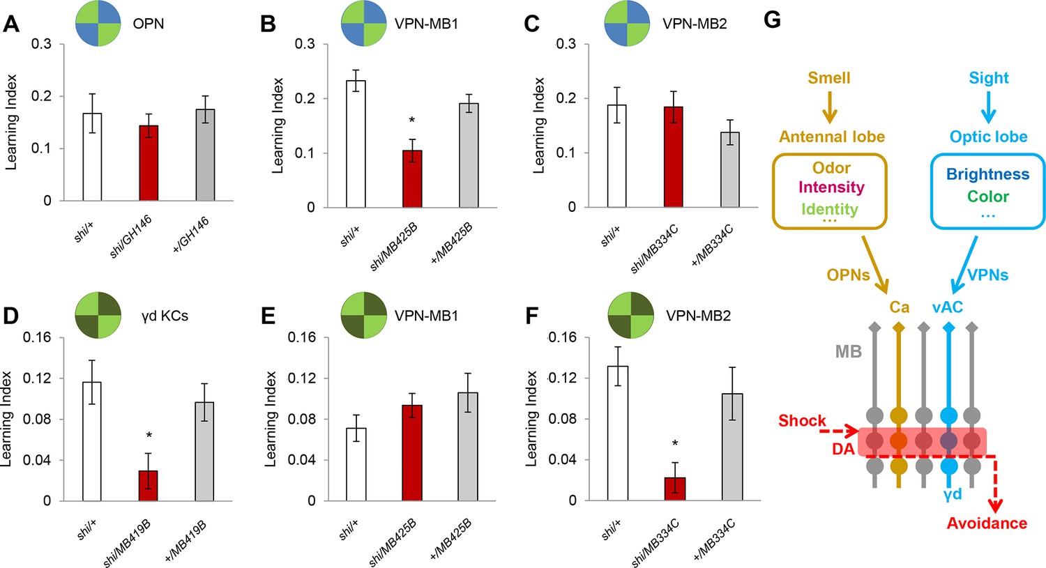
VPN-MB1 and VPN-MB2 convey distinct visual features.
(A) OPNs labeled by GH146-GAL4 are not required for visual color conditioning (one-way ANOVA, p>0.05), n = 8. (B–C) VPN-MB1 (MB425B-GAL4; B), but not VPN-MB2 (MB334C-GAL4; C), are required for color discrimination learning (one-way ANOVA, post-hoc pairwise comparison, p<0.01). n = 9–12. (D) γd neurons labeled by MB419B-GAL4 are required for green intensity learning (one-way ANOVA, post-hoc pairwise comparison, p<0.05), n = 9–11; these neurons are also required for color discrimination learning (Figure 1). (E–F) In contrast to the requirement in color discrimination learning, the blockade of VPN-MB2 (MB334C-GAL4; F), but not VPN-MB1 (MB425B-GAL4; E), significantly impaired intensity discrimination learning (one-way ANOVA, post-hoc pairwise comparison, p<0.05). n = 8–13. (G) Schematic of memory circuits in the MB. Visual and olfactory information is first processed in the optic lobe and antennal lobe, respectively. Components of sensory information (e.g. brightness and color) are separately processed there and conveyed to corresponding KC subtypes in the MB directly through distinct projection neurons Ca: calyx, vAC: ventral accessory calyx. These segregated representations of visual and olfactory information undergo the same dopaminergic (DA) valence modulation to operate acquired behavior (e.g. conditioned avoidance) via shared circuits.
Figure 4—figure supplement 1
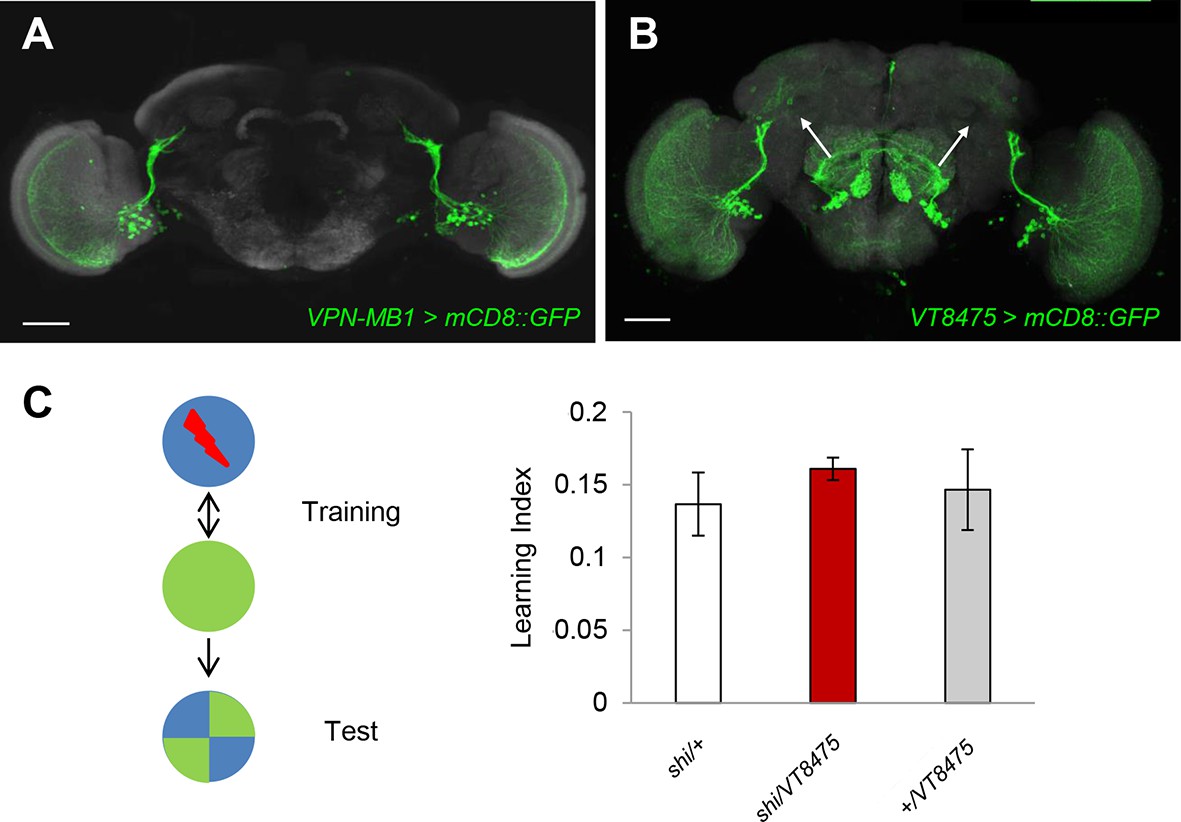
The blockade of similar VPNs without vAC connection does not impair color discrimination learning.
(A and B) MB425B-GAL4 (A) and VT008475-GAL4 label VPN-MB1 and similar VPNs that do not extend to the vAC (arrows). Scale-bars represents 50 µm. (C) Blocking output of neurons labeled by VT008475-GAL4 does not impair color learning (one-way ANOVA, p>0.05), n = 8.
Figure 4—figure supplement 2

The blockade of MBON-a1 does not impair intensity discrimination learning.
(A) MB331C-GAL4 labels MBON-a1 (arrows) in addition to VPN-MB2. (B) MB310C-GAL4 labels MBON-a1 (arrows) but not VPN-MB2. Scale-bars represents 50 µm. (C) Blocking output of neurons labeled by MB310C-GAL4 does not impair intensity learning (one-way ANOVA, p >0.05), n = 8–11.
Download links
A two-part list of links to download the article, or parts of the article, in various formats.
Downloads (link to download the article as PDF)
Open citations (links to open the citations from this article in various online reference manager services)
Cite this article (links to download the citations from this article in formats compatible with various reference manager tools)
Direct neural pathways convey distinct visual information to Drosophila mushroom bodies
eLife 5:e14009.
https://doi.org/10.7554/eLife.14009


