A feedback amplification loop between stem cells and their progeny promotes tissue regeneration and tumorigenesis
- National Institute of Biological Sciences, China
- Graduate School of Peking Union Medical College, China
Figures
Figure 1 with 1 supplement
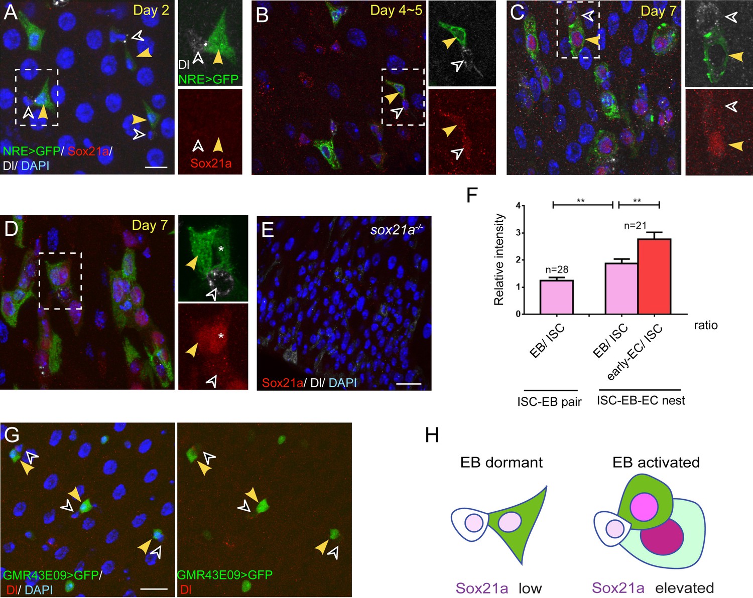
Sox21a is preferentially expressed in differentiating EBs.
(A–D) Sox21a (red) expression in midguts of 2 to 7 days old females co-stained with Dl (white) and NRE>GFP (green). Separate color channels for the insets were enlarged and displayed on the right side of each corresponding images. ISCs and EBs were indicated by white or yellow arrowheads, respectively. Sox21a expression was largely undetectable in the midgut of newly eclosed females (A) Weak Sox21a expression was detectable in ISCs and EBs in midgut of 5 days old females (B). Sox21a expression increased with age, but EB showed a relatively higher expression level than ISC (C). In each ISC-EB-early-EC cell nests, the early EC displayed the highest expression level than the EB or the ISC (D). (E) As a control, no signal was detected by anti-Sox21a staining (red) in Sox21a null midgut. (F) The relative fluorescence intensity of Sox21a expression in each ISC-EB pairs and ISC-EB-Early EC cell nests (also see method). Error bars represent s.e.m. n is as indicated. ** denotes student’s t test p<0.01. (G) Expression of GMR43E09>GFP (green) and Dl (red) in midgut of 5-day-old females. GFP levels were higher in EB (yellow arrowhead) than in adjacent ISC (black arrowhead). (H) A schematic model for dynamic Sox21a expression pattern: the Sox21a expression level may distinguish different state of progenitors. Sox21a expression is at the minimum in differentiation-arrested EBs which typically display a triangle shape with cell protrusions. We refer to this state of EBs as 'dormant'. Sox21a expression is elevated in EBs when EBs begin to differentiate and display oval-shaped morphology. We refer to this state of EBs as 'active'. Sox21a expression reaches its highest level in differentiating EC, and then gradually disappears as EC matures. Scale bars in A, B, 10 μm; in E, 20 μm.
Figure 1—figure supplement 1
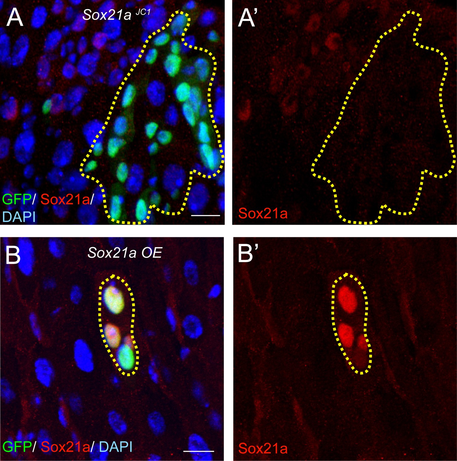
The Sox21a antibody is highly specific.
(A) Staining with anti-Sox21a antibody (red) in Sox21a mutant clones (green, dashed lines). The signal was always absent in Sox21a mutant clones. (B) Staining with anti-Sox21a antibody (red) in Sox21a overexpression (OE) clones (green, dashed lines). The signal was greatly upregulated in Sox21a OE clones. Scale bars: 10 μm
Figure 2
Sox21a mutation causes the development of intestinal tumors composed of differentiation-defective cells.
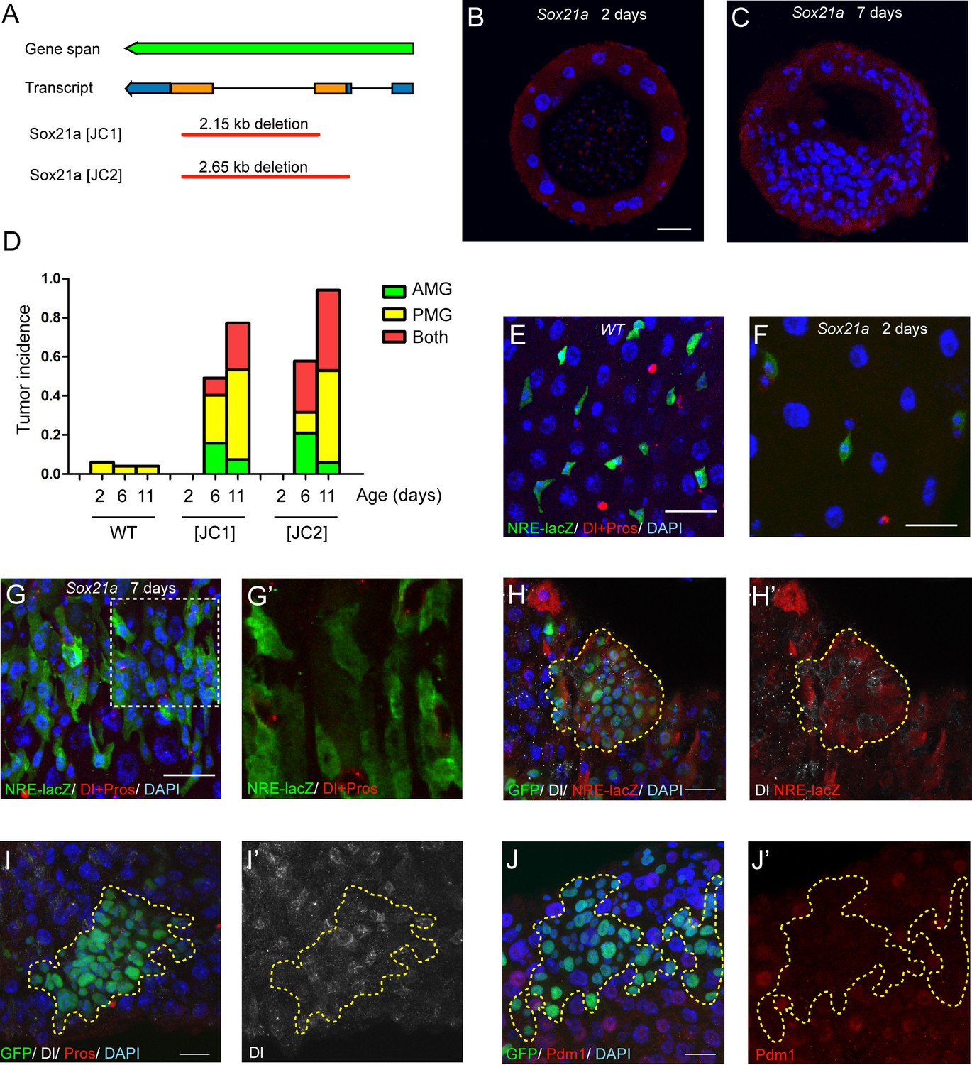
(A) A diagram describing the molecular lesions of two Sox21a mutant alleles. Both alleles carry a large DNA fragment deletion spanning the coding region of Sox21a. (B–C) Cross-section of midgut from 2 day old (B) and 7 day old (C) Sox21a mutant flies. With age, Sox21a mutants developed tumors with multilayered structure (C). (D) Quantitative analysis of the tumor incidence at different regions of midgut with age. Genotypes of flies analyzed: wild type (WT), Sox21a-/- [JC1] and Sox21a-/- [JC2]. n = 40–50 guts. (E–G’) Staining of NRE-lacZ (green) and Dl + Pros (red) in the midgut of WT (E), Sox21a mutant flies at day 2 (F) and day 7 (G, G’) after eclosion. Compared to WT guts, Sox21a mutant guts did not show EB accumulation at day 2 (E, F), but showed dramatic EB accumulation at day 7 (G, G’). Accumulated lacZ+ cells were negative for Dl or Pros expression (G’). (H, H’) Staining of NRE-lacZ (red) in Sox21a mutant clones on day 7 after clone induction. LacZ was cell-autonomously activated. Non-cell autonomous LacZ+ cell clusters were also observed (see text). (I–I’) Staining of Dl (white), Pros (red) in Sox21a mutant clones. The Dl+ cells were scatteredly distributed in the clones, and Pros+ cell was rarely found within the mutant clones. (J, J’) Staining of Pdm1 (red) in Sox21a mutant clones.. Pdm1 expression was absent in the entire mutant clones. Scale bars: 20 μm.
Figure 3
Sox21a functions to promote EB differentiation into EC.
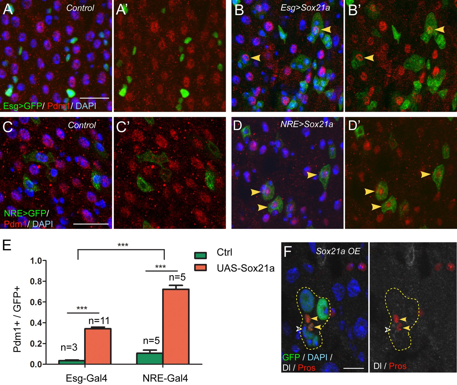
(A–B’) Expression of Esg>GFP (green) and Pdm1 (red) in control (A, A’) and Sox21a overexpressed guts(B, B’). Conditional expression of Sox21a in Esg+ cells for 7 days induced a part of Esg+ cells to turn on Pdm1 expression. (C–D’) Expression of NRE>GFP (green) and Pdm1 (red) in control (C,C’) and Sox21a overexpressed guts (D,D’). Conditional expression of Sox21a in NRE+ cells for 5 days induced most of the NRE+ cells to turn on Pdm1 expression. (E) Quantification of the percentage of Pdm1+ cells in GFP+ cells. Error bars represent s.e.m. n is as indicated. *** denotes student’s t test p<0.001. (F) Staining of Dl (white) and Pros (red) in MARCM clones with Sox21a overexpression (Sox21a OE) on day 5 after clone induction. Dl+ ISCs and Pros+ enteroendocrine cells could be detected in Sox21a OE clones. Scale bars in (A–D), 20 μm; in F, 10 μm.
Figure 4
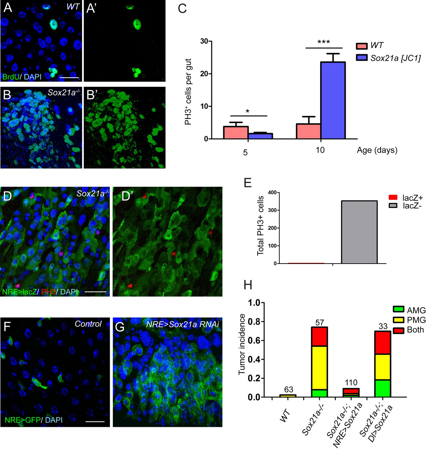
Sox21a mutant EBs are non-mitotic but are tumor-initiating cells.
(A–B’) BrdU (green) incorporation assay in WT control and Sox21a-/- gut. Compared to WT gut, Sox21a mutant gut showed increased BrdU incorporation in tumor regions. (C) Mitotic index of WT and Sox21a-/- midgut at day 5 and day 10 after eclosion. Error bars represent s.e.m. n = 40–50 guts. * denotes student’s t test p<0.05. *** denotes student’s t test p<0.001. (D–E) pH3 (red) staining in the tumor region of Sox21a mutant midgut (D,D’). Virtually all pH3+ cells were NRE>GFP- cells (D’,E). (F, G) Sox21a-RNAi driven by NRE-Gal4ts led to tumorous accumulation of GFP+ EBs. (H) Quantitative analysis of tumor incidence in the midgut of WT control, Sox21a-/-, Sox21a-/- ; NRE>Sox21a and Sox21a-/- ; Dl>Sox21a flies. Expression of Sox21a transgene in EB, but not in ISC, could effectively suppress tumor development in Sox21a mutant midgut. Total number of guts examined is as indicated. Scale bars: 20 μm.
Figure 5 with 2 supplements
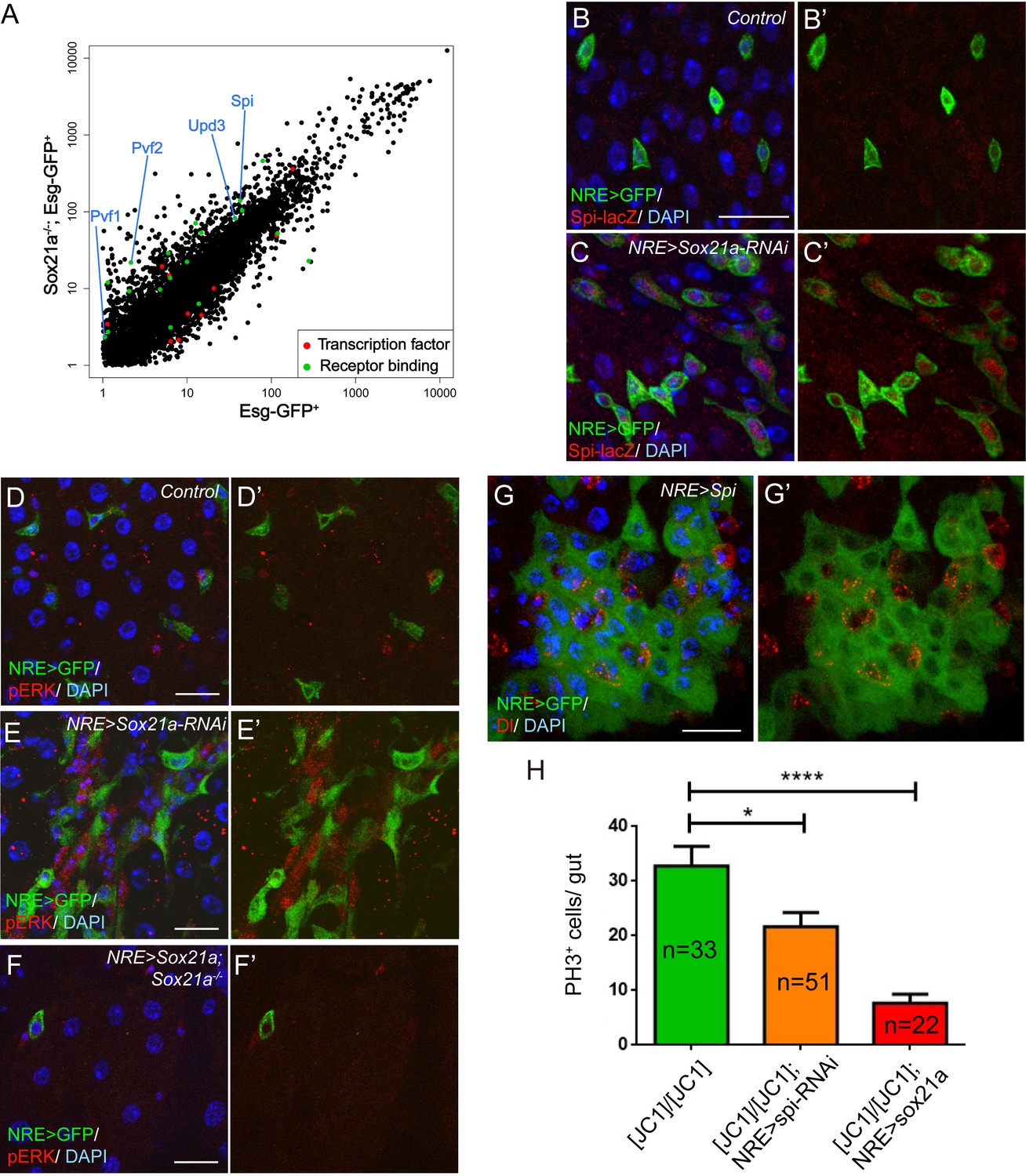
Spi/MAPK signaling is required for tumorigenesis driven by Sox21a mutant EBs.
(A) A scatter plot shows the comparison of gene expression profiles of Esg-GFP+ cells in wild type and Sox21a-/- midgut. Green dots depict genes of receptor binding proteins with 2-fold and higher changes, and red dots depict genes of transcription factors with 2-fold and higher changes in the mutant midgut. (B–C’) Spi-lacZ (red) and NRE>GFP (green) expression in control and NRE>Sox21a RNAi midguts of 7-day-old females. Spi-lacZ expression was undetectable in the wild type midgut (B, B’), but was elevated in EBs of NRE>Sox21a-RNAi midgut (C, C’). (D–F) pERK (red) staining in midguts of the following genotypes: NRE>GFP (control, D, D’), NRE>Sox21a RNAi (E, E’) and NRE>Sox21a; Sox21a-/- (F, F’). EB- specific Sox21a-RNAi caused dramatic enhancement of pERK signal in the diploid cells adjacent to EBs, and EB-specific expression of Sox21a suppressed pERK upregulation in Sox21a mutant midgut. (G, G’) Transgenic Spi expression driven by NRE-gal4ts led to tumorous accumulation of EBs. Those accumulated EBs were all negative for Dl expression (G’). (H) A plot showing that either EB- specific knockdown of spi or EB- specific expression of Sox21a transgene significantly reduced ISC overproliferation in Sox21a mutant intestine, and sox21a transgene expression had a much stronger effect. Error bars represent s.e.m. n is as indicated. * denotes student’s t test p<0.05. ***p<0.001. Scale bars: 20 μm.
Figure 5—figure supplement 1
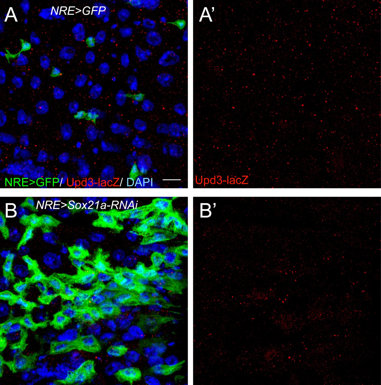
Upd3-lacZ was not significantly upregulated in Sox21a-depleted EBs.
(A) Staining of Upd3-lacZ (red) in midgut of NRE-Gal4; UAS-GFP (green). (B) Staining of Upd3-lacZ (red) in midgut of NRE-Gal4; UAS-GFP; UAS-Sox21a RNAi. GFP was in green. Upd3-lacZ was not obviously upregulated in Sox21a-depleted EBs. Weak upregulation of Upd3-lacZ was sometimes observed in the nearby GFP- cells. Scale bars: 10 μm
Figure 5—figure supplement 2
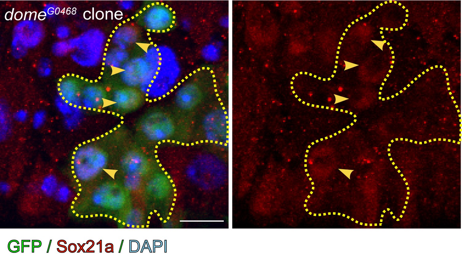
Sox21a was still expressed in JAK/STAT-compromised clones.
Staining of Sox21a (red) in dome468 clones, in which the cells are defective in differentiation. Expression of Sox21a could still be detected in dome mutant cells. Scale bar: 10 μm
Figure 6 with 1 supplement
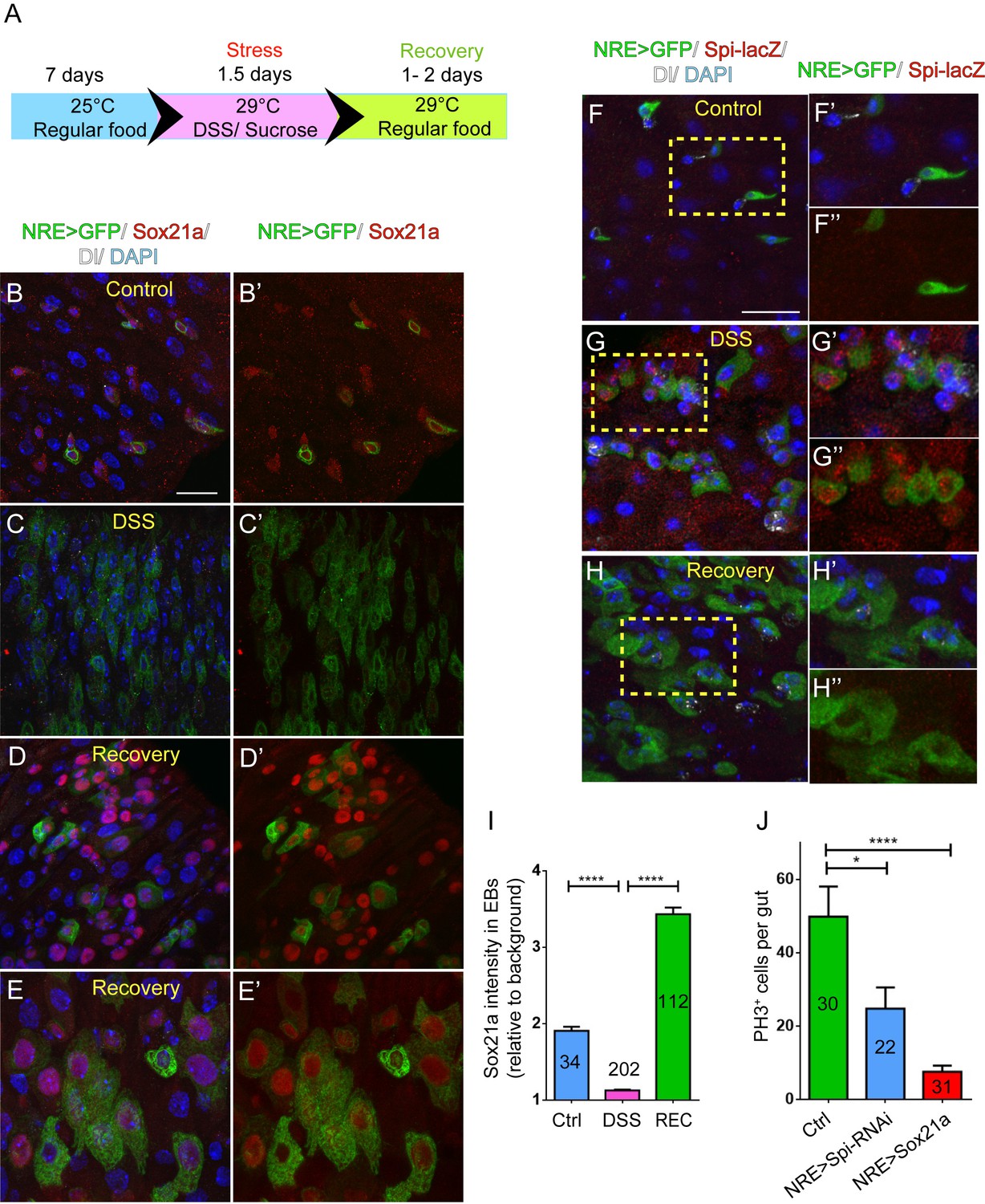
Sox21a-Spi- mediated ISC-EB amplification loop participates in damage-induced intestinal regeneration.
(A) The scheme of damage induction and recovery. (B) (B–E’) Sox21a (red) expression in the midgut of flies fed with sucrose-soaked diet (B, B’) and DSS-soaked diet (C, C’) as well as the flies of 2 day recovery after DSS feeding (D, D’, E, E’). Compared with sucrose-treated control gut (B, B’), DSS treatment showed rapid decline of Sox21a expression in EBs (C, C’). During the recovery phase, Sox21a was dramatically upregulated in EBs (D, D’) and differentiating ECs (E, E’). (F–H”) Spi-lacZ (red) expression in the midgut of flies treated with sucrose (F–F”) or DSS (G–G”) as well as the flies at the recovery phase after DSS treatment (H–H”). Compared with sucrose-treated midgut, in which Spi-lacZ expression was undetectable (F–F”), DSS treatment induced Spi-lacZ expression (G–G”). Spi-lacZ expression was shut down again at the recovery phase (H–H”). (I) Fluorescence intensity of Sox21a expression in EBs relative to background in the midgut of flies fed with sucrose, DSS and flies at the recovery phase after DSS treatment. Sox21a expression in EB was virtually reduced to background levels in DSS-induced damage phase, and then massively upregulated during the recovery phase (REC). Error bars represent s.e.m. n is as indicated. **** denotes student’s t test p<0.0001 (J) Quantification of pH3+ cells in midguts of indicated genotypes. EB- specific depletion of Spi could partially reduce DSS- induced mitosis, while EB-specific transgene expression of Sox21a could strongly inhibit DSS- induced mitosis. Error bars represent s.e.m. n is as indicated. * denotes student’s t test p<0.05. ***p<0.001. Scale bars: 20 μm.
Figure 6—figure supplement 1

GMR43E09>GFP (green) expression in the midgut of flies treated with sucrose (A–A’) or DSS (B, B’) as well as flies at the recovery phase after DSS treatment (C–C’).
Compared with sucrose-treated midgut (A, A’), DSS treatment caused rapid downregulation of GFP expression in EBs (B,B’). A temporal elevation of GFP expression was observed in the intestinal epithelium during the recovery phase (C, C’). Scale bar: 20 μm
Figure 7
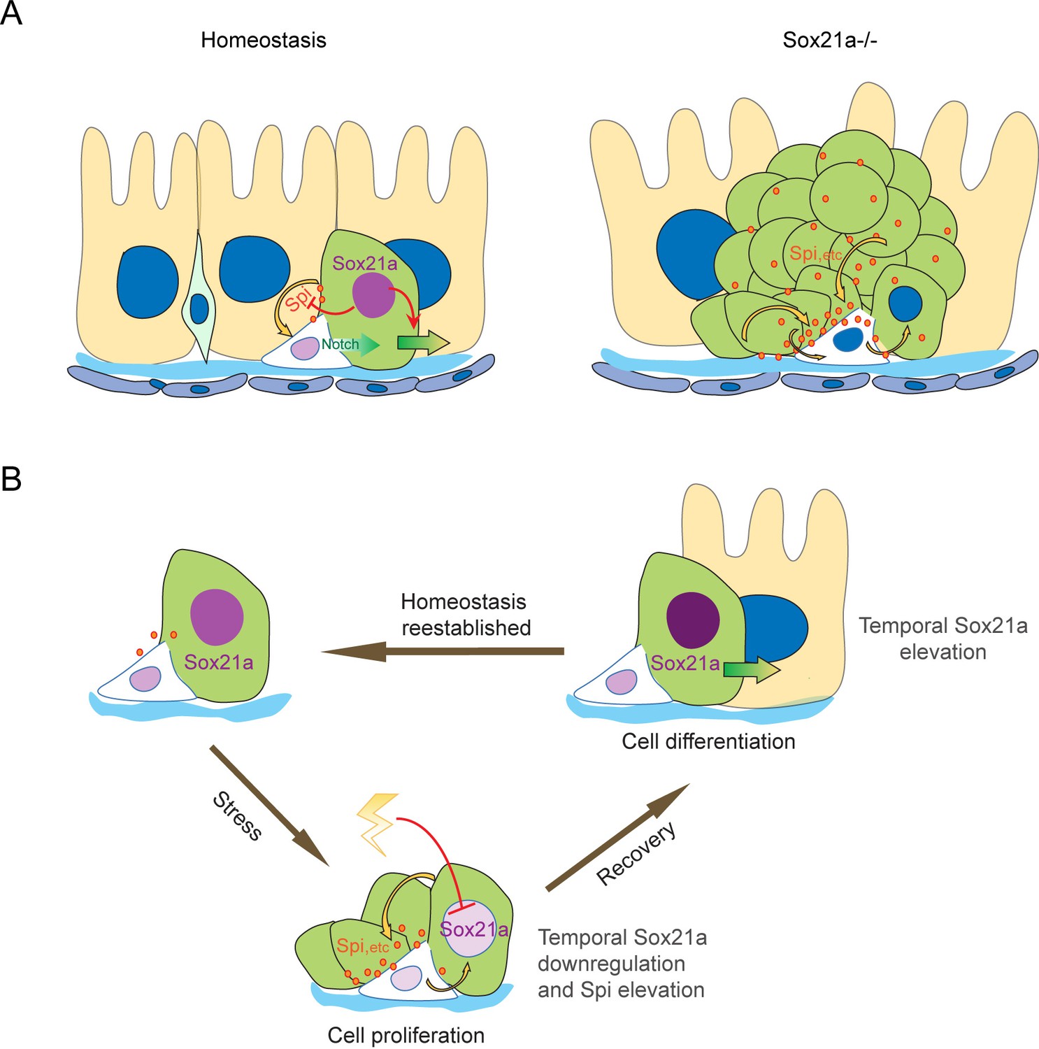
Schematic models of the ISC-EB feedback loop during normal homeostasis, regeneration and tumorigenesis.
(A) During normal homeostasis (left panel), ISC and EB typically exist as a progenitor pair. Sox21a is expressed in EB to promote EB differentiation and at the same time to restrict ISC activity by inhibiting paracrine signals, such as Spi, etc.. In Sox21a mutant midgut (right panel), continuous paracrine signals drives continuous activation of the ISC-EB loop, leading to massive production of differentiation-defective cells and tumorigenesis. (B) Sox21a-mediated amplification loop is employed in damage-induced intestinal regeneration. Tissue damage causes temporal downregulation of Sox21a in EB and consequently derepression of mitogenic factors, which act in a paracrine manner to promote ISC proliferation. This leads to a temporal activation of the ISC-EB amplification loop for rapid production of progenitor cells prepared for epithelial differentiation. During the recovery after the damage is withdrawn, Sox21a is temporally upregulated in EBs to promote epithelial differentiation and homeostasis reestablishment.
Download links
A two-part list of links to download the article, or parts of the article, in various formats.
Downloads (link to download the article as PDF)
Open citations (links to open the citations from this article in various online reference manager services)
Cite this article (links to download the citations from this article in formats compatible with various reference manager tools)
A feedback amplification loop between stem cells and their progeny promotes tissue regeneration and tumorigenesis
eLife 5:e14330.
https://doi.org/10.7554/eLife.14330


