Pro-death NMDA receptor signaling is promoted by the GluN2B C-terminus independently of Dapk1
- University of Edinburgh, United Kingdom
- Trinity College Dublin, Ireland
- University of Melbourne, Australia
- Wellcome Trust Sanger Institute, United Kingdom
Figures
Figure 1 with 1 supplement
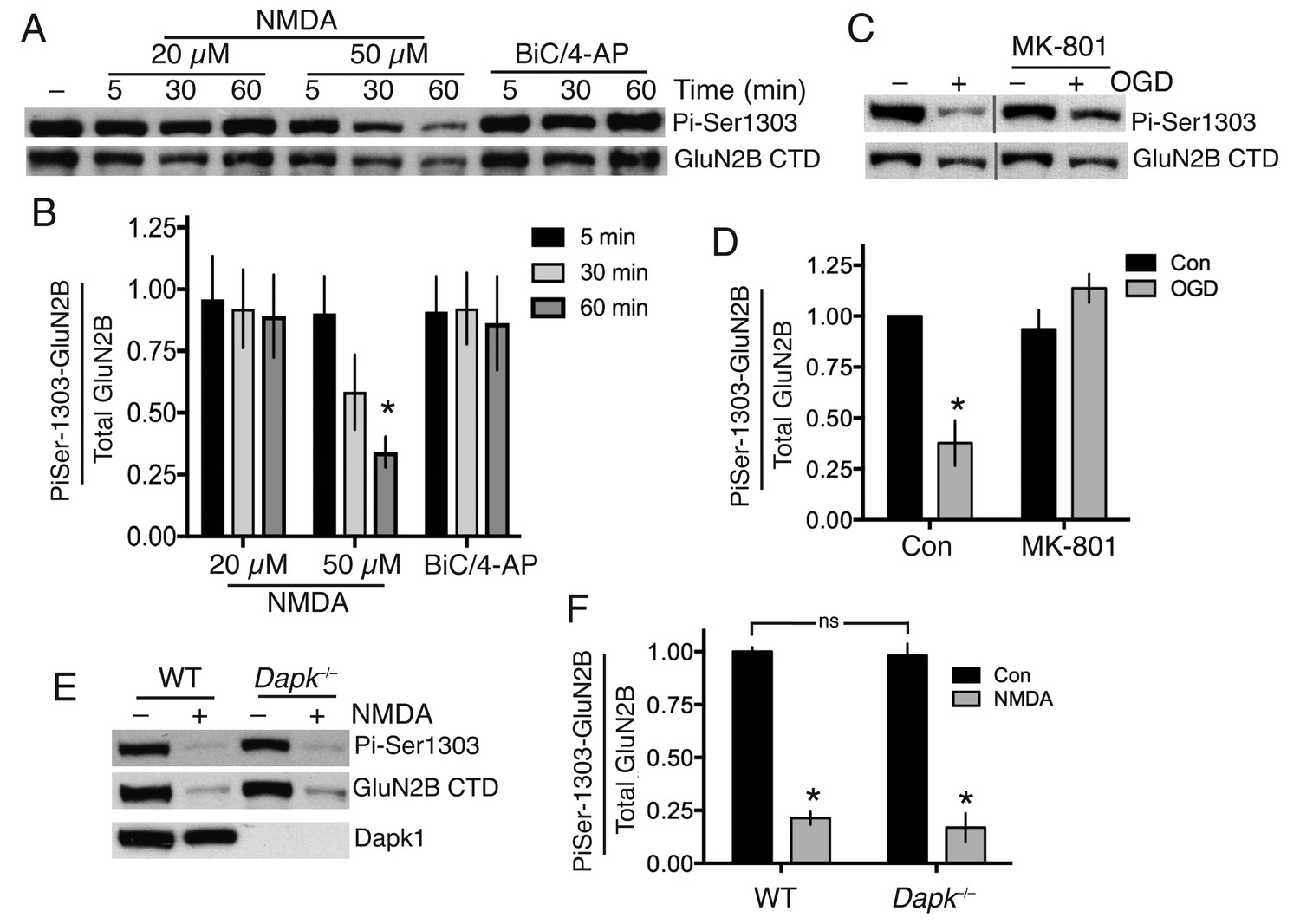
Neither Dapk1 nor excitotoxic insults increase GluN2B phosphorylation on Ser-1303.
(A,B) Strong excitotoxic insults induce GluN2B Ser-1303 dephosphorylation at later timepoints. Western analysis of extracts from cortical neurons treated as indicated with NMDA or bicuculline (50 µM) plus 4-amino pyridine (250 µM). (F(2,24)=3.904, p=0.034 (Two-way ANOVA). *p=0.0053 (Sidak's post-hoc test; 95% CI of diff: 0.1777 to 1.139, comparison to control without NMDA treatment, N = 3). (C,D) Mimicking ischemic conditions triggers dephosphorylation of GluN2B Ser-1303 in an NMDAR-dependent manner. Oxygen-glucose deprivation (OGD) applied for 120 min ±MK-801 (10 µM). F(1,12)=6.69, p=0.024 (Two-way ANOVA). *p=0.0003 (Sidak's post-hoc test, 95% CI of diff: 0.3289 to 0.9172, N = 4). (E,F) Dapk1 deficiency does not influence basal or NMDA-induced GluN2B Ser-1303 phosphorylation status. Neurons were treated ±50 µM NMDA for 60 min. F(1,10)=345.1, p<0.0001 (Two-way ANOVA, Con vs. NM). *p<0.0001 (both, compared to Con of that genotype, 95% CI of diff: 0.6384 to 0.9342, and 0.6411 to 0.9826 (reading left to right), N = 4 WT, N = 3 KO; with ‘N’ defined as a distinct culture from a distinct animal). ns: F(1,10)=0.5418, p=0.4786.
-
Figure 1—source data 1
Data relating to Figure 1—figure supplement 1a.
- https://doi.org/10.7554/eLife.17161.003
-
Figure 1—source data 2
Data relating to Figure 1b.
- https://doi.org/10.7554/eLife.17161.004
-
Figure 1—source data 3
Data relating to Figure 1—figure supplement 1f.
- https://doi.org/10.7554/eLife.17161.005
-
Figure 1—source data 4
Data relating to Figure 1—figure supplement 1h.
- https://doi.org/10.7554/eLife.17161.006
-
Figure 1—source data 5
Data relating to Figure 1d.
- https://doi.org/10.7554/eLife.17161.007
-
Figure 1—source data 6
Data relating to Figure 1f.
- https://doi.org/10.7554/eLife.17161.008
Figure 1—figure supplement 1
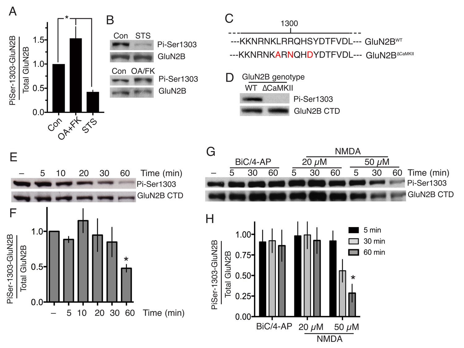
(A,B) Neurons were treated with staurosporine (STS, 1 µM) or FK-506 (FK, 5 µM)+okadaic acid (OA, 10 µM) for one hour, after which protein was harvested and western analysis for Phospho- (GluN2B Ser-1303) levels performed.
p=0.0023 (1-way ANOVA). Individual P-values left-to-right: 0.026, 0.047 (n = 8 (FK +OA); n = 4 (STS). (C) Schematic depicting the amino-acid changes resulting from mutations of the Grin2b gene in the GluN2B∆CaMKII allele. (D) Example Phospho- (GluN2B Ser-1303) western blot illustrating the lack of immunoreactivity of the mutated domain in extracts from GluN2B∆CaMKII/∆CaMKII neurons. (E,F) timecourse of GluN2B Ser-1303 phosphorylation status in response to NMDA treatment (50 µM). F(5,10)=4.019, p=0.023 (one-way ANOVA). *p=0.041 (Sidak's post-hoc test (n = 3), 95% CI of diff 0.01897 to 1.024). (G,H) Experiment performed as per Figure 1a except neurons were at DIV16 rather than DIV10. F (2, 24)=5.324, p=0.0122 (two-way ANOVA). *p=0.0022 (Sidak's post-hoc test (N = 3)).
Figure 2
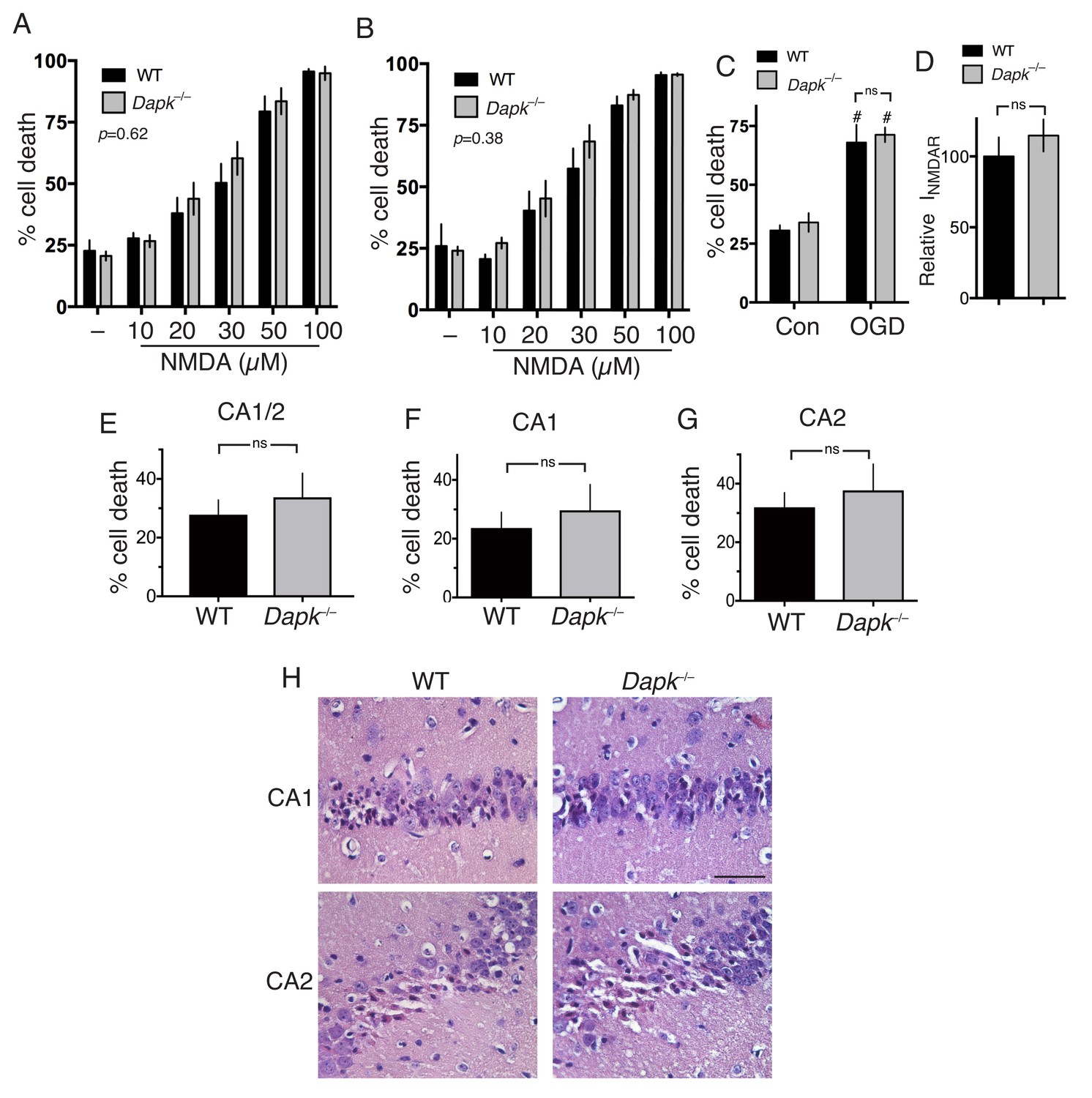
Excitotoxic and ischemic insults are not ameliorated by Dapk1 deficiency.
(A, B) NMDA-induced neuronal death is independent of Dapk1. Cortical neurons at DIV10 (A) or DIV16 (B) were treated as indicated for 1 hr, with neuronal death assessed at 24 hr. The p values relate to a two-way ANOVA test of differences between WT and Dapk–/– neurons (F(1,10) = 0.2676, n = 6 WT, 6 KO (DIV10); F(1,7)=0.8871, 2-way ANOVA, n = 4 WT, 5 KO (DIV16)). For each condition/genotype combination, 800–1000 cells were analysed per biological replicate. (C) OGD-induced neuronal death is independent of Dapk1. Cortical neurons at DIV10 were subjected to OGD for 120 min, before being returned to control medium. Neuronal death was assessed at 24 hr. No genotype-dependent difference was observed (F (1,12)=0.5062, p=0.490, but a strong influence of OGD was observed: F (1,12)=63.54, p<0.0001, two-way ANOVA. #p=0.0002, 0.0002 (reading left to right); Sidak's post-hoc test comparing control to OGD condition (n = 4 WT, n = 4 KO). (D) Dapk1 deficiency does not influence NMDAR currents. NMDAR currents were measured in n = 16 WT cells (from four separate cultures) and n = 25 KO cells (from six separate cultures). Currents were normalized to the mean current recorded from WT cells recorded on that precise day. p=0.411 (t = 0.831, df = 39), unpaired t-test. (E–G) Dapk1 deficiency does not influence vulnerability to ischemia in vivo. Adult male age-matched mice (n = 14 WT; n = 16 KO) were subjected to 20 min bilateral common carotid artery occlusion, sacrificed at 3 d, and pathology analysed. CA1/2 (E): p=0.555 (t = 0.598, df = 28); CA1 (F) :p=0.572 (t = 0.572, df = 28), CA2 p=0.592(G, t = 0.543, df = 28). Scale bar = 50 µm.
-
Figure 2—source data 1
Data relating to Figure 2a.
- https://doi.org/10.7554/eLife.17161.011
-
Figure 2—source data 2
Data relating to Figure 2b.
- https://doi.org/10.7554/eLife.17161.012
-
Figure 2—source data 3
Data relating to Figure 2c.
- https://doi.org/10.7554/eLife.17161.013
-
Figure 2—source data 4
Data relating to Figure 2d.
- https://doi.org/10.7554/eLife.17161.014
-
Figure 2—source data 5
Data relating to Figure 2e–g.
- https://doi.org/10.7554/eLife.17161.015
Figure 3 with 1 supplement

Both TAT-NR2BCT and TAT-sNR2BsCT are direct NMDAR antagonists.
(A–D) Both TAT-NR2BCT and TAT-sNR2BCT (scrambled version of TAT-NR2BCT) immediately antagonize NMDAR currents upon extracellular exposure. NMDA-induced currents were recorded under whole-cell voltage clamp, with the indicated peptides (at 5 µM) applied approximately 5 s after NMDA (to allow NMDAR currents to reach steady state). Arg-rich refers to the arginine-rich positively charged peptide; Neutral refers to the neutral peptide-see main text for sequences of these as well as TAT-NR2BCT and TAT-sNR2BCT. NMDA-induced NMDAR currents were monitored for a further 45 s and the percentage drop in currents calculated, compared to no peptide at all (Con) which represents a measure of natural desensitization over this period. p<0.0001 (one-way ANOVA). *p<0.0001, Sidak's post-hoc test (n = 8 of all conditions). Example traces shown in (B) (C) and (D). Scale bar: 15 s, 500 pA. (E) NMDAR antagonism by TAT-NR2BCT is inhibited by Mg2+ blockade. NMDAR currents were measured, after which neurons were incubated in TAT-NR2BCT (5 or 0.5 µM) for 60 s in the presence or absence of 1 mM Mg2+, after which NMDAR currents were measured again (in zero Mg2+, no peptide). p<0.0001 (one-way ANOVA, F(1,22)=47.16 (effect of [Mg2+])). *p<0.0001, p=0.0007, Sidak's post-hoc test (zero Mg2+: n = 6 (0.5 µM), n = 7 (5 µM); 1 mM Mg2+: n = 6 (0.5 µM), n = 7 (5 µM)). (F–H) NMDAR antagonism by TAT-NR2BCT is more effective on open channels. See main text for experimental details. p<0.0001 (two-way ANOVA, comparing initial current with subsequent measurements: F (2, 45)=25.22. p<0.0001 (two-way ANOVA, comparing 'closed-then-closed' protocol (grey bars, n = 8) with 'closed-then-open' protocol (black bars, n = 9): F (1, 45)=18.26. #p=0.003, 0.0009 (Sidak's post-hoc test), comparing to initial currents. *p<0.0001 (Sidak's post-hoc tests), comparisons indicated. (G) shows example recordings taken during the consecutive ‘closed’ then ‘closed’ channel protocol. (H) shows example recordings taken during the consecutive ‘closed’ then ‘closed’ channel protocol. Scale bar = 1 s, 250 pA.
-
Figure 3—source data 1
Data relating to Figure 3—figure supplement 1a.
- https://doi.org/10.7554/eLife.17161.017
-
Figure 3—source data 2
Data relating to Figure 3—figure supplement 1b.
- https://doi.org/10.7554/eLife.17161.018
-
Figure 3—source data 3
Data relating to Figure 3a.
- https://doi.org/10.7554/eLife.17161.019
-
Figure 3—source data 4
Data relating to Figure 3e.
- https://doi.org/10.7554/eLife.17161.020
-
Figure 3—source data 5
Data relating to Figure 3f.
- https://doi.org/10.7554/eLife.17161.021
Figure 3—figure supplement 1
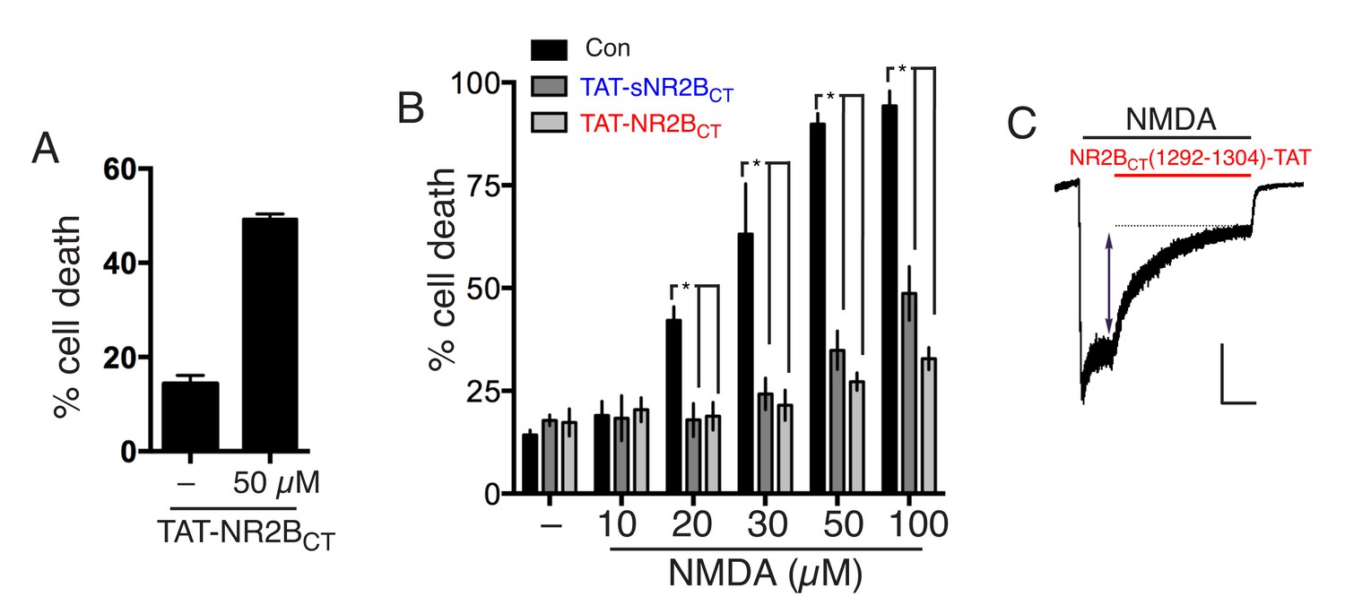
(A) Neurons were treated where indicated with 50 µM TAT-NR2BCT for 1 hr, with death assessed after 24 hr.
(B) Neurons were pre-treated where indicated with TAT-NR2BCT or TAT-sNR2BCT for 1 hr, prior to 1 hr NMDA treatment at the indicated concentrations, in the continued presence of the peptides where used. Subsequently, both NMDA and peptide were removed from the medium and death assessed after a further 23 hr. p<0.0001 (effect of peptide: two-way ANOVA, F(2,40)=76.13, n = 3–4). *p=0.0027, 0.0039 (20 µM),<0.0001,<0.0001 (30 µM),<0.0001,<0.0001 (50 µM),<0.0001,<0.0001 (100 µM), Sidak's post-hoc test. (C) Example trace from an experiment where NMDA-induced currents were recorded under whole-cell voltage clamp, with NR2BCT(1292–1304)-TAT (5 µM, Merck Millipore) applied approximately 5 s after NMDA (to allow NMDAR currents to reach steady state). NMDA-induced NMDAR currents were monitored to determine the degree of blockade. The trace is representative of 8 cells recorded this way, with the peptide blocking by 63 ± 3%. Scale bar = 5 s, 500 pA.
Download links
A two-part list of links to download the article, or parts of the article, in various formats.
Downloads (link to download the article as PDF)
Open citations (links to open the citations from this article in various online reference manager services)
Cite this article (links to download the citations from this article in formats compatible with various reference manager tools)
Pro-death NMDA receptor signaling is promoted by the GluN2B C-terminus independently of Dapk1
eLife 6:e17161.
https://doi.org/10.7554/eLife.17161


