Symbiont-induced odorant binding proteins mediate insect host hematopoiesis
- University of Cincinnati, United States
- Yale School of Public Health, United States
- University of Connecticut, United States
- Yale University, United States
Figures
Figure 1 with 1 supplement
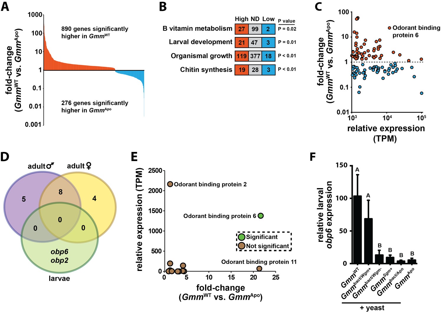
Symbiont-mediated differential expression of odorant binding protein 6 in tsetse larvae.
(A) Number of genes exhibiting significant differential expression, and a relative transcript abundance [in transcripts per million (TPM)] over 3, in GmmWT compared to GmmApo larvae. Significance is based on a Baggerly’s test followed by a false detection rate correction (p<0.01). (B) Significantly enriched gene ontology categories, determined using a Fisher’s exact test. (C) Genes exhibiting significant differential expression (measure as fold-change in gene expression) in GmmWT compared to GmmApo larvae, and a minimum TPM value of 1000. Significance was determined as in (A). (D) Enrichment analysis of odorant binding protein-encoding genes expressed in GmmWT adult males (purple) and females (yellow), and GmmWT larvae (green). (E) Relative expression (TPM) of tsetse odorant binding protein-encoding genes in GmmWT larvae, and their differential expression (measure as fold-change in gene expression) in GmmWT compared to GmmApo larvae. Significance was determined as in (A). (F) Relative obp6 expression in GmmWT larvae, as well as larvae derived from symbiont-cured moms fed a diet supplemented with yeast and Wigglesworthia-containing bacteriome extracts (Gmmbact/Wgm+), Wigglesworthia-free bacteriome extracts (Gmmbact/Wgm-), Sodalis cell extracts (GmmSgm+), and bacteriome extracts harvested from GmmApo females (Gmmbact/Apo). GmmWT and GmmApo flies served as controls. n = 6 biological replicates for groups GmmWT, Gmmbact/Wgm+ and GmmSgm+ samples, and n = 5 biological replicates for Gmmbact/Wgm-, Gmmbact/Apo and GmmApo samples. Replicates for all groups contain a mixture of four first and second instar larvae. Data are presented as mean ± SEM. Bars with different letters indicate a statistically significant difference (specific p values are listed in Figure 1—source data 1) between samples. Statistical analysis = ANOVA followed by Tukey’s HSD post-hoc analysis.
-
Figure 1—source data 1
Obp6 expression in aposymbiotic tsetse larvae following supplementation.
- https://doi.org/10.7554/eLife.19535.004
Figure 1—figure supplement 1
Developmental stage-specific enrichment analysis of tsetse orthologues that putatively cluster within the ‘hematopoiesis’ COG.
Ontology enrichment analyses were performed as described in the Materials and methods, under the ‘Transcriptomics’ sub-heading. Refer to Supplementary file 2 for a description of the enriched hematopoiesis-associated gene expressed specifically in GmmWT and GmmApo tsetse larvae.
Figure 2
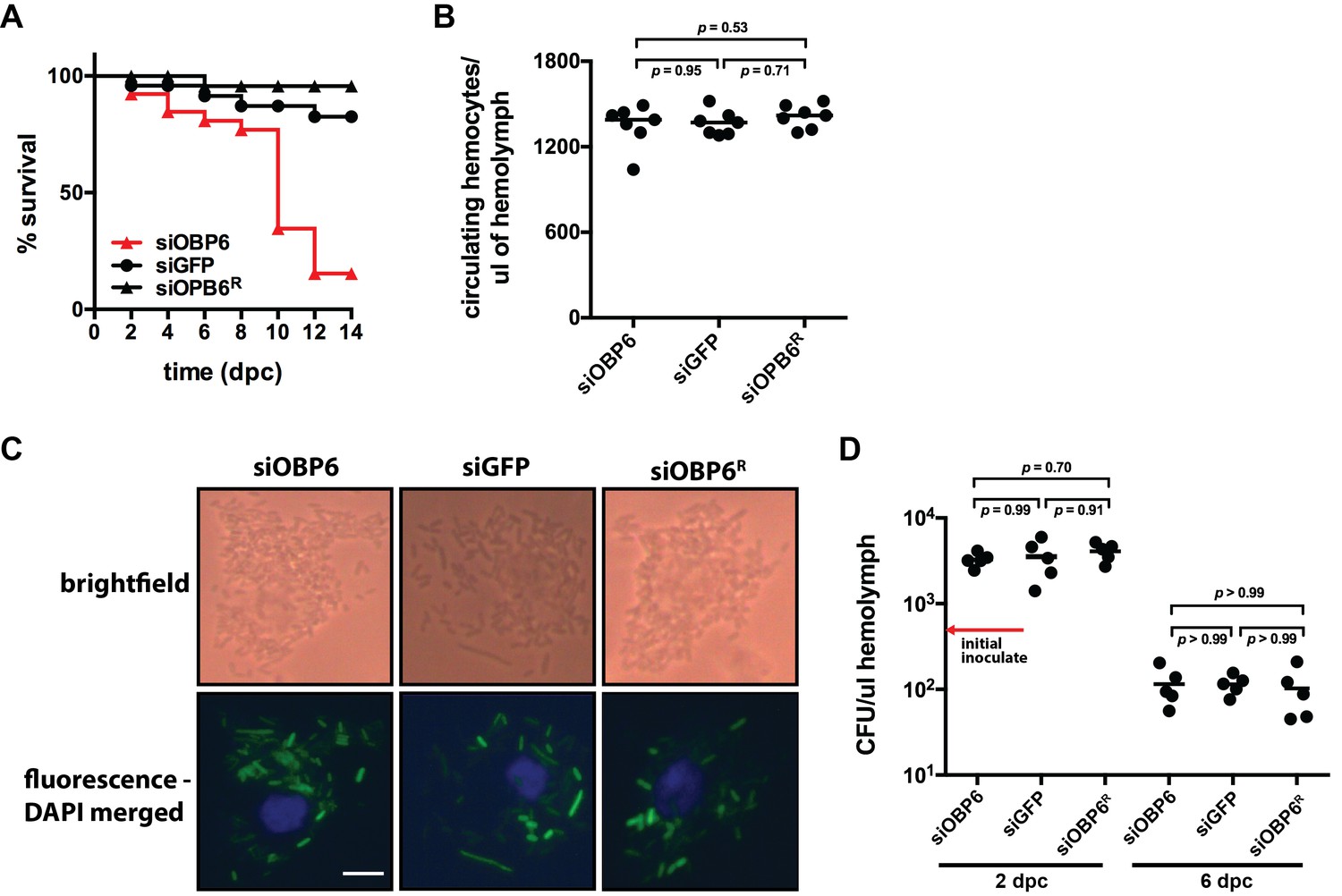
Tsetse odorant binding protein 6 does not mediate the development and function of phagocytic hemocytes.
(A) Survival following systemic challenge of siOBP6 and siGFP adults with 5 × 10² CFU of E. coli K12. Fly survival was monitored every other day for the duration of the 14 day experimental period. Survival assays were performed in triplicate, using 25 flies per replicate. Red curve depicts a statistically significant difference in infection outcome (p<0.0001, log-rank test). (B) Hemocyte abundance in siOBP6 and siGFP adults was quantified microscopically using a hemocytometer (Figure 2—source data 1). (C) A representative micrograph of hemocyte-engulfed recE. coliGFP from siOBP6, siGFP and siOBP6R adults. Experiment was performed using hemolymph collected from four distinct flies per (Figure 2—source data 2). Hemolymph was collected 12 hpc and fixed on glass slides using 2% paraformaldehyde. Magnification is x400. (D) E. coli densities (CFU/μl of hemolymph) in the hemolymph of siOBP6, siGFP and siOBP6R adults at 2 and 6 dpc (Figure 2—source data 3). In (B) and (D), symbols represent one hemolymph sample per group, and bars represent the median hemocyte quantity (B) or bacterial density (D) per sample. Statistical analysis = ANOVA followed by Tukey’s HSD post-hoc analysis.
-
Figure 2—source data 1
Circulating hemocytes per microliter of hemolymph.
- https://doi.org/10.7554/eLife.19535.007
-
Figure 2—source data 2
Phagocytosis by tsetse hemocytes.
- https://doi.org/10.7554/eLife.19535.008
-
Figure 2—source data 3
Colony forming units (CFU) per microliter of hemolymph.
- https://doi.org/10.7554/eLife.19535.009
Figure 3
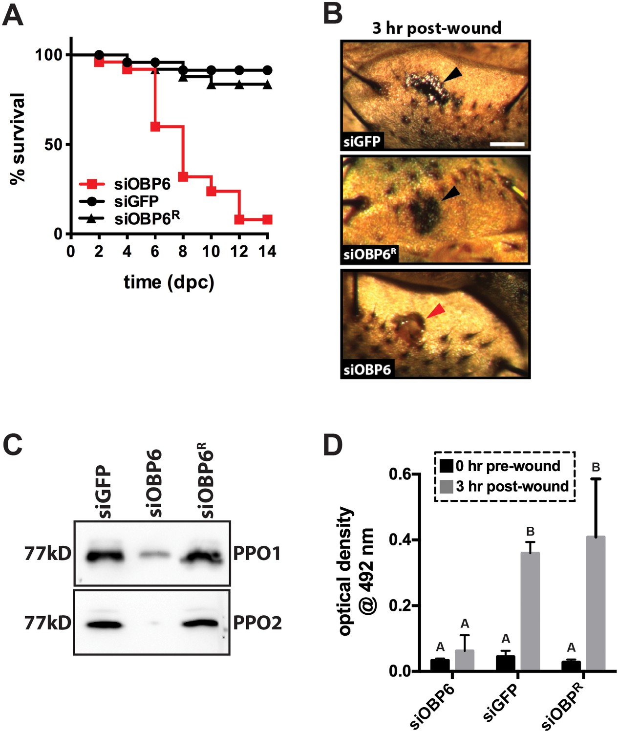
Obp6 mediates the melanization cascade in adult tsetse.
(A) Survival following administration of clean wounds to the thoracic cuticle of siOBP6, siGFP and siOBP6R adults. Survival assays were performed in triplicate, using 25 flies per replicate. Red curve depicts a statistically significant difference in infection outcome (p<0.0001, log-rank test). (B) A representative micrograph of the cuticle of siRNA treated adults 3 hr post-wounding (hpw) with a clean needle. Melanin deposited at the wound site of siGFP and siOBP6R controls, and hemolymph exudate from a siOBP6 treatment individual, are identified by black and red arrowheads, respectively. Scale bar = 500 μm. Experiment was performed using four distinct flies per group (Figure 3—source data 1). (C) Quantitation of PPO1 and PPO2 in the hemolymph of siOBP6, siGFP and siOBP6R adults three hpw with a clean needle. Shown is a representative Western blot analysis using Drosophila anti-PPO1 and anti-PPO2 antibodies. 8 μl of pooled hemolymph was run per gel lane. Hemolymph was collected and pooled from four individuals from each group. Western blots were repeated in triplicate [Figure 3—source data 2 (for PPO1 westerns) and Figure 3—source data 3 (for PPO2 westerns)]. (D) PO activity in the hemolymph of siOBP6, siGFP and siOBP6R adults at 0 and 3 hpw with a clean needle. n = 5 biological replicates per group per time point for pre-wound readings, and n = 8 biological replicates per group per time point for post-wound readings. Data are presented as mean ± SEM. Bars with different letters indicate a statistically significant difference between pre- and post-wound values (specific p values are listed in the Figure 3—source data 4). Statistical test = 2 way ANOVA followed by Tukey’s HSD post-hoc analysis.
-
Figure 3—source data 1
Melanin deposition at tsetse cuticular wound sites.
- https://doi.org/10.7554/eLife.19535.011
-
Figure 3—source data 2
Tsetse prophenoloxidase 1 (PPO1) western blots.
- https://doi.org/10.7554/eLife.19535.012
-
Figure 3—source data 3
Tsetse prophenoloxidase 2 (PPO2) western blots.
- https://doi.org/10.7554/eLife.19535.013
-
Figure 3—source data 4
Tsetse phenoloxidase (PO) assays.
- https://doi.org/10.7554/eLife.19535.014
Figure 4
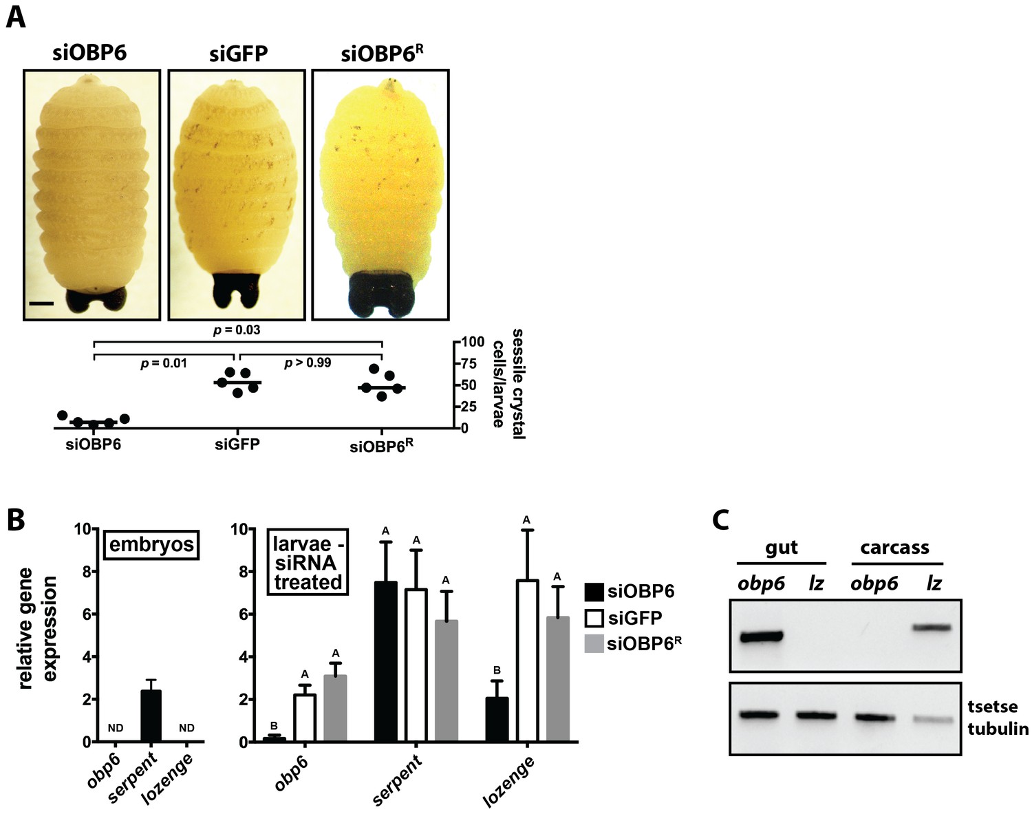
Obp6 expression in the gut of larval tsetse is an integral component of the systemic pathway that actuates crystal cell production.
(A) Representative micrograph depicting spontaneous PPO activation in early third instar siGFP, siOBP6 and siOBP6R tsetse larvae following subjection to a 10 min heat shock at 65°C. Experiment was repeated using one larvae from five distinct moms from each group. Melanotic spots were quantitated microscopically. Statistical analysis = Kruskal-Wallis test followed by Dunn’s post-hoc analysis (Figure 4—source data 1). (B) RT-qPCR analysis of obp6, serpent and lozenge expression in embryos prior to maternal treatment with siRNA, and in siOBP6, siGFP and siOBP6R tsetse larvae from siRNA treated moms. Embryo replicates (n = 5) contain three embryos, larval replicates (n = 7 for siOBP6, n = 5 for siGFP and n = 6 for siOBP6R) contain a mixture of four first and second instar larvae. ND, not detectable. Data are presented as mean ± SEM. Bars with different letters indicate a statistically significant difference between samples (specific p values for larval samples are listed in the Figure 4—source data 2). Statistical analysis = 2 way ANOVA followed by Tukey’s HSD post-hoc analysis. (C) Representative image of obp6 and lozenge spatial expression patterns, determined using semi-quantitative RT-PCR, in the gut and carcass of second instar GmmWT larvae. Experiment was repeated using guts and carcasses from five distinct individuals (Figure 4—source data 3).
-
Figure 4—source data 1
Sessile crystal abundance in larval tsetse.
- https://doi.org/10.7554/eLife.19535.016
-
Figure 4—source data 2
Relative obp6, serpent and lozenge gene expression in tsetse embryoes and larvae.
- https://doi.org/10.7554/eLife.19535.017
-
Figure 4—source data 3
Tissue distribution of obp6 and lozenge expression in tsetse larvae.
- https://doi.org/10.7554/eLife.19535.018
Figure 5 with 1 supplement
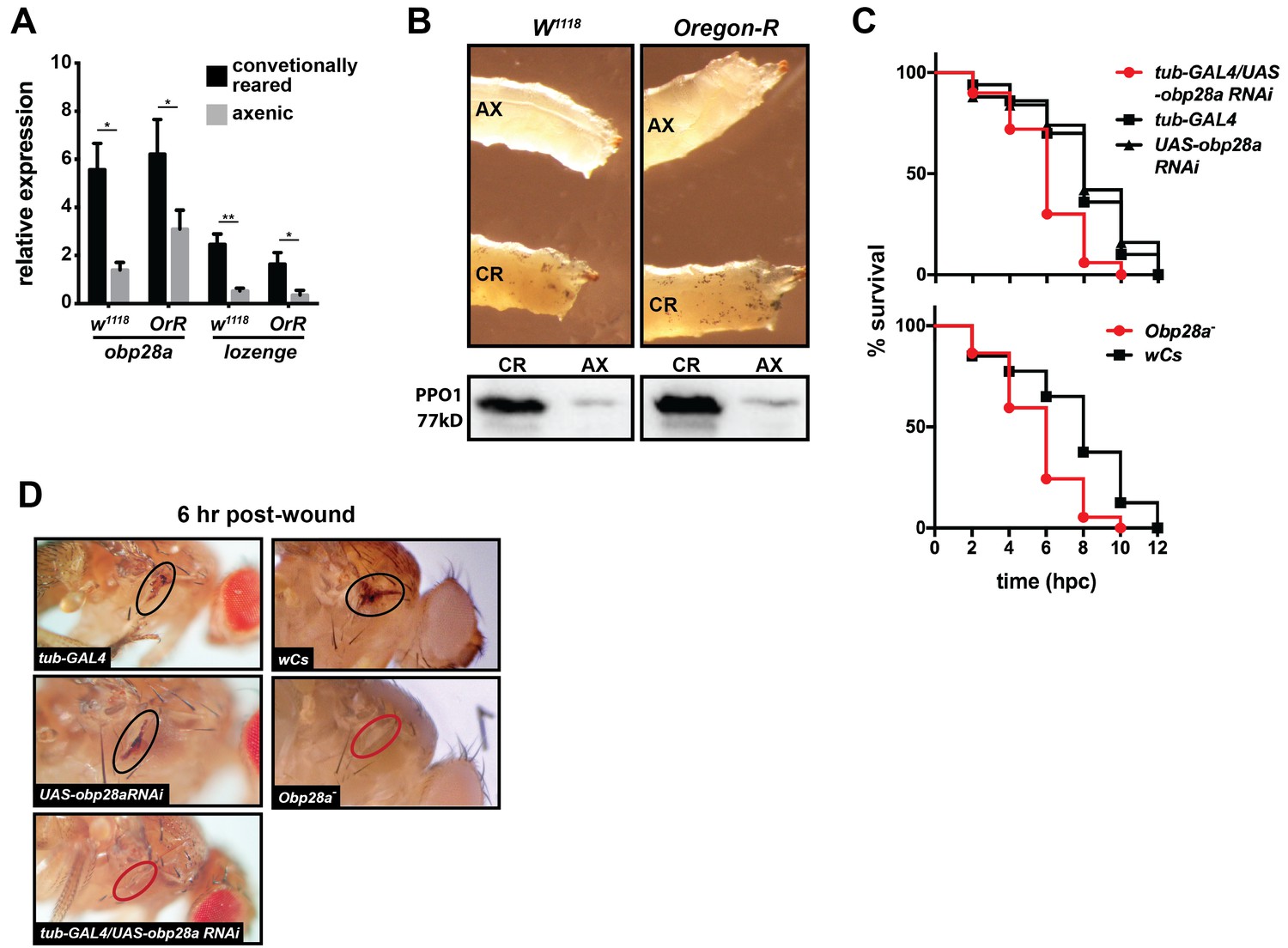
Drosophila’s indigenous microbiota actuates larval hematopoietic pathways and thus functionality of the adult melanization response.
(A) Relative expression of obp28a and lozenge in conventionally reared (CR) and axenic (AX) Oregon-R and w1118 Drosophila larvae. n = 9 (Oregon-R) and 6 (w1118) biological replicates per group, each containing a mixture of thirty second and early-3rd instar larvae. Data are presented as mean ± SEM. Asterisks indicate statistical significance (specific p values are listed in the Figure 5—source data 1). Statistical analysis = unpaired t-tests, corrected for multiple comparisons using the Holm-Sidak method. (B) AX larvae house fewer sessile crystal cells, and produce less PPO, than do and CR individuals. Top panels are representative micrographs depicting spontaneous PPO activation in AX and CR w1118 and Oregon-R larvae following subjection to a 10 min heat shock at 65°C. n = 5 larvae per group [Figure 5—source data 2 (for Oregon-R larvae) and Figure 5—source data 3 (for w1118 larvae)]. Bottom panels are representative Western blots using Drosophila anti-PPO1 antibodies. 8 μl of pooled hemolymph was run per gel lane. Hemolymph was collected and pooled from 15 individuals from each group. Western blots were repeated in triplicate (Figure 5—source data 4). (C) Survival of obp28a RNAi knockdown Drosophila (tub-GAL4/UAS-obp28a RNAi) and knockout (Obp28a-) adults compared to controls (tub-GAL4, UAS-obp28a RNAi and wCs) following wounding with a clean needle. Experiment was performed in triplicate, n = 50 (RNAi) and n = 45 (knockout) per group per replicate. Red curve depicts a statistically significant difference in infection outcomes [p<0.0001 (RNAi) and p=0.0002 (knockout), log-rank test]. (D) A representative micrograph of the cuticle of obp28a knockdown, knockout and control Drosophila adults six hpw with a clean needle. Experiment was performed using two distinct experimental fly cohorts [n = 4 flies per group per experiment; Figure 5—source data 5 (for RNAi flies) and Figure 5—source data 6 (for deletion mutants)]. Wounds on the cuticle of control (melanized) and obp28a knockdown and knockout individuals (not melanized) are identified with black and red ovals, respectively.
-
Figure 5—source data 1
Obp28a and lozenge expression in conventionally reared and axenic w1118 and Oregon-R Drosophila.
- https://doi.org/10.7554/eLife.19535.020
-
Figure 5—source data 2
Sessile crystal cells in conventionally reared and axenic Oregon-R Drosophila larvae.
- https://doi.org/10.7554/eLife.19535.021
-
Figure 5—source data 3
Sessile crystal cells in conventionally reared and axenic w1118 Drosophila larvae.
- https://doi.org/10.7554/eLife.19535.022
-
Figure 5—source data 4
Drosophila prophenoloxidase 1 (PPO1) western blots.
- https://doi.org/10.7554/eLife.19535.023
-
Figure 5—source data 5
Melanin deposition at Drosophila cuticular wound sites following RNAi-mediated knockdown of obp28a.
- https://doi.org/10.7554/eLife.19535.024
-
Figure 5—source data 6
Melanin deposition at Drosophila cuticular wound sites in obp28a deletion mutants.
- https://doi.org/10.7554/eLife.19535.025
Figure 5—figure supplement 1

Axenic and conventionally reared w1118 and Oregon-R larvae following subjection to a 10 min heat shock at 65°C.
White ellipses demarcate an area of the larval cuticle that contains a high density of ruptured crystal cells.
Figure 6
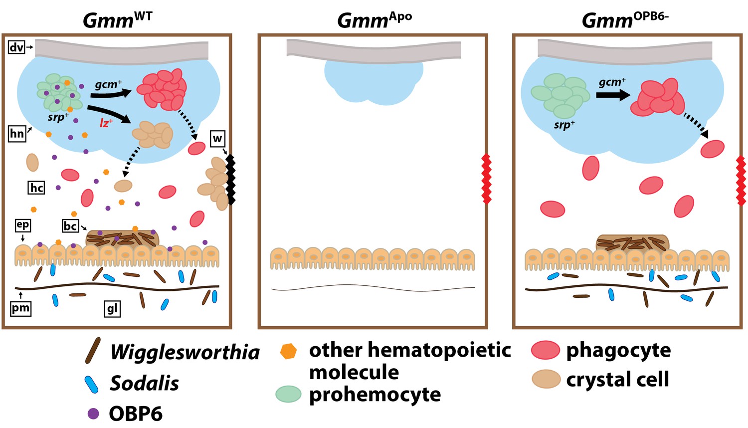
Model illustrating the functional relationship between maternally-transmitted enteric symbionts and melanization in tsetse.
GmmWT larvae imbibe enteric symbiotic-containing milk gland secretions throughout their intrauterine developmental program. These bacteria colonize larval gut-associated tissues, including the bacteriome, and in doing so, induce the expression of obp6. OBP6 is either secreted directly into the hemolymph, or acts locally to induce expression of another unknown, (also secreted) protein. One of these molecules then acts systemically in the larval hematopoietic niche (hn) to stimulate lozenge (lz) expression in a small proportion of serpent (srp) expressing prohemocytes. These cells then become PPO-producing crystal cells [remaining prohemocytes become phagocytes after expressing glial cells missing (gcm)]. Finally, crystal cells are expelled from the hn, where they circulate in the hemolymph and are available to produce wound-healing melanin. Larvae that develop in the absence of symbiotic bacteria (GmmApo) fail to produce any hemocytes, while those that develop in the presence of reduced obp6 transcript abundance (GmmOBP6-) fail to express lozenge and thus likely fail to generate crystal cells. dv, dorsal vessel; hc, hemocoel; w, wound; ep, epithelial cells of midgut; bc, bacteriome; pm, peritrophic matrix; gl, gut lumen.
Additional files
-
Supplementary file 1
Results of RNA-seq analysis, indicating genes that are expressed at significantly different levels (Baggerly’s test followed by a false detection rate at p<0.01), in GmmWT and GmmApo larvae.
- https://doi.org/10.7554/eLife.19535.028
-
Supplementary file 2
Ontogeny analysis of hematopoiesis associated genes in GmmWT compared to GmmApo larvae.
- https://doi.org/10.7554/eLife.19535.029
-
Supplementary file 3
Cartoon summarizing RNAi and subsequent functional experiments.
All relevant experimental details are described in the Materials and methods, under the ‘RNA interference’ sub-heading.
- https://doi.org/10.7554/eLife.19535.030
-
Supplementary file 4
Trans-generational RNAi-based depletion of obp6 expression in intrauterine GmmWT larvae.
(A) Pregnant females were intra-thoracically injected with Cy3 tagged anti-obp6 short interfering (si) RNAs (siOBP6Cy3). A representative micrograph showing that siOBP6Cy3 had disseminated throughout the maternal hemocoel by three days post-treatment (dpt; top left panel), and was present in second instar larvae from the first gonotrophic cycle (GC1; middle left panel) of these females as visualized using fluorescent illumination (bottom left panel). By 26 dpt, siOBP6Cy3 was absent from treated moms (top right panel) and their third gonotrophic cycle (GC3) larvae (middle and bottom right panels). Five pregnant treatment and recovered females were visualized to observe siOBP6Cy3 dissemination and transfer to larvae. (B) Effectiveness of siRNA-based obp6 knockdown in intrauterine second instar tsetse larvae. Relative expression of obp6 in second instar siGFP, siOBP6 and siOBP6R intrauterine larvae. RT-qPCR analysis was performed using larvae from two distinct experiments, each of which included 4 (siGFP and siOBP6) or 3 (siOBP6) biological replicates (each consisting of a mixture of four first and second instar larvae). All RT-qPCR results were normalized relative to tsetse's constitutively expressed β-tubulin gene (determined from each corresponding sample). Data are presented as mean of all replicates from both experiments, ± SEM. Bars with different letters indicate a statistically significant difference (p<0.05) between treatments. Statistical analysis = 2 way ANOVA.
- https://doi.org/10.7554/eLife.19535.031
-
Supplementary file 5
DsiRNAs used in this study.
- https://doi.org/10.7554/eLife.19535.032
-
Supplementary file 6
PCR primers used in this study.
- https://doi.org/10.7554/eLife.19535.033
Download links
A two-part list of links to download the article, or parts of the article, in various formats.
Downloads (link to download the article as PDF)
Open citations (links to open the citations from this article in various online reference manager services)
Cite this article (links to download the citations from this article in formats compatible with various reference manager tools)
Symbiont-induced odorant binding proteins mediate insect host hematopoiesis
eLife 6:e19535.
https://doi.org/10.7554/eLife.19535


