Dpp from the anterior stripe of cells is crucial for the growth of the Drosophila wing disc
- Biozentrum der Universität Basel, Switzerland
Figures
Figure 1 with 1 supplement
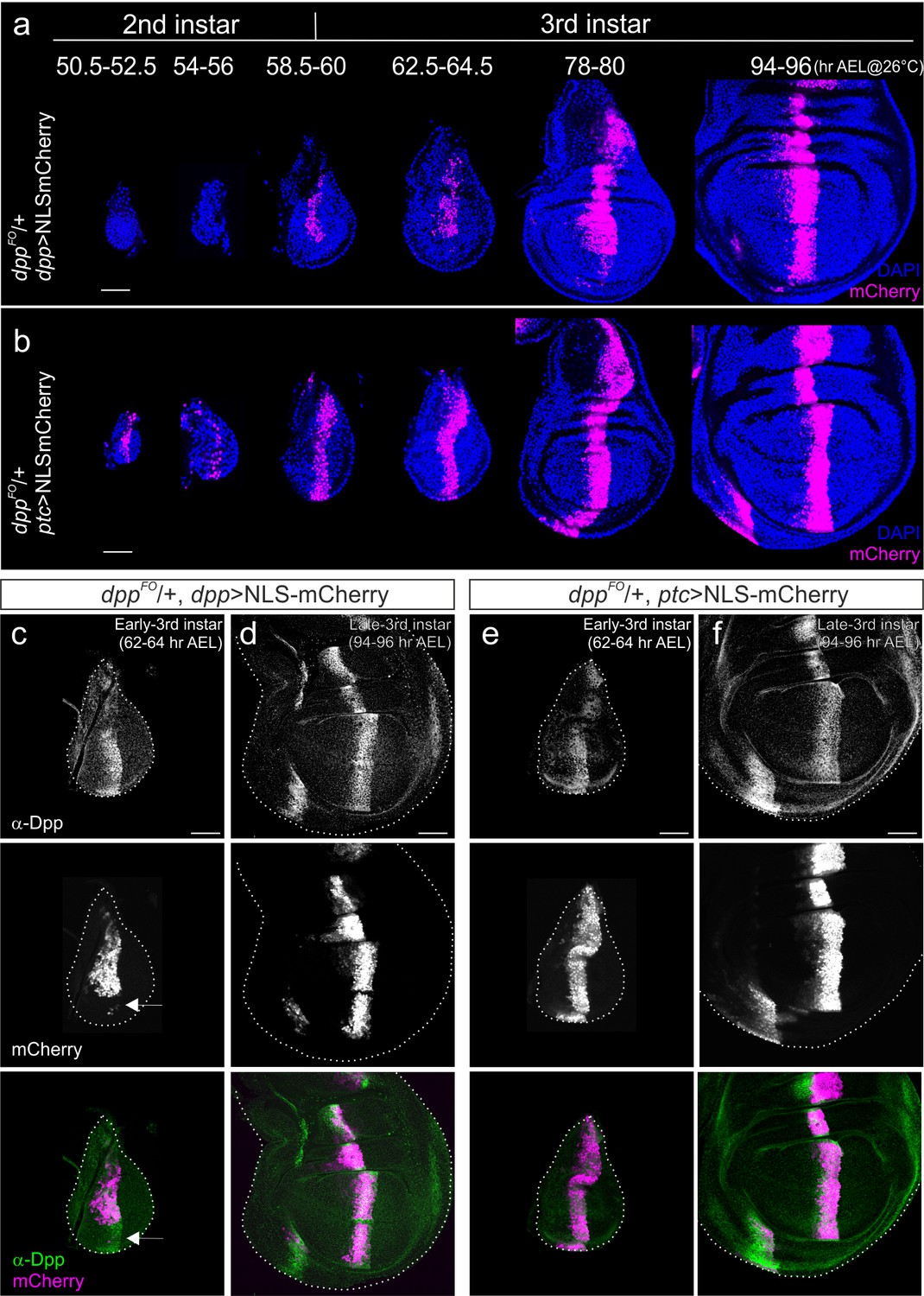
Comparison of dpp-Gal4 and ptc-Gal4 expression pattern with Dpp expression in the Drosophila wing disc.
(a) Temporal expression pattern of dpp-Gal4 (dppFO/+; dpp-Gal4/UAS-NLS-mCherry) (b) temporal expression pattern of ptc-Gal4 (dppFO, ptc-Gal4/+; UAS-NLS-mCherry). Single confocal images except 50.5–52.5 hr AEL by maximum intensity projection. (c, d) Comparison of anti-Dpp staining and dpp-Gal4 expression (NLS-mCherry) in the early (c) and late (d) third instar wing disc of a dppFO/+; dpp-Gal4/UAS-NLS-mCherry larva. (e, f) Comparison of anti-Dpp staining and ptc-Gal4 expression (NLS-mCherry) in the early (e) and late (f) third instar wing disc of a dppFO/+; ptc-Gal4/UAS-NLS-mCherry larva. Average intensity projection from 5 sequential confocal images. Scale bars 50 μm. Anterior is to the left in all figures.
Figure 1—figure supplement 1
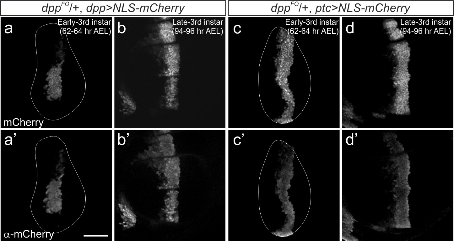
Comparison of mCherry fluorescent signal and anti-mCherry staining.
(a–d) Comparison of mCherry fluorescent signal (a–d) and anti-mCherry staining (a’–d’) in the early (a, b) and late (c, d) third instar wing disc of a dppFO/+; dpp-Gal4/UAS-NLS-mCherry larva (a,b) and a dppFO/+; ptc-Gal4/UAS-NLS-mCherry larva (c,d). Average intensity projection from 3 sequential confocal images. Scale bars 50 μm. Anterior is to the left in all figures.
Figure 2
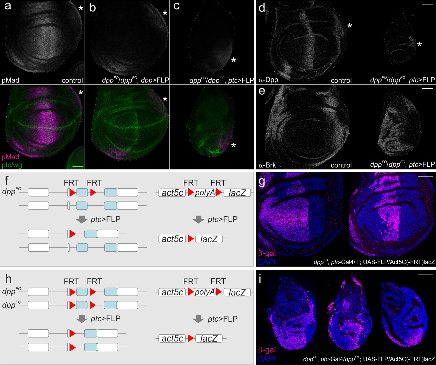
Defects in wing disc growth by removing dpp using ptc-Gal4.
(a–c) Anti-pMad and anti-ptc/wg staining in a dppFO/+; UAS-FLP/+ (control) late third instar wing disc (a), in a dppFO/dppFO; dpp-Gal4/UAS-FLP late third instar wing disc (b), and in a dppFO, ptc-Gal4/dppFO; UAS-FLP/+ late third instar wing disc (c). (d) anti-Dpp staining in a dppFO, ptc-Gal4/+; UAS-FLP/+ (control) late third instar wing disc (left), and in a dppFO, ptc-Gal4/dppFO; UAS-FLP/+ late third instar wing disc (right). (e) anti-Brk staining in a dppFO, ptc-Gal4/+; UAS-FLP/+ (control) late third instar wing disc (left), and in a dppFO, ptc-Gal4/dppFO; UAS-FLP/+ late third instar wing disc (right). (*) marks the future alula region. (a–e) Average intensity projection from 5 sequential confocal images. (f, h) an experimental setup to test the efficiency of FLP/FRT mediated recombination. (g) anti-β-gal staining in a dppFO, ptc-Ga4/+; UAS-FLP/act5C(-FRT)lacZ late third instar wing disc (control). (i) anti β-gal staining in a dppFO, ptc-Gal4/dppFO;UAS-FLP/act5C(-FRT)lacZ late third instar wing disc. (g, i) A single confocal image. Scale bars 50 μm. Anterior is to the left in all figures.
Figure 3 with 4 supplements
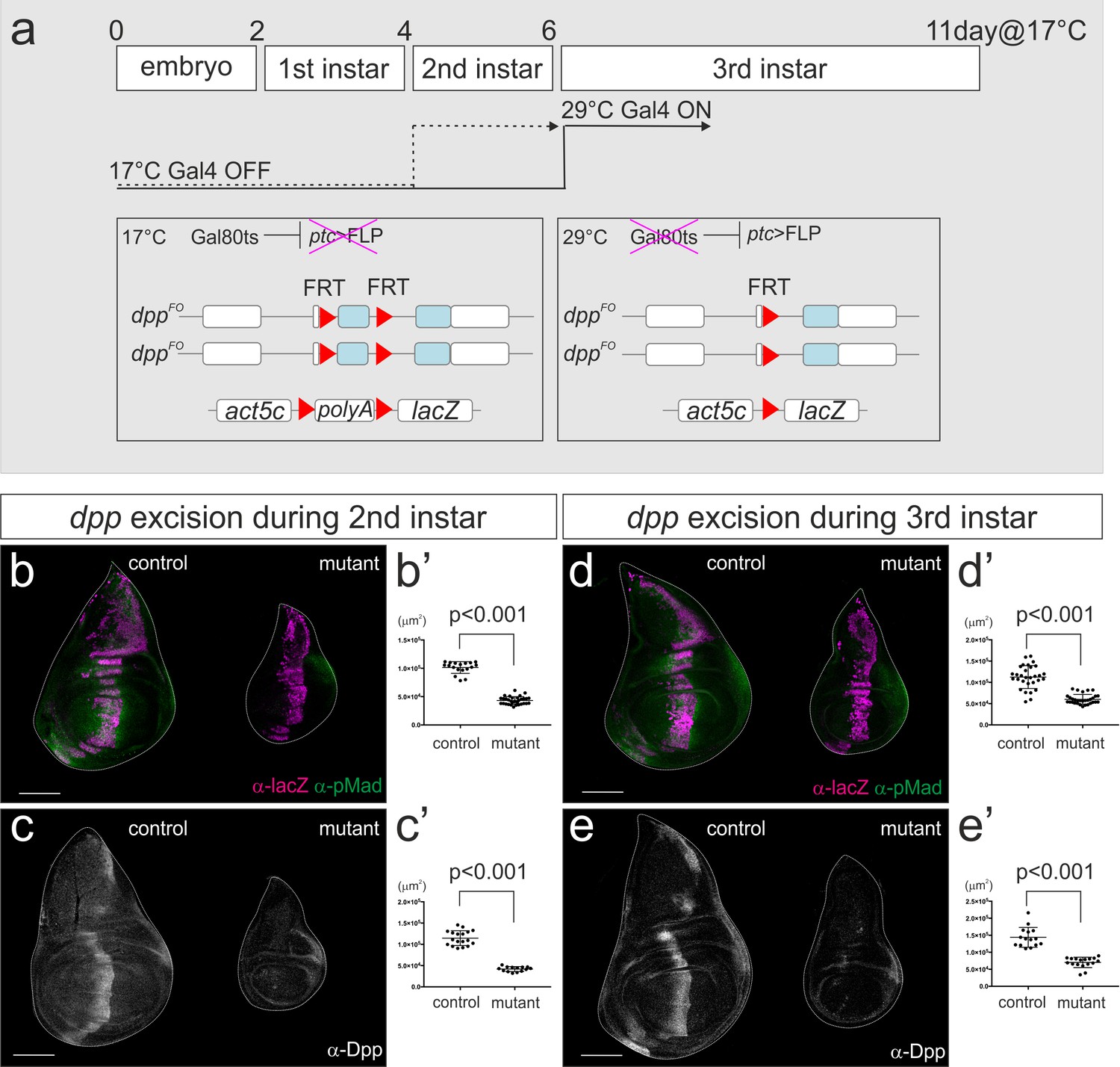
dpp stripe is required for wing disc growth during second and third instar larval stages.
(a) A scheme to genetically remove dpp from the second or third instar larval stages using Gal80ts system. At 17°C, Gal4 activity is blocked by Gal80ts. At 29°C, Gal80ts is inactivated and Gal4 starts to induce FLP expression in the anterior stripe of cells. After embryo collection for 2–4 hr at room temperature, the embryos were incubated at 17°C until temperature shift. Temperature was shifted after 4 days (second instar) or 6 days (third instar) at 17°C, and late third instar wing discs were dissected after 53 hr or 43 hr later respectively. (b–e) Removal of dpp using ptc-Gal4 during second instar larval stages (b, c) or third instar larval stages (d, e). (b, d) anti-pMad staining and anti-β-gal staining (lineage tracing) in a control male wing disc (dppFO, ptc-Gal4/CyO; tub-Gal80ts/UAS-FLP, act5C(-FRT)lacZ) (left), and a male wing disc removing dpp during the specified time point (dppFO, ptc-Gal4/dppFO; tub-Gal80ts/UAS-FLP, act5C(-FRT)lacZ) (right). (c, e) anti-Dpp staining in a control male wing disc (dppFO, ptc-Gal4/+; tub-Gal80ts/ +) (left), and a male wing disc removing dpp during the specified time point (dppFO, ptc-Gal4/dppFO; tub-Gal80ts/UAS-FLP) (right). (b’–e’) Quantification of the wing disc size of (b–e). Mean ± s.d. p<0.001 by two sided Student’s t-test. Scale bars 100 μm.
-
Figure 3—source data 1
Quantification of wing disc size for Figure 3b–e.
- https://doi.org/10.7554/eLife.22319.007
Figure 3—figure supplement 1
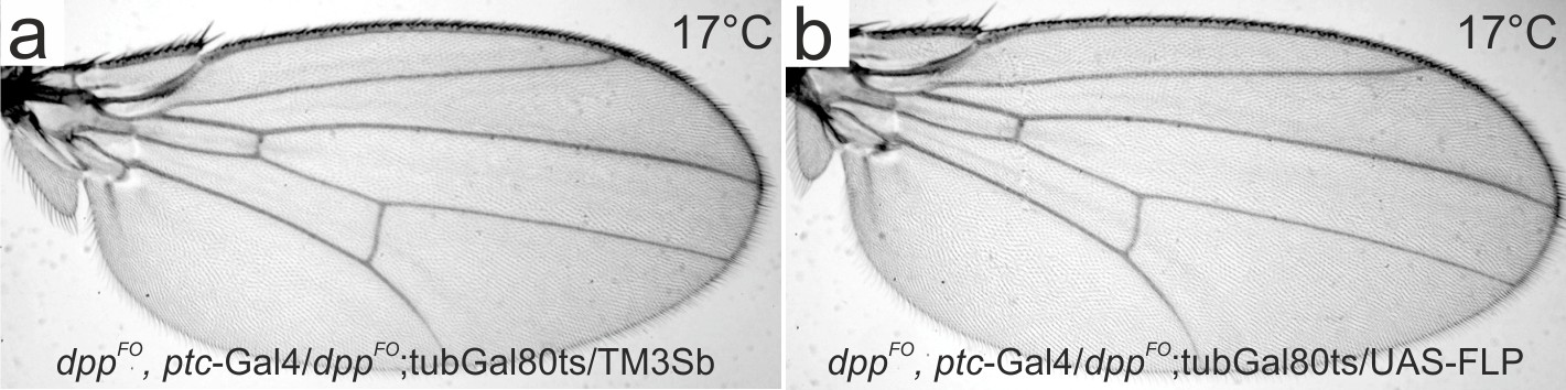
A control experiment under permissive temperature (17°C) for Figure 3.
(a) A control male wing of dppFO, ptc-Gal4/dppFO; tub-Gal80ts/TM3Sb. (b) a male wing of dppFO, ptc-Gal4/dppFO; tub-Gal80ts/UAS-FLP.
Figure 3—figure supplement 2
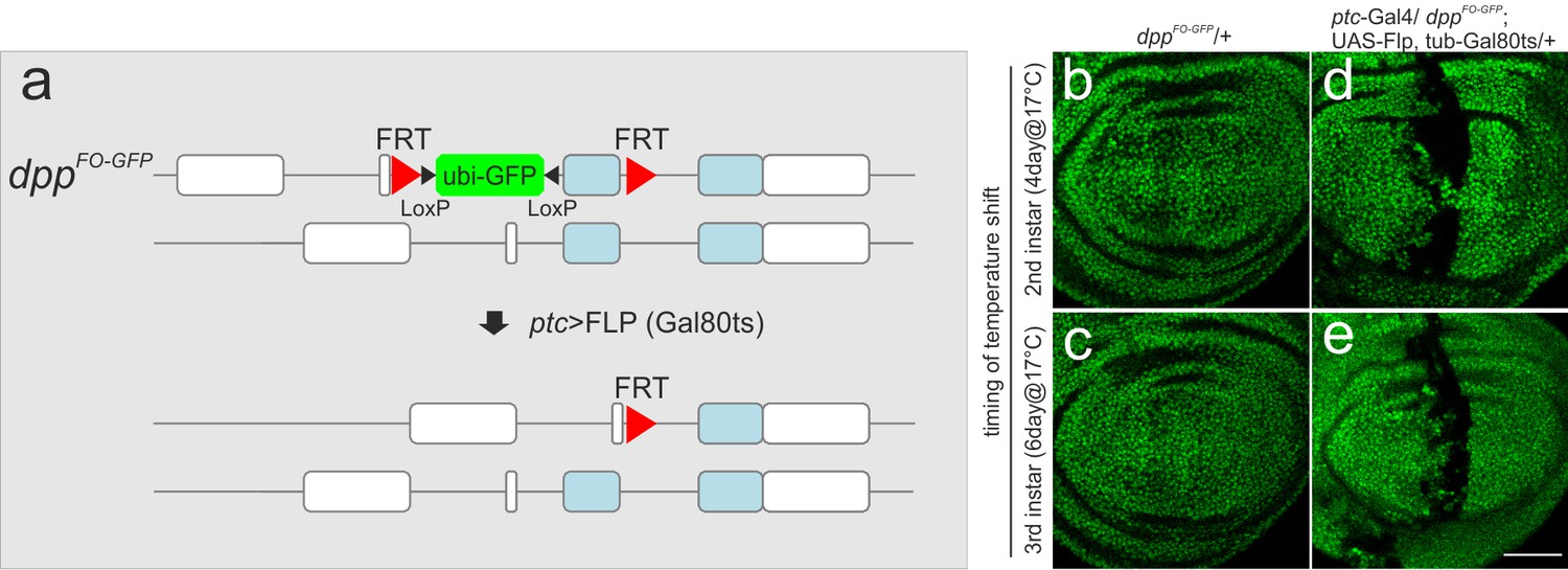
Visualization of the regions where dpp is removed using ptc-Gal4 in the wing imaginal disc.
(a) A scheme to mark the regions where dpp is removed using ptc-Gal4. dppFO-GFP contains ubi-GFP within the FRT cassette. Upon FLP/FRT mediated recombination, Ubi-GFP would be removed. Thus the regions where GFP signal is eliminated correspond to the regions where dpp is removed. (b, c) a dppFO-GFP/+ late third instar wing disc (control) after temperature shift at the beginning of second (b) or third (c) instar larval stages. (d, e) a ptc-Gal4/dppFO-GFP; UAS-FLP, tub-Gal80ts/+ late third instar wing disc after temperature shift at the beginning of second (d) and third (e) instar larval stages. Single confocal images. Scale bars 50 μm. Anterior is to the left in all figures.
Figure 3—figure supplement 3
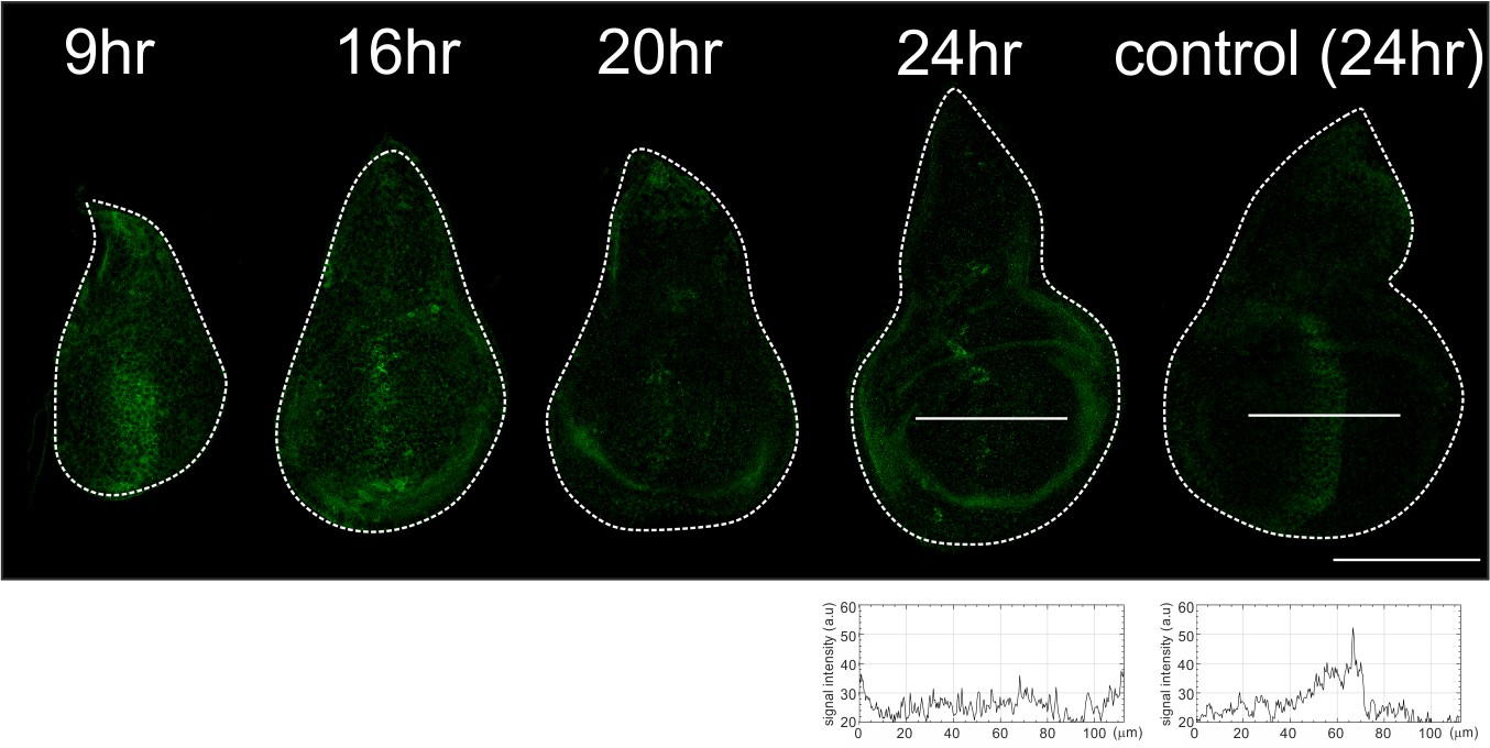
Temporal resolution of dpp removal using ptc-Gal4.
Anti-Dpp staining in dppFO, ptc-Gal4/dppFO; tub-Gal80ts/UAS-FLP wing discs at specified time (9, 16, 20, 24 hr) after temperature shift at the beginning of third instar larval stage (17°C), and in a dppFO, ptc-Gal4/dppFO; tub-Gal80ts/TM6C wing disc (control) at 24 hr after temperature shift. Average intensity projection from 3 sequential confocal images. Scale bars 100 μm. Anterior is to the left in all figures.
Figure 3—figure supplement 4
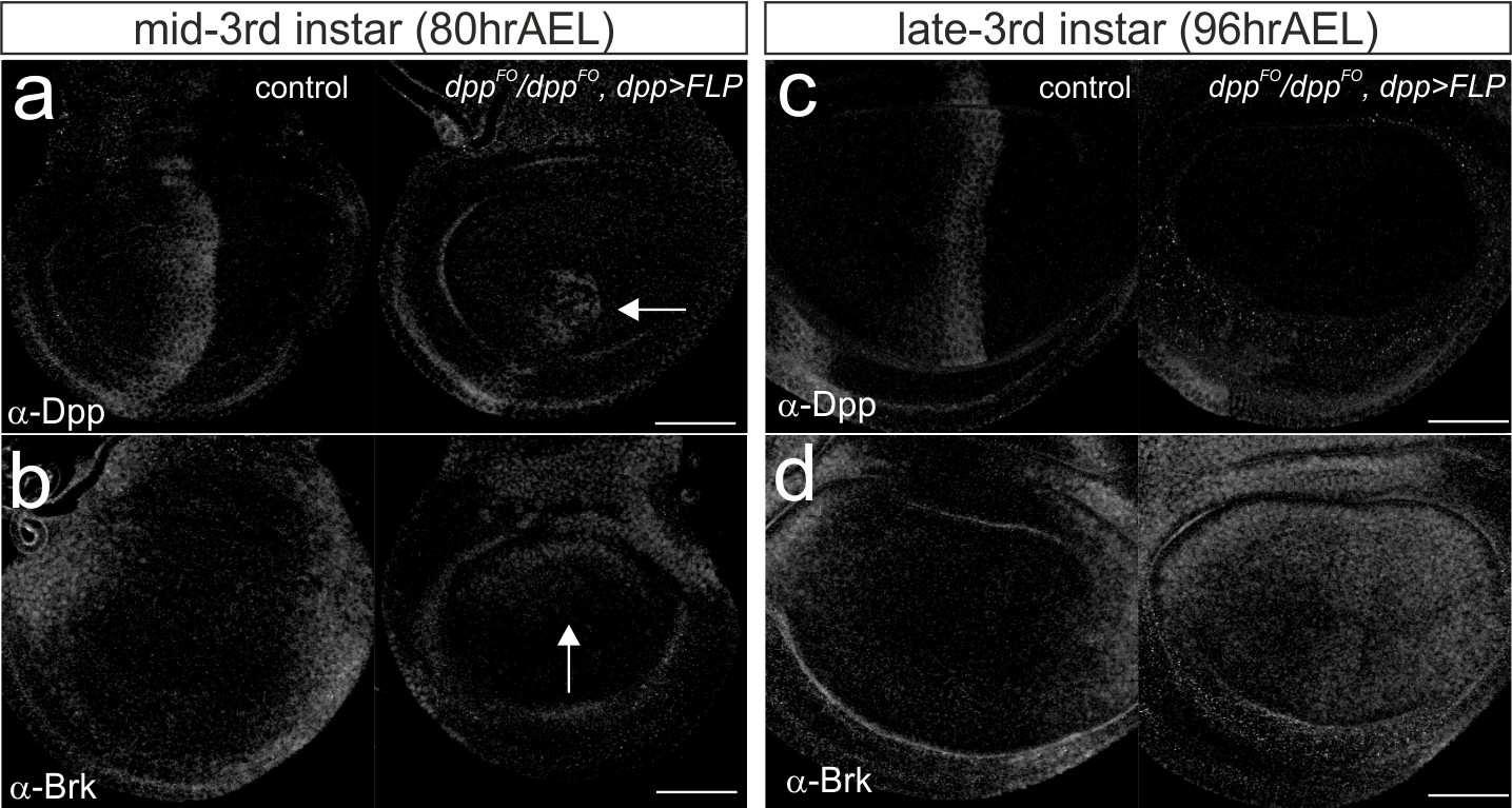
Temporal changes in Dpp and Brk expression by removing dpp using dpp-Gal4.
(a–b) Anti-Dpp staining (a) and anti-Brk staining (b) in a dppFO/+; UAS-FLP/+ (control) mid-third instar wing disc (left), and in a dppFO/dppFO; dpp-Gal4/UAS-FLP mid-third instar wing disc (right). (c–d) anti-Dpp staining (c) and anti-Brk staining (d) in a dppFO/+; UAS-FLP/+ (control) late third instar wing disc (left), and in a dppFO/dppFO; dpp-Gal4/UAS-FLP late third instar wing disc (right). Average intensity projection from 3 sequential confocal images. Scale bars 50 μm. Anterior is to the left in all figures.
Download links
A two-part list of links to download the article, or parts of the article, in various formats.
Downloads (link to download the article as PDF)
Open citations (links to open the citations from this article in various online reference manager services)
Cite this article (links to download the citations from this article in formats compatible with various reference manager tools)
Dpp from the anterior stripe of cells is crucial for the growth of the Drosophila wing disc
eLife 6:e22319.
https://doi.org/10.7554/eLife.22319


