Distinct roles of ATM and ATR in the regulation of ARP8 phosphorylation to prevent chromosome translocations
- Hiroshima University, Japan
- Tohoku University, Japan
- Radiation Biology Center, Kyoto University, Japan
- Oncode Institute, Netherlands
Figures
Figure 1 with 2 supplements
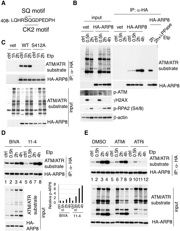
Identification of etoposide-induced ARP8 phosphorylation and the possible responsible kinase.
(A) Amino acid sequence 408 through 420 of ARP8. The Ser412 residue, within the ATM/ATR substrate motif and the CK2 substrate motif, is indicated. (B) Immunoprecipitation analysis of ARP8 phosphorylation. U2OS cells transiently expressing an empty HA vector or a vector encoding HA-tagged ARP8 were treated with DMSO (ctrl) or etoposide (Etp) for 15 min, then washed twice and cultured in complete medium for the indicated times. The nuclear extracts were incubated with anti-HA-conjugated anti-mouse IgG Dynabeads. The precipitates were electrophoresed through a gel and probed by western blotting with an anti-ATM/ATR substrate antibody or an anti-HA antibody. λPPase treatment identified the band of phosphorylated HA-ARP8. The blot of input was probed by antibodies against phospho-ATM (p-ATM), γ H2AX or phospho-RPA2 at Ser4/8 (p-RPA2). β-actin was used as a loading control. (C) Identification of the ARP8 phosphorylation site by an immunoprecipitation analysis. U2OS cells were transfected with an empty HA vector (vet), or a vector encoding HA-tagged wild-type ARP8 (WT) or HA-ARP8 S412A (S412A) for 48 hr. The cells were washed after treatment with etoposide or DMSO for 15 min, cultured in fresh medium, and harvested at the indicated time points. Whole cell extracts were used for the immunoprecipitation analysis. (D) Etoposide-induced ARP8 phosphorylation in ATM-deficient BIVA and ATM-proficient 11–4 cells. Immunoprecipitation analysis of cell extracts of BIVA or 11–4 cells transfected with HA-tagged wild-type ARP8 using anti-HA antibodies. The cells were treated with DMSO (ctrl) or etoposide (Etp) for 15 min, cultured in fresh medium, and harvested at the indicated time points. Whole cell extracts were used for the immunoprecipitation analysis, which was performed as described in (B). The amounts of phosphorylated ARP8 and HA-ARP8 were quantified, using the Image J software. The results of the quantitative analysis are shown as the relative values to the DMSO controls. Source data are presented in Figure 1—source data 1. (E) Immunoprecipitation analysis of cell extracts from 11 to 4 cells expressing HA-tagged ARP8. The cells were treated with DMSO, 10 μM ATMi (KU55933), or 10 μM ATRi (VE821) for 2 hr before etoposide treatment, and then the inhibitors (5 μM) were added after the cells were washed.
-
Figure 1—source data 1
Source raw data for Figure 1D.
- https://doi.org/10.7554/eLife.32222.005
Figure 1—figure supplement 1
ARP8 contains an ATM/ATR substrate SQ motif at Ser412 and Q413.
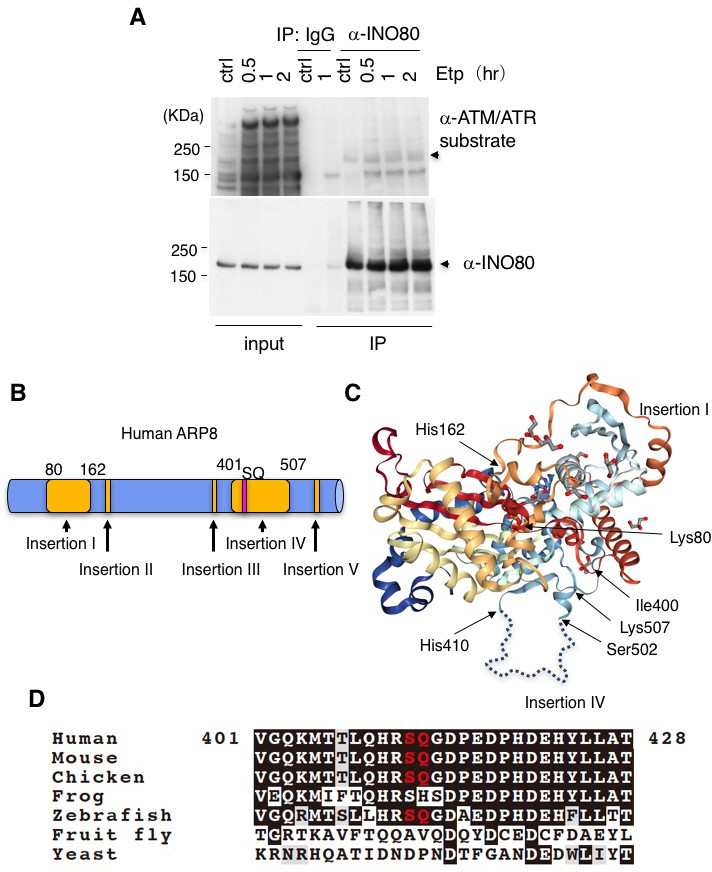
(A) Damage-induced INO80 phosphorylation was not detected by an ATM/ATR antibody. Immunoprecipitation analysis of INO80 phosphorylation. After treatment with or without etoposide for 15 min, cells were cultured in fresh medium for 0.5, 1 or 2 hr. The whole cell l extracts from U2OS cells were incubated with anti-INO80 or normal IgG-conjugated Dynabeads. The precipitates were electrophoresed through a gel and probed by western blotting with an anti-ATM/ATR substrate antibody or an anti-INO80 antibody. The arrows indicate the position of INO80. (B) Schematic diagram of full-length human ARP8. The insertions of ARP8 are shown in yellow, and the SQ motif in insertion IV is pink. (C) A 3D structure model of ARP8. Insertion I, insertion IV, and the locations of important amino acid residues are indicated. This structure was obtained from the Protein Database (PDB ID: 4FO0), and was modified according to the report by Gerhold et al. and the description for PDB ID: 4FO0. (D) Alignment of multiple ARP8 sequences. The partial amino acid sequences containing the SQ motif or the corresponding amino acids, which are colored red. The homologous sequence corresponding to human ARP8 was obtained from the NCBI Protein Database.
Figure 1—figure supplement 2
ATM, but not CK2 is responsible for ARP8 phosphorylation after etoposide treatment.
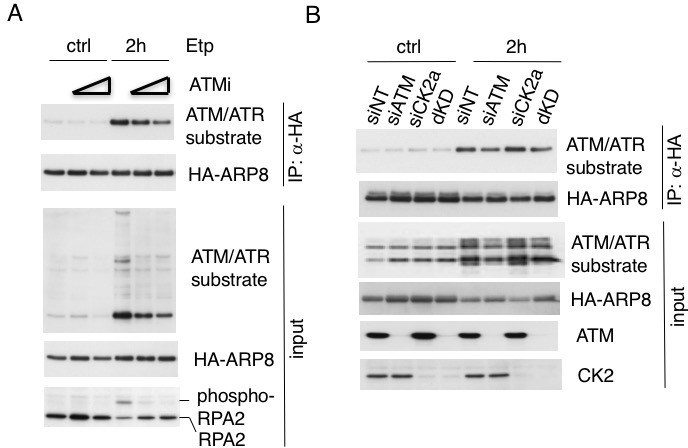
(A) Immunoprecipitation analysis of U2OS cells transiently expressing HA-tagged ARP8, treated with or without an ATM inhibitor (ATMi). ATM inhibitor (10 or 20 μM) was added as indicated for 2 hr before etoposide (Etp) treatment. (B) Immunoprecipitation analysis of ARP8 phosphorylation, using U2OS cells cotransfected with HA-tagged ARP8 and either siATM, siCK2, or the double siRNAs (dKD) for 48 hr. The cells were cultured for 2 hr in fresh medium after etoposide (Etp) treatment. Immunoprecipitation was performed with anti-HA-conjugated beads. The precipitated proteins were detected by western blotting, using the indicated antibodies.
Figure 2 with 2 supplements
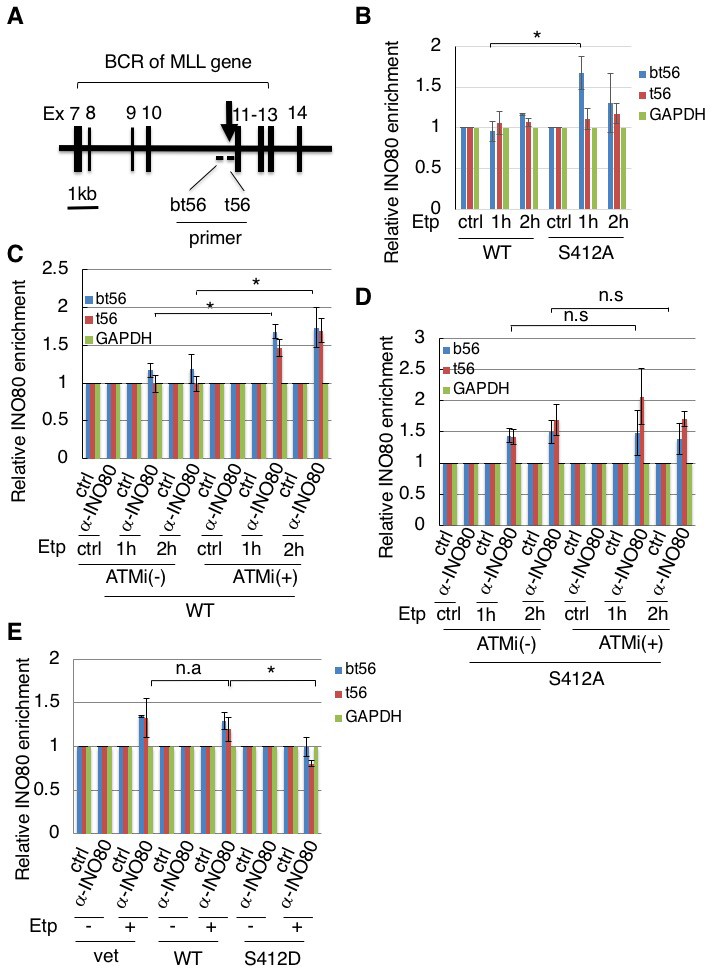
Phosphorylation of ARP8 negatively regulates the etoposide-induced enrichment of INO80.
(A) Schematic representation of the BCR in the MLL gene. The locations of the primers used in the real-time PCR analyses are shown. The arrow indicates 11q23 chromosome translocation breakpoint hotspot identified in treatment-related leukemia. Ex: Exon. (B) ChIP analysis of the INO80 loading onto the MLL BCR in endogenous ARP8-depleted11-4 Flp-In cells expressing either the siRNA-resistant wild-type (WT) or phospho-deficient ARP8 (S412A) after tetracycline treatment. The cells were treated with DMSO (ctrl) or etoposide for 15 min, washed, and then cultured in fresh medium for 1 or 2 hr. GAPDH is shown as the control region. Values represent the means ± SE from three independent experiments. *: p<0.05. Source data are presented in Figure 2—source data 1. (C) ChIP analysis of the INO80 loading onto the MLL BCR in wild-type ARP8 expressing11-4 Flp-In cells. The cells were treated with/without an ATM inhibitor (KU55933) for 2 hr before etoposide treatment, and then the inhibitors (5 μM) were added after the cells were washed. The experiment was performed as described in (B). Values represent the means ± SE from three independent experiments. *: p<0.05. The level of ATM phosphorylation or expression of INO80 was shown in Figure 2—figure supplement 2B. Source data are presented in Figure 2—source data 1. (D) ChIP analysis of the INO80 loading onto the MLL BCR in S412A ARP8 expressing 11–4 Flp-In cells. The cells were treated with/without 10 μM ATM inhibitor for 2 hr before etoposide treatment, and then the inhibitors (5 μM) were added after the cells were washed. The experiment was performed as described in (B). Values represent the means ± SE from three independent experiments. n.s: no significant difference. The levels of ATM phosphorylation and INO80 expression are shown in Figure 2—figure supplement 2C. Source data are presented in Figure 2—source data 1. (E) ChIP analysis of the INO80 loading onto the MLL BCR in endogenous ARP8-depleted BIVA cells transfected with either the siRNA-resistant wild-type (WT) or phospho-mimetic ARP8(S412D). The control cells were transfected with an empty vector and a non-targeting siRNA (vet). The cells were treated with DMSO (ctrl) or etoposide for 15 min, washed, and then cultured in fresh medium for 1 hr. Values represent the means ± SE from three independent experiments. *p<0.05, n.a: not analyzed. Source data are presented in Figure 2—source data 1.
-
Figure 2—source data 1
Source raw data for Figure 2B-E.
- https://doi.org/10.7554/eLife.32222.009
-
Figure 2—source data 2
Source raw data for Figure 2—figure supplement 1A and B.
- https://doi.org/10.7554/eLife.32222.010
Figure 2—figure supplement 1
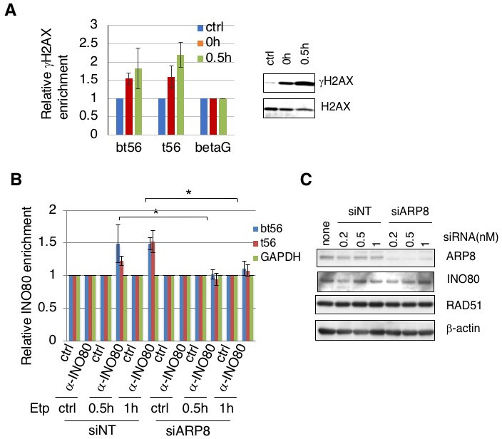
(A) Etoposide treatment induced H2AX phosphorylation on BCR of MLL gene. ChIP analysis of γH2AX enrichment on the MLL BCR in ATM proficient 11–4 cells. The cells were treated with DMSO (ctrl) or etoposide for 15 min, washed, and then fixed immediately (0 hr) or after culture in fresh medium for 0.5 hr (0.5 hr). β-globin (betaG) is shown as the control region. Values represent the means ± SE from four independent experiments. The results of immunoblotting analyses of γH2AX and H2AX are shown. The cells were treated with DMSO (ctrl) or etoposide for 15 min, washed, and then harvested immediately (0 hr) or after culture in fresh medium for 0 (Etp) or 0.5 hr (0.5 hr). Source data are presented in Figure 2—source data 2. (B) Depletion of ARP8 reduced the etoposide-induced enrichment of INO80 onto the BCR of the MLL gene. ChIP analysis of INO80 loading onto the MLL BCR in AT5BIVA cells depleted by either a non-targeting siRNA (siNT) or siARP8. The cells were treated with DMSO (ctrl) or etoposide for 15 min, washed, and then cultured in fresh medium for 30 or 60 min. GAPDH is shown as the control region. Values represent the means ± SE from four independent experiments. *: p<0.05. Source data are presented in Figure 2—source data 2. (C) ARP8 depletion did not affect INO80 or RAD51 expression. Immunoblotting analysis of non-targeting siRNA(NT) or siARP8 transfected cells. The final concentration of each siRNAs is indicated. β-actin was used as the loading control. In further studies, we used 0.2–0.3 nM of siRNA. The data showed that the depletion of ARP8 did not disturb INO80 and RAD51 levels.
Figure 2—figure supplement 2
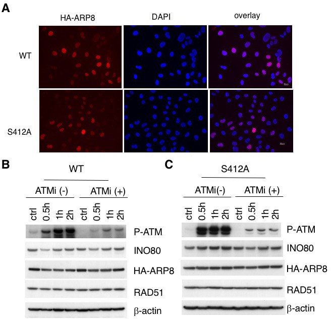
Establishment of stable and inducible ARP8 expressing 11–4 Flp-In cells.
(A) Immunofluorescence staining of 11–4 Flp-In cells expressing HA-tagged wild-type ARP8 (WT) or S412 phospho-deficient ARP8 (SA). For induction of HA-ARP8 expression, tetracycline was added for 24 hr. The anti-HA antibody was used to identify the expression of HA-ARP8. DAPI was used for DNA staining. (B and C) Immunoblotting analysis of phosphorylated-ATM, and expression level of INO80 and RAD51 in 11–4 Flp-In cells. The endogenous ARP8-depleted11-4 Flp-In cells with tetracycline inducible expression of siRNA-resistant wild-type (WT) (B) or S412A ARP8 (S412A) (C), were treated with/without 10 μM ATM inhibitor for 2 hr, before DMSO (ctrl) or etoposide treatment. The cells were harvested at the indicated time point after etoposide removal. β-actin is shown as a loading control.
Figure 3 with 2 supplements
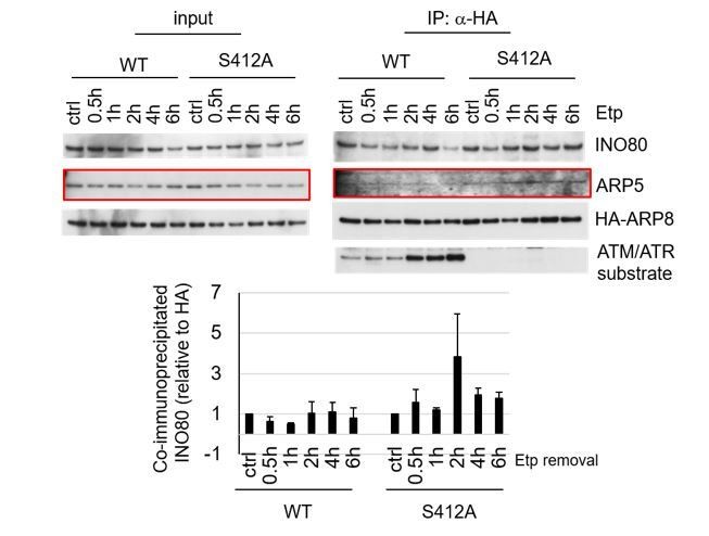
ARP8 phosphorylation deficiency increased its interaction with INO80.
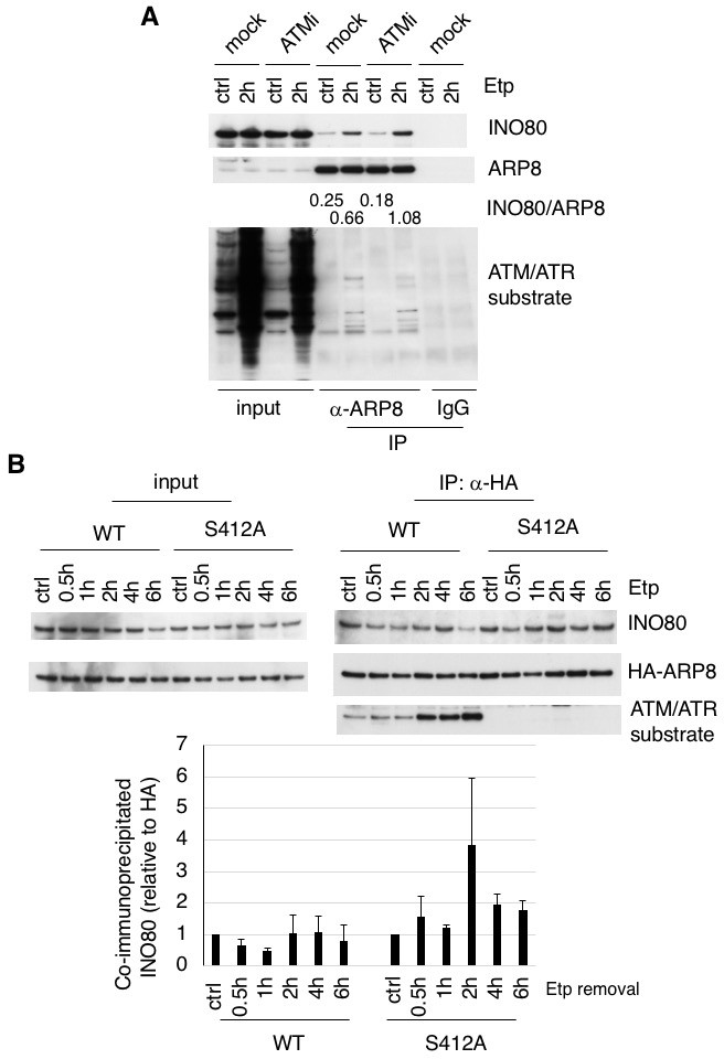
(A) Immunoprecipitation analysis of the interaction between INO80 and ARP8 in ATM inhibitor treated U2OS cells. The cells were treated with 10 μM KU55933 (ATMi) or equal amounts of DMSO (mock) for 2 hr, and then treated with DMSO (ctrl) or etoposide for 15 min, washed, and then cultured in fresh medium with or without 5 μM KU55933 for 2 hr. Immunoprecipitation analysis was performed with either anti-ARP8 antibodies or normal IgG. The relative immunoprecipitated amounts of INO80 are shown. Quantitative analysis was performed using the Image J software. (B) Examination of the interaction between INO80 and ARP8 in U2OS cells expressing HA-tagged wild-type or S412A ARP8. The endogenous ARP8-depleted cells were treated with etoposide for 15 min. After the cells were washed, they were placed in fresh medium and harvested at the indicated time points. The nuclear extracts were incubated with anti-HA-conjugated anti-mouse IgG Dynabeads. The precipitates were electrophoresed through a gel and probed by western blotting with an anti-INO80 or an anti-HA or an anti-ATM/ATR substrate antibody. The amounts of INO80 and HA-ARP8 were quantified, using the Image J software. The results of quantitative analysis are shown as the relative values as compared to the DMSO control from three independent experiments. Source data are presented in Figure 3—source data 1.
-
Figure 3—source data 1
Source raw data for Figure 3B.
- https://doi.org/10.7554/eLife.32222.014
-
Figure 3—source data 2
Source raw data for Figure 3—figure supplement 1B.
- https://doi.org/10.7554/eLife.32222.015
-
Figure 3—source data 3
Source raw data for Figure 3—figure supplement 2B.
- https://doi.org/10.7554/eLife.32222.016
Figure 3—figure supplement 1
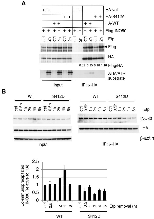
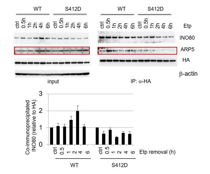
Examination of the interaction between ARP8 and INO80.
(A) Examination of the interaction between INO80 and wild-type ARP8 or phospho-deficient ARP8. U2OS cells were co-transfected with Flag-INO80 and HA (vet), HA-ARP8 (WT), or HA-ARP8 S412A vectors, respectively, for 48 hr and subsequently treated with DMSO (ctrl) or etoposide for 15 min, washed, and then cultured in fresh medium for 2 hr. The immunoprecipitation analysis was performed using anti-HA antibodies. The precipitates were electrophoresed through a gel and probed by western blotting with an anti-Flag or an anti-HA or anti-ATM/ATR substrate antibody. The co-immunoprecipitated amounts of Flag-INO80 relative to HA-ARP8 are shown. Quantitative analysis was performed using the Image J software. (B) Co-immunoprecipitation analysis of ARP8 and INO80. ATM-deficient BIVA cells were co-transfected with the siARP8 and siARP8-resistant HA-tagged wild-type ARP8 or phospho-mimetic S412D ARP8 mutant. After etoposide removal, the cells were recovered at the indicated time points. The nuclear extracts were incubated with anti-HA-conjugated anti-mouse IgG Dynabeads. The precipitates were electrophoresed through a gel and probed by western blotting with either an anti-INO80 antibody or an anti-HA antibody. The amounts of INO80 and HA-ARP8 were quantified, using the Image J software. The results of quantitative analysis are shown as the relative values as compared to the DMSO control, from three independent experiments. Source data are presented in Figure 3—source data 2.
Figure 3—figure supplement 2
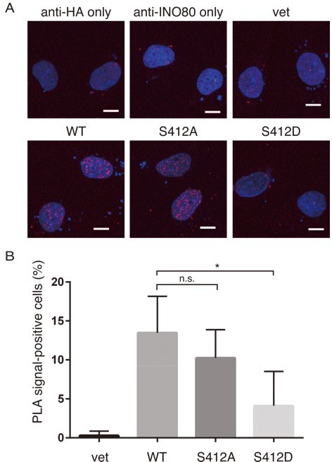
Examination of ARP8-INO80 interactions using Proximity ligation assay.
(A) Proximity ligation assay (PLA) signals (in red) indicate ARP8-INO80 interactions. GM0637 cells were transfected with the HA-tagged wild-type, S412 A or S412D mutant of ARP8 or empty vector (vet). Nuclei were stained with DAPI (in blue). Scale bar: 10 μm. (B) Percentages of cells with more than five PLA signals. All results are the means of four independent experiments, and error bars show the standard deviation of the mean. *p<0.05. n.s.: no significant difference. Source data are presented in Figure 3—source data 3.
Figure 4 with 1 supplement
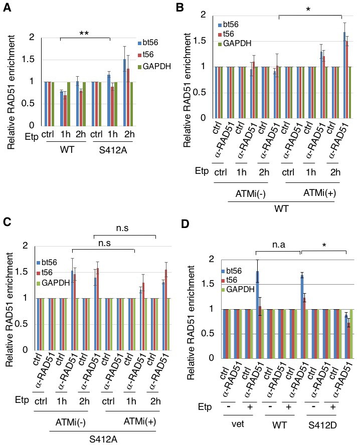
ARP8 phosphorylation prevents the excessive RAD51 loading onto MLL BCR.
(A) ChIP analysis of the RAD51 loading onto the MLL BCR in endogenous ARP8-depleted11-4 Flp-In cells expressing the siRNA-resistant wild-type (WT) or phospho-deficient ARP8 (S412A) after tetracycline treatment. The cells were treated with DMSO (ctrl) or etoposide for 15 min, washed, and then cultured in fresh medium for 1 or 2 hr. Values represent the means ± SE from three independent experiments. **: p<0.01. Source data are presented in Figure 4—source data 1. (B) ChIP analysis of the RAD51 loading onto the MLL BCR in wild-type ARP8 expressing 11–4 cells. Following a treatment with/without 10 μM ATM inhibitor (KU55933) for 2 hr, the cells were treated with DMSO (ctrl) or etoposide for 15 min, washed, and then cultured in fresh medium with or without 5 μM KU55933 for 1 or 2 hr. Values represent the means ± SE from three independent experiments. *p<0.05. The levels of ATM phosphorylation and expression of RAD51 are shown in Figure 2—figure supplement 2B. Source data are presented in Figure 4—source data 1. (C) ChIP analysis of the RAD51 loading onto the MLL BCR in S412A ARP8 mutant expressing 11–4 Flp-In cells. The experiment was performed as described in (B). Values represent the means ± SE from three independent experiments. n.s: no significant difference. The levels of ATM phosphorylation and RAD51 expression are shown in Figure 2—figure supplement 2C. Source data are presented in Figure 4—source data 1. (D) ChIP analysis of the RAD51 loading onto the MLL BCR in endogenous ARP8-depleted BIVA cells transfected with either the siRNA-resistant wild-type (WT) or phospho-mimetic ARP8 (S412D). GAPDH is shown as the control region. The control cells were transfected with the empty vector and the non-targeting siRNA (vet). Values represent the means ± SE from three independent experiments. *p<0.05. n.a: not analyzed. Source data are presented in Figure 4—source data 1.
-
Figure 4—source data 1
Source raw data for Figure 4A-D.
- https://doi.org/10.7554/eLife.32222.019
-
Figure 4—source data 2
Source raw data for Figure 4—figure supplement 1B and C.
- https://doi.org/10.7554/eLife.32222.020
Figure 4—figure supplement 1
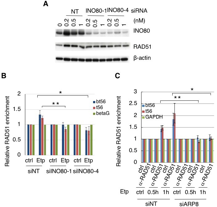
Requirement of INO80 and ARP8 for RAD51 binding to the BCR of the MLL gene.
(A) Immunoblotting analysis of INO80 depletion efficiency. Two kinds of siRNAs against INO80 were used for the detection of the INO80 knockdown efficiency. RAD51 expression did not affect the INO80 knockdown. β−actin was used as the loading control. (B) ChIP analysis of the enrichment of RAD51 in AT5BIVA cells treated with non-targeting siRNA (siNT) or the siRNAs against INO80 (siINO80-1 and siINO80-4). The cells were treated with etoposide for 15 min, washed, and then cultured in fresh medium for 1 hr (Etp). Vehicle treatment was performed as a control. The DNA was analyzed by real-time PCR, using the MLL BCR gene primers. The β−globin gene (betaG) was used as a control region. Values represent the means ± SE from six independent experiments. *p<0.05. Source data are presented in Figure 4—source data 2. (C) ChIP analysis of RAD51 loading onto the MLL BCR, in AT5BIVA cells transfected with the non-targeting siRNA (siNT) or siARP8. The cells were treated with DMSO (ctrl) or etoposide for 15 min, washed, and then cultured in fresh medium for 30 or 60 min. GAPDH is shown as the control region. Values represent the means ± SE from four independent experiments. *p<0.05, **p<0.01. Source data are presented in Figure 4—source data 2.
Figure 5 with 1 supplement

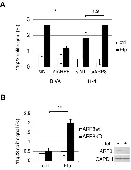
ARP8 phosphorylation averts 11q23 chromosome translocations.
(A) Dual-color FISH analysis of chromosome 11q23. Representative FISH images using etoposide treated BIVA cells are shown. Arrows indicate the split signals (separated by >1 µm). Scale bar: 5 μm. (B) The percentages of AT5BIVA or 11–4 cells with split chromosome 11q23 signals are shown. The non-targeting control siRNA (siNT) or siARP8-depleted cells were treated with DMSO (ctrl) or etoposide for 15 min, washed, and cultured for 6 hr in fresh medium. At least 2,000 cells were analyzed in every experiment. The average percentages of cells with split signals from four independent experiments are shown. Values represent the means ± SE. ***p<0.001 as determined by the Z test. The ARP8 knockdown is shown in the gel image on the right. Source data are presented in Figure 5—source data 1. (C, D) Dual-color FISH analyses of chromosome 11q23 using ARP8-depleted AT5BIVA (C) and 11–4 (D) cells expressing the siARP8-resistant ARP8 wild-type (WT), S412D, or S412A. The average percentages of the cells with split signals from three independent experiments are shown. Values represent the means ± SE. ***p<0.001 as determined by the Z test. Source data are presented in Figure 5—source data 1. (E) Dual-color FISH analyses of chromosome 11q23 using 11–4 cells expressing the siARP8-resistant ARP8 S412A. The cells were treated with/without 10 μM ATM inhibitor (KU55933) for 2 hr before etoposide treatment. After the cells were washed, KU55933 (5 μM) was added until the cells were harvested. The average percentages of the cells with split signals from three independent experiments are shown. Values represent the means ± SE from three independent experiments. n.s.: no significant difference. Source data are presented in Figure 5—source data 1.
-
Figure 5—source data 1
Source raw data for Figure 5B-E.
- https://doi.org/10.7554/eLife.32222.023
-
Figure 5—source data 2
Source raw data for Figure 5—figure supplement 1A and B.
- https://doi.org/10.7554/eLife.32222.024
Figure 5—figure supplement 1
Dual-color FISH analysis of chromosome 11q23 using a different DNA probe set.
(A) Dual-color FISH analysis of chromosome 11q23 in AT5BIVA and 11–4 cells, using the probe from Vysis. The non-targeting control siRNA (siNT) or siARP8-depleted cells were treated with etoposide for 15 min, washed, and cultured for 6 hr in fresh medium. Values represent the means ± SE from three independent experiments. *p<0.05. n.s: no significant difference. Source data are presented in Figure 5—source data 2. (B) Dual-color FISH analysis of chromosome 11q23 in wild-type or ARP8-knockout Nalm-6 cells. The cells were treated with 20 μM of etoposide for 15 min, and then cultured for 4 hr in fresh medium. In every experiment, 200 cells were analyzed. The average percentages of the cells with split signals are shown from four independent experiments. Values represent the means ± SE. **p<0.01, as determined by the Z test. The tetracycline-induced ARP8 knockout is shown in the gel image on the right. Source data are presented in Figure 5—source data 2.
Figure 6
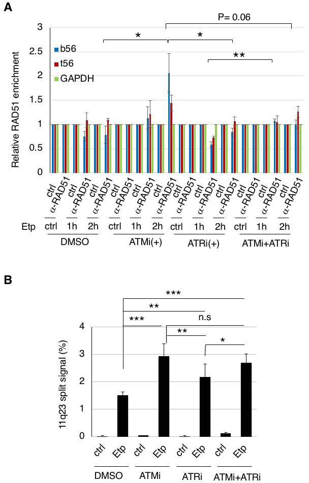
ATM, but not ATR, negatively regulates RAD51 loading onto the BCR after etoposide treatment to repress 11q23 chromosome translocations.
(A) ChIP analysis of the RAD51 loading onto the MLL BCR in ATMi or ATRi or a combination of ATMi and ATRi treated 11–4 cells. 11–4 cells were treated with ATMi (10 µM), ATRi (10 µM), or a combination of ATMi and ATRi for 2 hr before etoposide treatment. After washing the cells, the inhibitors (5 μM) were added until the cells were harvested. The ChIP analysis was performed as described in Figure 4. Values represent the means ± SE from three independent experiments. *p<0.05. **p<0.01. Source data are presented in Figure 6—source data 1. (B) The percentages of 11–4 cells with split chromosome 11q23 signals are shown. 11–4 cells were treated with ATMi (10 µM), ATRi (10 µM), or a combination of ATMi and ATRi for 2 hr before etoposide treatment. After the cells were washed, the inhibitors (5 μM) were added until the cells were harvested. Dual-color FISH analyses of chromosome 11q23 were performed as described in Figure 5. Values represent the means ± SE from three independent experiments. *p<0.05. **p<0.01, ***p<0.001, n.s.: no significant difference.
-
Figure 6—source data 1
Source raw data for Figure 6A and B.
- https://doi.org/10.7554/eLife.32222.026
Author response image 1
Author response image 2
Tables
Key resources table
| Reagent type (species) or resource | Designation | Source or reference | Identifiers | Additional information |
|---|---|---|---|---|
| Gene (Homo Sapiens) | arp8 | NA | GenBank: GeneID 93973 | |
| Gene (Homo Sapiens) | ino80 | NA | GenBank: GeneID 54617 | |
| Gene (Homo Sapiens) | atm | NA | GenBank: GeneID 472 | |
| Gene (Homo Sapiens) | atr | NA | GenBank: GeneID 545 | |
| Gene (Homo Sapiens) | rad51 | NA | GenBank: GeneID 5888 | |
| Gene (Homo Sapiens) | rpa2 | NA | GenBank: GeneID 6118 | |
| Cell line (Homo Sapiens) | AT5BIVA | PMID:21048951 | RRID: CVCL_7442 | Cell line maintained in S. Matsuura lab; |
| Cell line (Homo Sapiens) | 11–4 | PMID:21048951 | Cell line maintained in S. Matsuura lab; | |
| Cell line (Homo Sapiens) | U2OS | ATCC | RRID: CVCL_0042 | |
| Cell line (Homo Sapiens) | GM0637 | other | RRID: CVCL_7297 | Cell line maintained in T. Cremer lab; |
| Cell line (Homo Sapiens) | Tet-Off ARP8 Nalm-6 | PMID:25299602 | Cell line maintained in M. Harata lab; | |
| Transfected construct (Homo Sapiens) | Flp-In T-REx 11–4 | this paper | Progenitor = 11–4; constructed by use of Flp-In T-REx core kit (Invitrogen) | |
| Transfected construct (Homo Sapiens) | Flp-In T-REx 11–4 HA-ARP8-WT | this paper | Progenitor = Flp In T-REx 11–4; Addition of tetracyclin induces expression of HA-tagged recombinant ARP8 wild-type protein | |
| Transfected construct (Homo Sapiens) | Flp-In T-REx 11–4 HA-ARP8-S412A | this paper | Progenitor = Flp In T-REx 11–4; Addition of tetracyclin induces expression of HA-tagged recombinant ARP8-S412A mutant protein | |
| Transfected construct (Homo Sapiens) | U2OS HA-ARP8-WT | this paper | Progenitor = U2 OS; stably expressing HA-tagged recombinant ARP8 wild-type protein | |
| Transfected construct (Homo Sapiens) | U2OS HA-ARP8-S412A | this paper | Progenitor = U2 OS; stably expressing HA-tagged recombinant ARP8-S412A protein | |
| Antibody | anti-phospho ATM/ATR substrate motif (rabbit monoclonal) | Cell Signaling Technology | Cell Signaling Technology:Cat# 6966S; RRID:AB_10949894 | |
| Antibody | anti-ATM protein kinase pS1981 (mouse monoclonal) | Rockland | Rockland:Cat# 200-301-400; RRID:AB_217868 | |
| Antibody | anti-HA Tag (mouse monoclonal) | Merck Millipore | Merck Millipore:Cat# 05–904; RRID:AB_417380 | |
| Antibody | anti-Histone H2A.X, phospho (Ser139) (mouse monoclonal) | Merck Millipore | Merck Millipore:Cat# 05–636; RRID:AB_309864 | |
| Antibody | anti-human RAD51 (rabbit polyclonal) | Bio Academia | Bio Acdemia:Cat# 70–001; RRID:AB_2177110 | |
| Antibody | anti-phospho RPA32 (S4/S8) (rabbit polyclonal) | Bethyl Laboratories | Bethyl Laboratories:Cat# A300-245A; RRID:AB_210547 | |
| Antibody | anti-INO80 (rabbit polyclonal) | Bethyl Laboratories | Bethyl Laboratories:Cat# A303-371A; RRID:AB_10950580 | |
| Antibody | anti-Human RPA/p34 (Replication Protein A) Ab-1 (mouse monoclonal) | Lab Vision | Lab Vision:Cat# MS-691-P0; RRID:AB_143149 | |
| Antibody | anti-GAPDH (mouse monolconal) | Santa Cruz Biotechnology | Santa Cruz Biotechnology: sc-32233; RRID:AB_627679 | |
| antibody | anti-Histone H2A.X | Abcam | Abcam: Cat# ab124781; RRID_AB_10971675 | |
| antibody | anti-beta-actin (mouse monoclonal) | Sigma-Aldrich | Sigma-Aldrich:Cat # A5441; RRID:AB_476744 | |
| antibody | anti-ARP8 (rabbit monoclonal) | PMID:25299602 | ||
| antibody | Cy3-secondary | Invitrogen | ||
| Recombinant DNA reagent | pME18FL-hARP8 | PMID:18163988 | ||
| Recombinant DNA reagent | pcDNA3.1/Myc-His(-) | Invitrogen | Invitrogen:Cat # V855-20 | |
| Recombinant DNA reagent | pcDNA5/FRT/TO | Invitrogen | Invitrogen:Cat # K650001 | |
| Recombinant DNA reagent | HA-arp8 | this paper | Progenitor = pME18FL-hARP8; PCR, HA tag was fused; mutagenized in the Ambion Silencer Select s41201 siRNA target site for resistance; inserted into pcDNA3.1/Myc-His(-) or pcDNA5/FRT/TO | |
| Recombinant DNA reagent | HA-arp8-S412A | this paper | Progenitor = HA-arp8; PCR, mutagenized; inserted into pcDNA3.1/Myc-His(-) or pcDNA5/FRT/TO | |
| Recombinant DNA reagent | HA-arp8-S412D | this paper | Progenitor = HA-arp8; PCR, mutagenized; inserted into pcDNA3.1/Myc-His(-) | |
| Sequence-based reagent | oligonucleotide for construction of siRNA-resistant HA-ARP8 | this paper | 5'-CTCAACAAAATGCCACCATCGTTCAGACGTATAATTGAAAATGTGGATG-3' | |
| Sequence-based reagent | oligonucleotide for construction of siRNA-resistant HA-ARP8-S412A | this paper | 5'-TTGCAGCACAGAGCTCAGGGCGATCCTG-3' | |
| Sequence-based reagent | oligonucleotide for construction of siRNA-resistant HA-ARP8-S412D | this paper | 5'-TTGCAGCACAGAGATCAGGGCGATCCTG-3' | |
| Sequence-based reagent | primer for RT-PCR of bt56 forward | PMID:21048951 | 5'-TACTCTGAATCTCCCGCA-3' | |
| Sequence-based reagent | primer for RT-PCR of bt56 reverse | PMID:21048951 | 5'-CGCTCGTTCTCCTCTAA-3' | |
| Sequence-based reagent | primer for RT-PCR of t56 forward | PMID:21048951 | 5'-TTGCCAAGTCTGTTGTGAG-3' | |
| Sequence-based reagent | primer for RT-PCR of t56 reverse | PMID:21048951 | 5'-CAGAGGCCCAGCTGTAGTTC-3' | |
| Sequence-based reagent | primer for RT-PCR of GAPDH forward | this paper | 5'-TCTCCCCACACACATGCACTT-3' | |
| Sequence-based reagent | primer for RT-PCR of GAPDH reverse | this paper | 5'-CCTAGTCCCAGGGCTTTGATT-3' | |
| Sequence-based reagent | primer for RT-PCR of beta-globin forward | PMID:21048951 | 5'-TTGGACCCAGAGGTTCTTTG-3' | |
| Sequence-based reagent | primer for RT-PCR of beta-globin reverse | PMID:21048951 | 5'-GAGCCAGGCCATCACTAAAG-3' | |
| Commercial assay or kit | Duolink PLA | Sigma-Aldrich | Sigma-Aldrich:Cat # DUO92002, DUO92004, DUO92008 | Proximity Ligation Assay |
| Chemical compound, drug | etoposide | Sigma-Aldrich | Sigma-Aldrich:Cat # E1383 | |
| Chemical compound, drug | ATM inhibitor (KU55933) | Merck Millipore | Merck Millipore:Cat# 118500 | |
| Chemical compound, drug | ATR inhibitor IV | Merck Millipore | Merck Millipore:Cat# 504972 | |
| Software, algorithm | Metefer 4 MetaCyte | Metasystems | v 3.11.4 | software for FISH analysis |
| Software, algorithm | Image J | NIH | ||
| Other | XL MLL Plus | Metasystems Probes | Metasystems Probes:Cat# D5060-100-OG | probe for FISH analysis |
| Other | LSI MLL Dual Color, BreakApart Rearrangement Probe | Vysis, Abbott Molecular Inc. | Vysis, Abbott Molecular Inc.: 32–190083 | probe for FISH analysis |
Additional files
-
Transparent reporting form
- https://doi.org/10.7554/eLife.32222.027
Download links
A two-part list of links to download the article, or parts of the article, in various formats.
Downloads (link to download the article as PDF)
Open citations (links to open the citations from this article in various online reference manager services)
Cite this article (links to download the citations from this article in formats compatible with various reference manager tools)
Distinct roles of ATM and ATR in the regulation of ARP8 phosphorylation to prevent chromosome translocations
eLife 7:e32222.
https://doi.org/10.7554/eLife.32222


