Defective RNA polymerase III is negatively regulated by the SUMO-Ubiquitin-Cdc48 pathway
- Salk Institute for Biological Studies, United States
- The Scripps Research Institute, United States
Figures
Figure 1 with 1 supplement
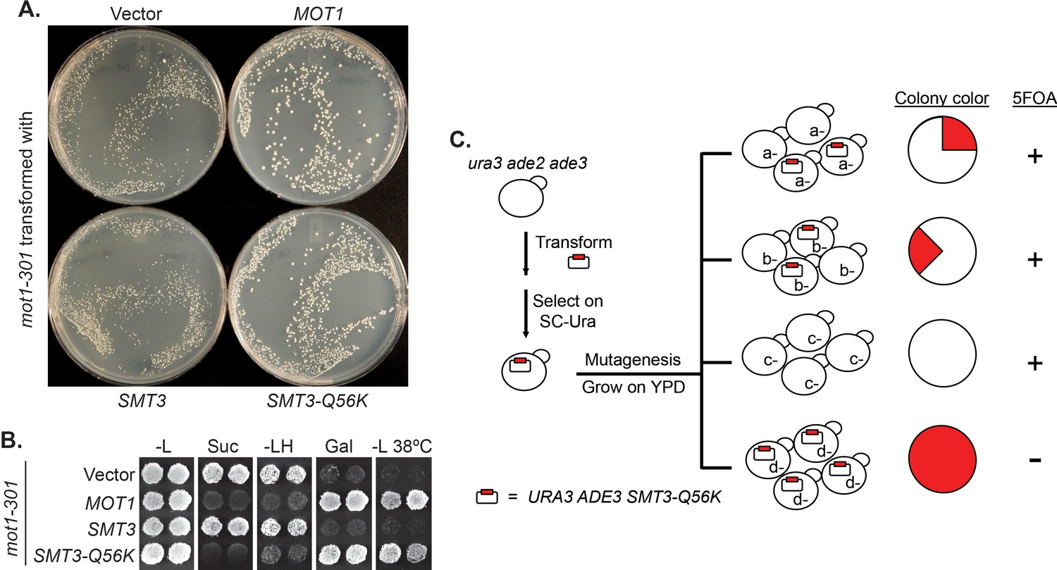
A reverse suppressor screen using the dominant SMT3-Q56K mutant.
(A) A mot1-301 strain was transformed with CEN LEU2 vectors carrying indicated genes, then selected for transformants on SC-Leu plates. Wild-type MOT1 or SMT3-Q56K made mot1-301 cells grow faster. (B) Transformants from (A) were patched on SC-Leu (-L) then replica plated to SC-Leu-His (-LH), YPSucrose (Suc), SC-Galactose (Gal) plates, or a SC-Leu plate incubated at elevated temperature 38°C (-L 38°C). mot1-301 is His+ (Spt-), Suc+ (Bur-), Gal-, and Ts-, whereas SMT3-Q56K reversed all four phenotype. The suppression is dominant because the wild-type genomic copy of SMT3 was present in all the strains. (C) Design of the screen. The starting strain is an ura3 ade2 ade3 triple mutant. ura3 is used for URA3 plasmid selection and 5FOA-sensitivity test. ade2 ade3 double mutant colonies are white, but the wild-type ADE3 on the plasmid complements ade3 and turns the cells red. A mutant (d-) that requires the plasmid for viability will form a uniformly red and 5FOA-sensitive colony.
Figure 1—figure supplement 1
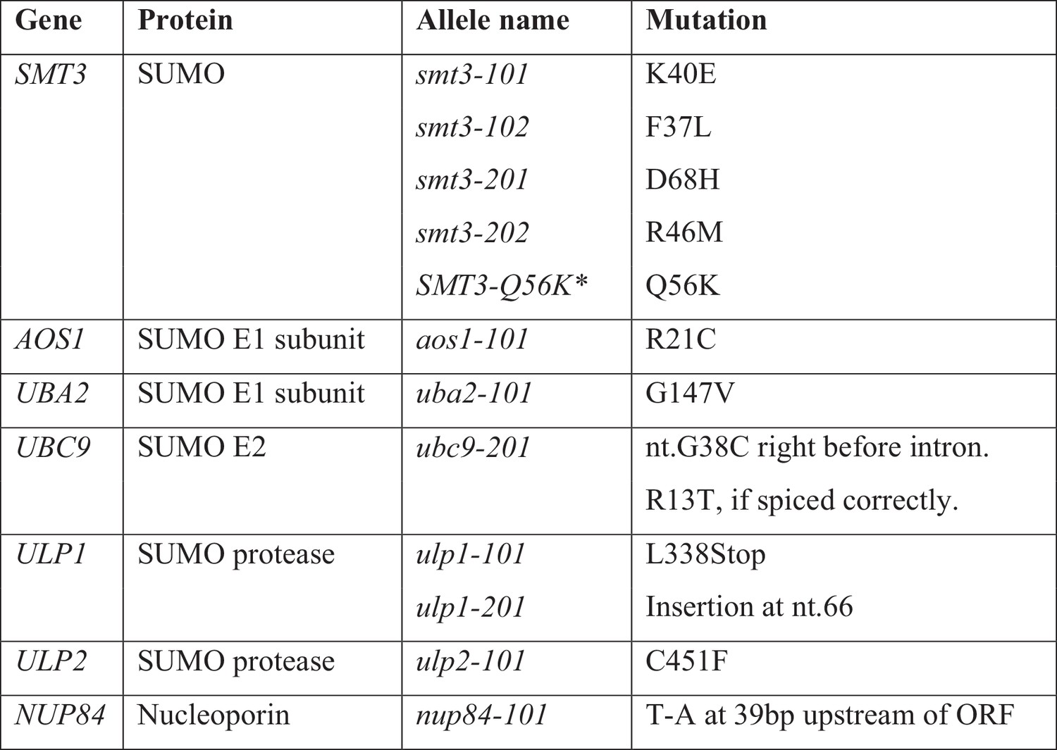
mot1-301 suppressor mutations.
* Dominant mutation.
-
Figure 1—figure supplement 1—source data 1
Source data for Figure 1—figure supplement 1
- https://doi.org/10.7554/eLife.35447.004
Figure 2 with 3 supplements
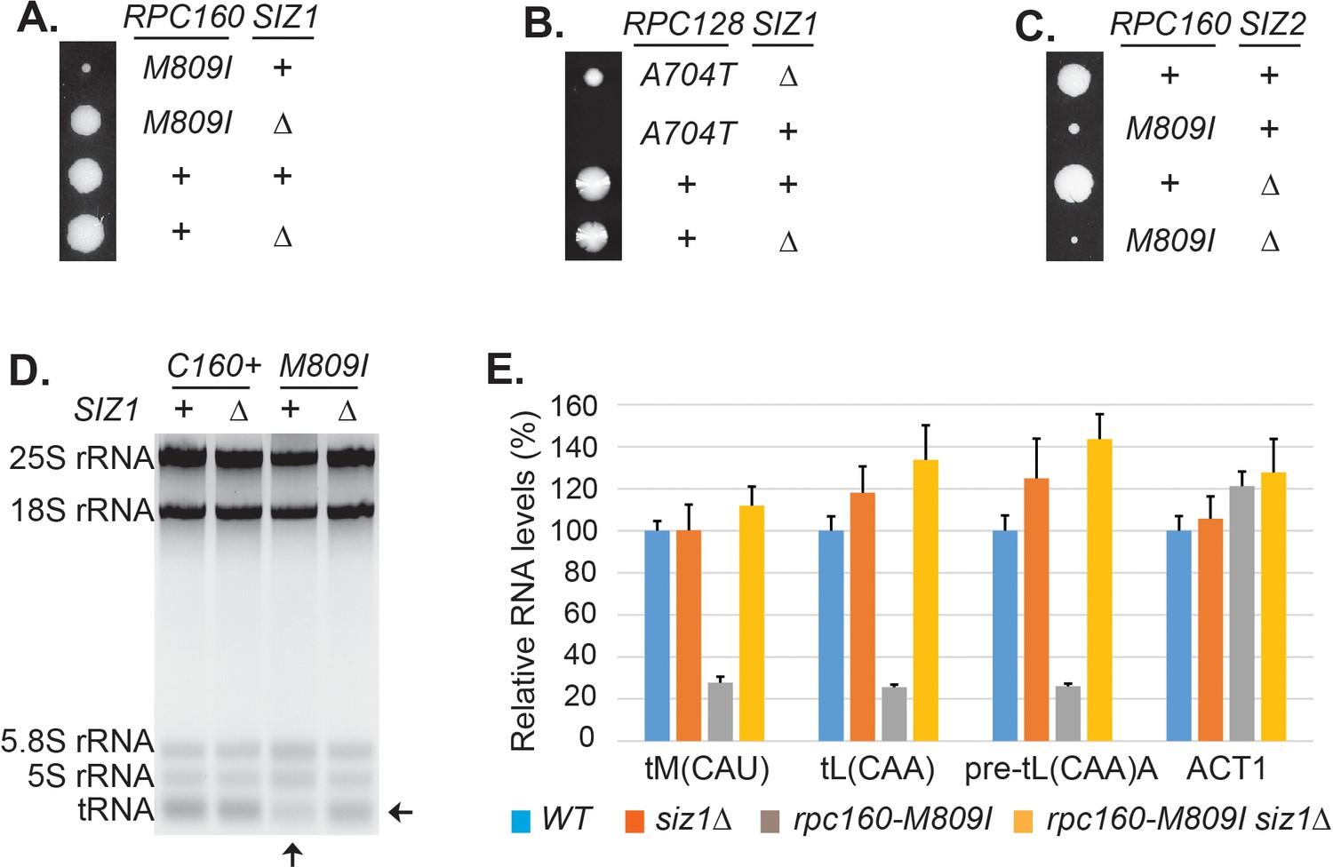
Disrupting sumoylation rescues Pol III mutations.
(A) Tetrad analysis of a cross between rpc160-M809I and siz1Δ. Tetrads were dissected on YPD, then incubated at 30°C for 4 days. The offspring of one representative tetrad was shown with genotypes labeled. (B) Similar tetrad analysis for rpc128-A704T and siz1Δ. (C) Similar tetrad analysis for rpc160-M809I and siz2Δ. (D) 2 μg of RNA extracted from the indicated strains was run on a 2.8% agarose gel containing ethidium bromide, then visualized with UV. (E) RNA from (C) was reverse transcribed into cDNA, followed by real-time PCR analysis. GAPDH transcripts were used as loading control. Data are mean ± standard deviation calculated from six data points (two biological replicates and three technical replicates), presented as relative amount compared to wild type. The intron-containing pre-mature tRNA (pre-tL(CAA)A) is short-lived, so its abundance reflects the Pol III transcriptional activity.
-
Figure 2—source data 1
Raw Ct values for Figure 2E.
- https://doi.org/10.7554/eLife.35447.012
Figure 2—figure supplement 1
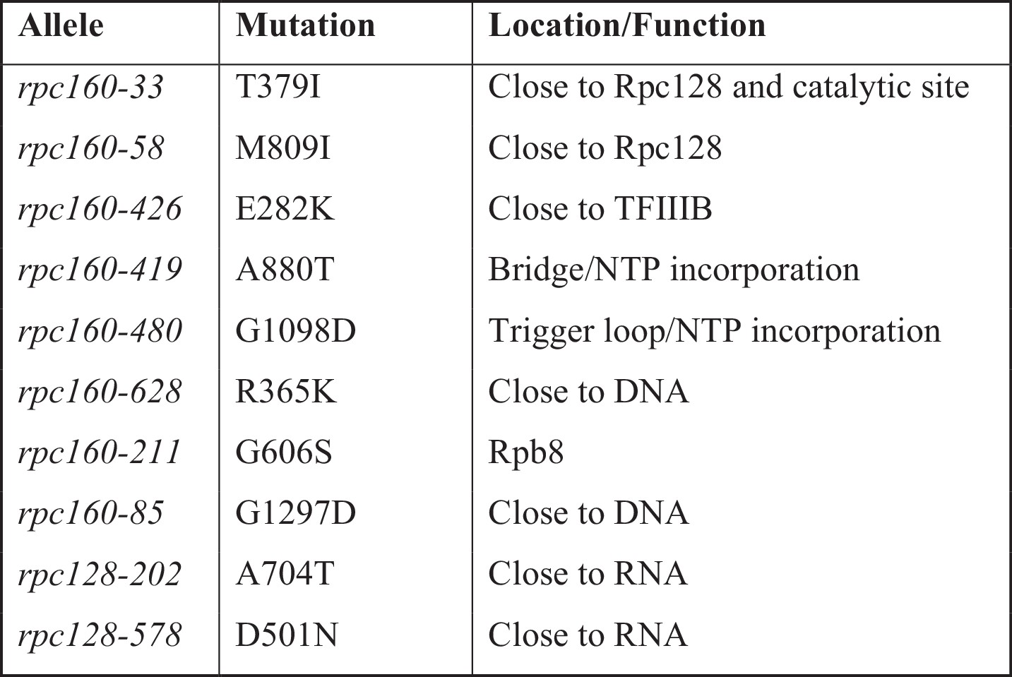
Position of the mutated residues in Pol III structure.
https://doi.org/10.7554/eLife.35447.007-
Figure 2—figure supplement 1—source data 1
Source data for Figure 2—figure supplement 1
- https://doi.org/10.7554/eLife.35447.008
Figure 2—figure supplement 2

siz1Δ rescued a wide spectrum of Pol III mutations.
(A) Plasmid shuffle experiments to test the growth phenotype of known rpc160 and rpc31 mutants in wild-type SIZ1 or siz1Δ background. The RPC160 or RPC31 alleles were on LEU2 vectors. The parental rpc160Δ and rpc31Δ strains contain a URA3 plasmid carrying wild-type RPC160 or RPC31, respectively. (B) Wild-type RPC160 was placed under control of the GAL1 promoter on a LEU2 vector then transformed into an rpc160 strain carrying a URA3 RPC160 plasmid, which was lost in the presence of 5FOA. The 5FOA plate contained glucose as the only carbon source, which strongly repressed RPC160 expression, making the cells grow extremely slowly. siz1Δ partially alleviated this growth defect. (C–E) Similar plasmid shuffle experiments showing the rescue effect of siz1Δ on disease causing mutations. rpc160-DN: D384N, N789I.
Figure 2—figure supplement 3
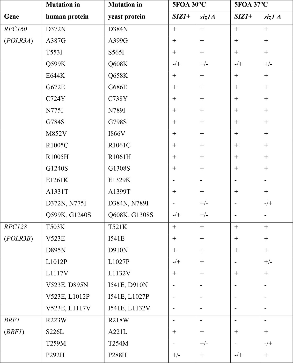
Growth phenotype of Pol III disease mutations in yeast.
CEN LEU2 plasmids containing the indicated mutant alleles (e.g. rpc160 mutants) were transformed into a corresponding null strain in wild-type SIZ1 or siz1Δ background (e.g. rpc160Δand rpc160Δ siz1Δ strains) covering by a wild-type URA3 plasmid (e.g. a URA3 RPC160 plasmid). Transformants were then patched on a SC-Leu plate, replica plated to 5FOA plates, and incubated at 30°C or 37°C. Growth was scored after 2 days of incubation. The human gene names were shown in parenthesis underneath the yeast gene names.
-
Figure 2—figure supplement 3—source data 1
Source data for Figure 2—figure supplement 3
- https://doi.org/10.7554/eLife.35447.011
Figure 3

Specificity of the rescue effect.
(A) The rpa190Δ, rpb1Δ, or rpc160Δ strain carries a URA3 plasmid carrying wild-type RPA190, RPB1, and RPC160 gene, respectively, in order to maintain viability. These strains were then transformed with LEU2 plasmids carrying the indicated wild type or mutant alleles, and selected on synthetic media lacking leucine (SC-L). Transformants were spotted in fivefold serial dilutions onto a 5FOA plate to assess the growth phenotype of the mutant allele, as the original URA3 plasmids were shuffled out of the cell in the presence of 5FOA. (B–D) Tetrad analysis between rpc160-M809I (shown as rpc160) and kns1Δ, mck1Δ, and maf1Δ. (E) Tetrad analysis between rpc160-M809I siz1Δ and maf1Δ. (F) The indicated strains from the cross in (E) were plated in fivefold dilutions onto YPD (glucose) or YPG (glycerol) plates and incubated at 30°C or 37°C as indicated.
Figure 4 with 1 supplement

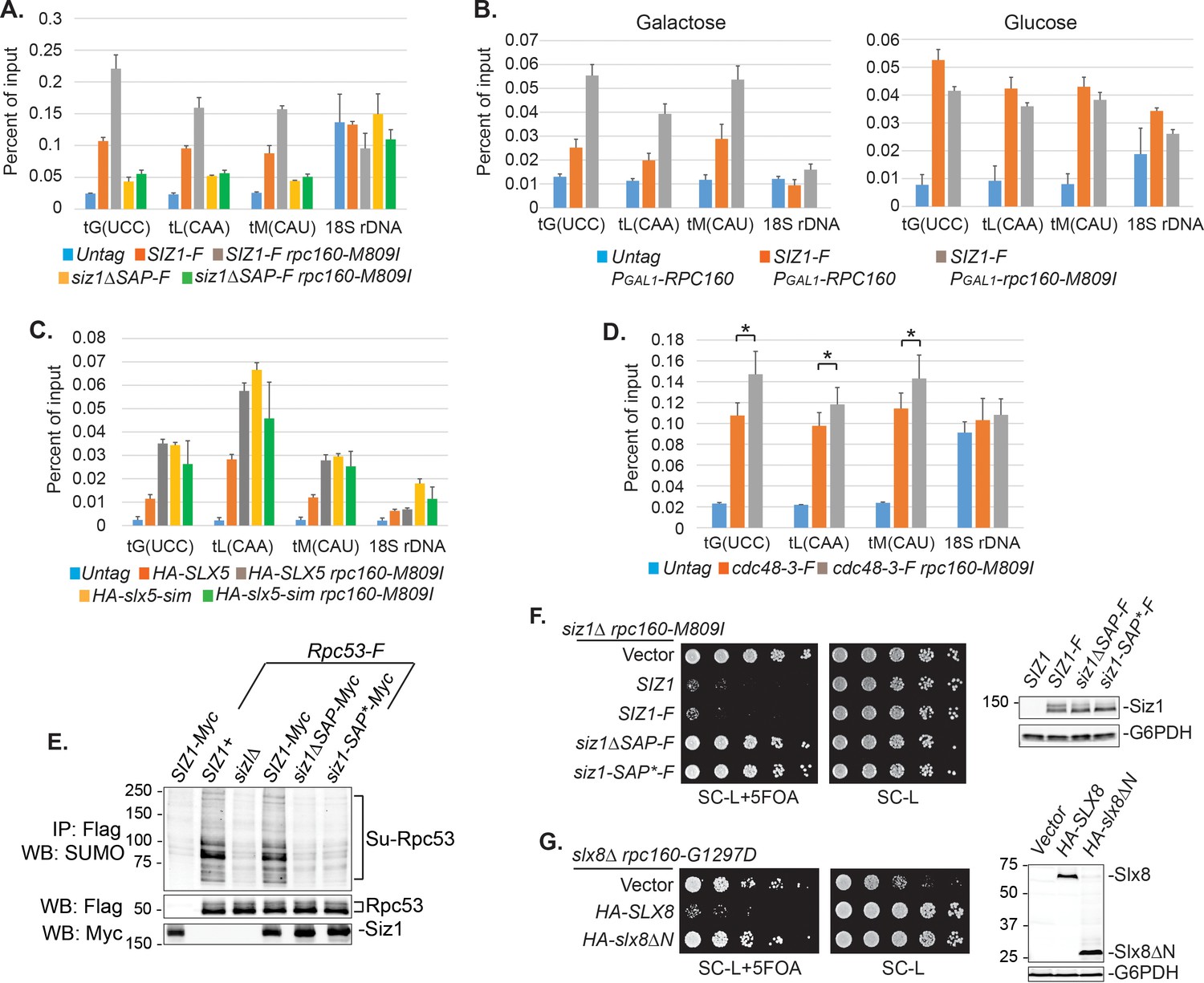
SUMO represses Pol III by modifying Rpc53.
(A) Total protein extracted from the indicated strains was subjected to anti-Flag IP to purify Flag-tagged Rpc160 Pol III complexes and associated proteins. Precipitated proteins were eluted with Flag peptide, followed by SDS-PAGE and immunoblot analysis with an anti-Flag or anti-SUMO antibody. (B) The eluant from (A) was subjected to anti-GFP IP using GFP-Trap beads to isolate the sumoylated species from Pol III. The beads were subsequently washed with PBS containing 8M urea and 1% SDS to remove Rpc160-associated unmodified proteins, then incubated with 2× Laemmli’s buffer at 100°C to elute sumoylated proteins. The success of the IP was confirmed by anti-SUMO immunoblot. The purified materials were subjected to tryptic digestion and analyzed by mass-spectrometry. (C) Flag-tagged Rpc53 was IP-ed from the indicated strains using anti-Flag beads, and detected by an anti-Flag antibody (bottom). Sumoylated Rpc53 (Su-Rpc53) was detected by anti-SUMO antibody (top). An untagged RPC53 strain was used as a negative control. (D) Similar experiment as in (C) showing Rpc53 sumoylation in wild type RPC160 cells versus rpc160-M809I mutant cells. (E) Mapping Rpc53 sumoylation sites by mutagenesis analysis. CEN plasmids carrying wild type or mutant Flag-tagged RPC53 were co-transformed with a 2μ SMT3 plasmid into a wild-type yeast strain. Rpc53-Flag proteins were purified with anti-Flag IP, followed by SDS-PAGE and immunoblot analysis with anti-Flag (bottom) or anti-SUMO antibody (top). (F) An rpc128-A704T strain carrying a URA3 RPC128 plasmid was transformed with LEU2-based RPC128 or RPC53 plasmids, then grown on 5FOA medium, which forces the cells to lose the URA3 RPC128 plasmid. rpc53-3KR (K51,115,236R) rescued the growth of rpc128-A704T, whereas N-terminal SUMO fusion (Su-rpc53-3KR) abolished the rescue effect. The rescue effect is dominant because all the cells in this experiment contain wild type RPC53 in the genome. (G) Similar plasmid shuffle experiment as in Figure 2A. The LEU2 plasmids carrying the indicated RPC53 alleles were transformed into an rpc53Δ strain containing a URA3 RPC53 plasmid. The transformants were then plated onto a 5FOA plate to lose the URA3 RPC53 plasmid, and the results showed that the N-terminally SUMO-fused Rpc53 protein (Su-rpc53-3KR) fully supports cell viability.
Figure 4—figure supplement 1
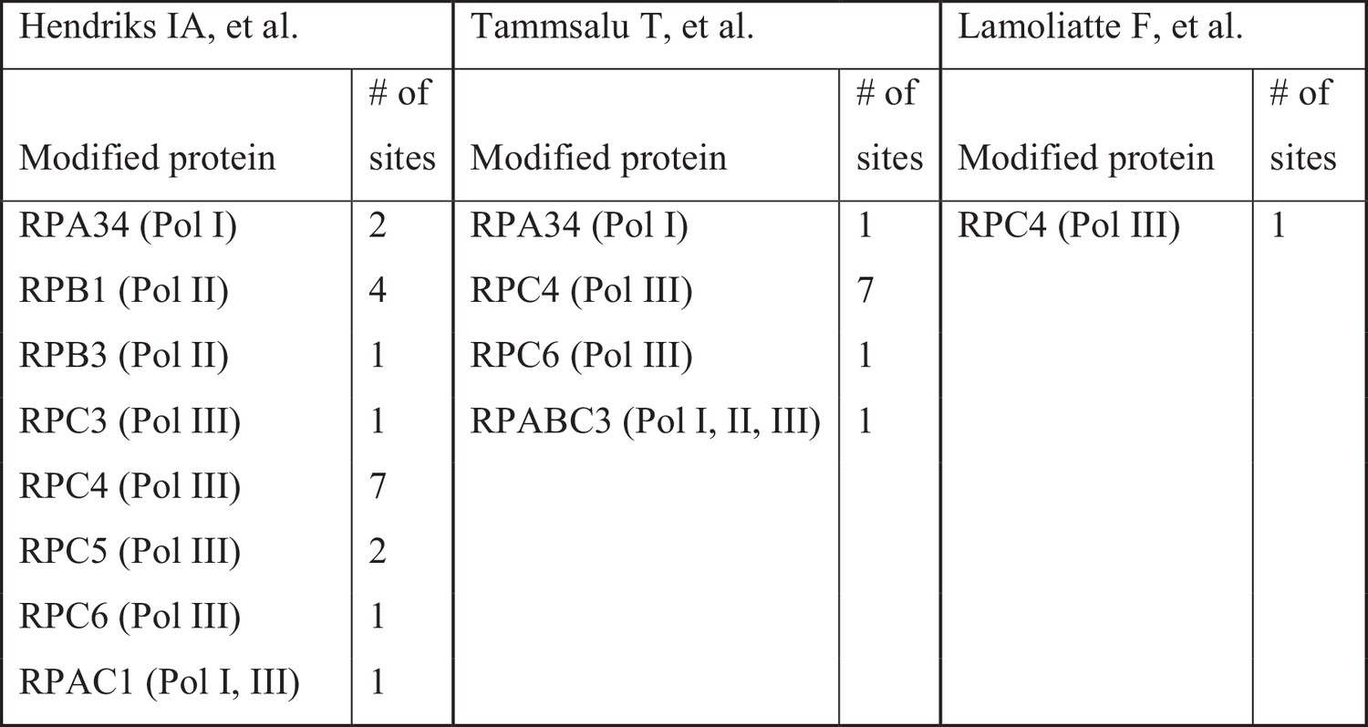
Summary of sumoylated polymerase subunits.
https://doi.org/10.7554/eLife.35447.015-
Figure 4—figure supplement 1—source data 1
Source data for Figure 4—figure supplement 1
- https://doi.org/10.7554/eLife.35447.016
Figure 5 with 2 supplements
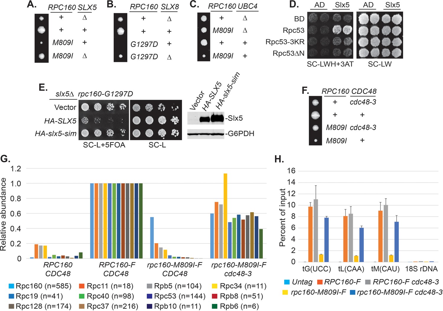
Pol III is repressed by ubiquitylation and p97/Cdc48.
(A–C) The indicated rpc160 mutant strains were crossed with slx5Δ, slx8Δ, or ubc4Δ strain, respectively, followed by tetrad analysis. The cross between slx8Δ and rpc160-G1297D was shown, because slx8Δ caused obvious growth defect by itself, so the rescue effect was more obvious on rpc160-G1297D, which is a sicker mutant than rpc160-M809I. (D) Yeast two-hybrid interactions between Slx5 and Rpc53. SLX5 and RPC53 were cloned into a 2μ LEU2 Gal4 activation-domain (AD) vector and a 2μ TRP1 DNA-binding domain (BD) vector, respectively, and co-transformed into yeast strain PJ69-4A. Transformants were selected on synthetic media lacking leucine and tryptophan (SC-LW), then patched and replica plated to selective media lacking histidine to test for interactions. The histidine-lacking media was supplement with 3-aminotriazole (SC-LWH + 3AT) for a more stringent phenotype. (E) LEU2 plasmids carrying HA-tagged wild type or SIM-defective SLX5 (HA-slx5-sim) were transformed into an rpc160-G1297D slx5Δ strain containing a URA3 RPC160 plasmid. Transformants were selected on SC-L then spotted onto an SC-L + 5 FOA plate to lose wild-type RPC160. HA-SLX5 complemented slx5Δ so the cells became sicker compared to the empty vector control transformants, while HA-slx5-sim did not complement, indicating that the SIMs are essential for the function of SLX5 in this assay. The lost Slx5 function by the SIM mutations was not caused by insufficient proteins, since there were comparable levels of Slx5 proteins, as determined by an anti-HA immunoblot on total cell lysates (right, top panel). G6PDH served as a loading control (right, bottom panel). (F) Tetrad analysis between rpc160-M809I and cdc48-3. (G) Determination of Rpc160 association with other Pol III subunits. Pol III complexes containing Flag-tagged wild type or mutant Rpc160 in wild-type CDC48 or cdc48-3 cells were isolated using anti-Flag agarose beads, followed by TMS labeling and mass-spec analysis to quantify the relative amounts of Rpc160-interacting proteins. Signals for 12 of the 17 Pol III subunits, including Rpc160, were detected, and normalized to the signals from RPC160-Flag cells. n = Number of times when a unique peptide for the indicated protein is measured. An untagged RPC160 strain was used as a negative control. (H) ChIP analysis of Rpc160. Flag-tagged RPC160 or rpc160-M809I was expressed from a plasmid in wild-type CDC48 or cdc48-3 cells, as indicated. The Flag-tagged proteins were purified using anti-Flag agarose beads. An untagged strain was used as negative control. Three tRNA gene loci, as well as 18S rDNA (negative control), were examined. Chromatin association was determined by real-time PCR of the indicated genomic loci, using the percent of input method. Data are mean ± standard deviation calculated from six data points (two biological replicates and three technical replicates).
-
Figure 5—source data 1
Raw Ct values for Figure 5G.
- https://doi.org/10.7554/eLife.35447.020
-
Figure 5—source data 2
Raw Ct values for Figure 5H.
- https://doi.org/10.7554/eLife.35447.021
Figure 5—figure supplement 1
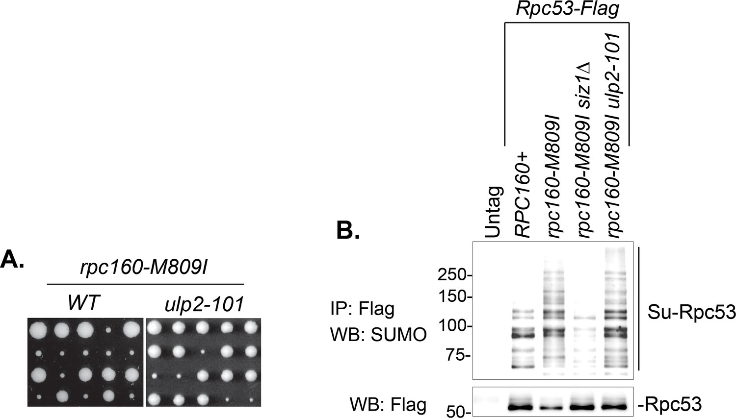
ulp2-101 rescued rpc160-M809I without abolishing Rpc53 sumoylation.
(A) An rpc160-M809I strain was crossed with a wild type (left) or an ulp2-101 mutant strain (right), followed by tetrad analysis. The growth of five representative tetrads were shown. The cross between rpc160-M809I and wild-type ULP2 always yielded two large colonies and two small colonies, indicating rpc160-M809I caused severe growth defect. The cross between rpc160-M809I and ulp2-101 in most cases yielded three large colonies and one small colony, indicating rpc160-M809I was rescued by ulp2-101. (B) Flag-tagged Rpc53 proteins from the indicated strains were purified using anti-Flag beads, followed by SDS-PAGE and immunoblotting using an anti-SUMO antibody (top) or an anti-Flag antibody (bottom). siz1Δ abolished Rpc53 sumoylation, while ulp2-101 did not.
Figure 5—figure supplement 2
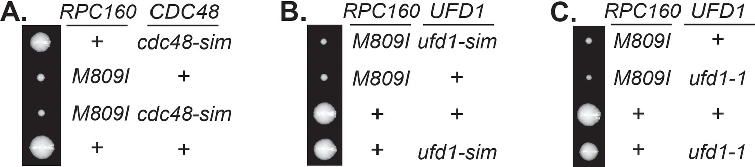
Genetic relationship between rpc160, cdc48, and ufd1.
(A–C) Tetrad analysis between rpc160-M809I and cdc48-sim, ufd1-sim, and ufd1-1, respectively, showing none of these mutations rescued rpc160-M809I.
Figure 6

Pol III repression by ubiquitylation is partially mediated through Rpc160.
(A) CEN URA3 plasmids expressing HA-tagged wild type or mutant Rpc160, as indicated, were transformed into a wild-type strain, and their stabilities were assayed during a cycloheximide (CHX) chase time course. Rpc160 was detected by an anti-HA antibody, and G6PDH was used as a loading control. Quantification of the bands was shown below the immunoblot. (B) Tetrad analysis of the diploid strains, RPC160+/rpc160-M809I (top) and RPC160+/rpc160-M809I-3KR (bottom). Tetrads from these two diploids were dissected and plated on the same YPD plate at the same time, in order to compare the growth of rpc160-M809I and rpc160-M809I-3KR cells. The growth of three dissected tetrads were shown. The large colonies are wild-type RPC160 cells, and the small colonies are rpc160-M809I (top) or rpc160-M809I-3KR (bottom) cells. The rpc160-M809I-3KR cells grew slightly faster than the rpc160-M809I cells. (C) Left: 2 μg of RNA extracted from the indicated strains was run on a 2.8% agarose gel containing ethidium bromide, then visualized with UV. Two colonies were picked for each strain. Right: RNA from left was reverse transcribed into cDNA, followed by real-time PCR analysis, as described in Figure 2E. GAPDH transcripts were used as loading control. (D) Similar tetrad analysis as in (B) of the diploid strains, RPC160+/rpc160-G1297D (top) and RPC160+/rpc160-G1297D-3KR (bottom). Large colonies are wild-type RPC160 cells, while the missing colonies (top) are rpc160-G1297D cells, and the small colonies (bottom) are rpc160-G1297D-3KR cells. (E) An rpc128-A704T strain was crossed with an rpc160-3KR strain, followed by tetrad analysis. (F) A CEN URA3 rpc160-M809I-HA plasmid was transformed into the indicated strains, and the stabilities of the Rpc160-M809I-HA proteins were determined by CHX chase time course, as described in (A). (G) A CEN URA3 rpc160-M809I-HA plasmid was transformed into a wild-type strain, and protein stabilities were determined by CHX chase experiment in the presence of DMSO or MG132.
-
Figure 6—source data 1
Raw Ct values for Figure 6C.
- https://doi.org/10.7554/eLife.35447.023
Figure 7
DNA is involved in Pol III repression.
(A) Chromatin IP of Siz1-Flag. An untagged strain was used as negative control. Chromatin association was determined by real-time PCR of the indicated genomic loci, using the percent of input method. Data are mean ± standard deviation calculated from six data points (two biological replicates and three technical replicates). (B) Plasmids carrying GAL1 promoter-driven RPC160 or rpc160-M809I were transformed in untagged or Flag-tagged SIZ1 cells as indicated. Cells were grown in galactose (left) or glucose (right) media to activate or inhibit transcription from the GAL1 promoter, respectively, followed by ChIP analysis of Siz1-Flag. All strains also express wild-type RPC160 from its endogenous promoter. (C–D) Similar ChIP analysis of HA-Slx5 and Cdc48-3-Flag as in (A). * p-value<0.05. (E) Flag-tagged Rpc53 proteins were purified from the indicated wild type or siz1 mutant strains, using anti-Flag beads, followed by SDS-PAGE and immunoblotting using an anti-SUMO antibody. Rpc53 was detected by an anti-Flag antibody, and Siz1 was detected by an anti-Myc antibody. Either truncation (ΔSAP) or point mutation (SAP*) of the SAP domain resulted in loss of Rpc53 sumoylation. (F) Left: LEU2 plasmids carrying wild type or mutant SIZ1 alleles were transformed into an rpc160-M809I siz1Δ strain containing a URA3 RPC160 plasmid. Transformants were selected on SC-L plate, then spotted in fivefold dilution onto a SC-L + 5 FOA plate. Wild type SIZ1 complemented siz1Δ so the cells became sick on SC-L + 5 FOA plate, while the SAP mutants did not complement. Right: Comparable amounts of wild type and mutant Siz1 proteins were determined by anti-Flag immunoblotting on whole cell lysate, using G6PDH as loading control. (G) Similar plasmid shuffle experiment as in (F). LEU2 plasmids carrying HA-tagged wild-type SLX8 or slx8ΔN (Δ2–163) were transformed into an rpc160-G1297D slx8Δ strain containing a URA3 RPC160 plasmid. Wild-type SLX8 complemented slx8Δ, while slx8ΔN did not. Comparable amounts of Slx8 proteins were determined by an anti-HA immunoblot.
-
Figure 7—source data 1
Raw Ct values for Figure 7A–D.
- https://doi.org/10.7554/eLife.35447.025
Figure 8
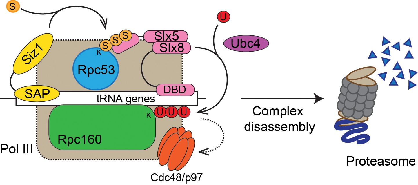
Model of Pol III regulation by SUMO, ubiquitin, and Cdc48.
A stable interaction between chromatin and the SAP domain of Siz1 stimulates its activity to modify Rpc53 with SUMO (S). Rpc53 sumoylation triggers ubiquitin (U) modification of Rpc160 and potentially other proteins by the Slx5-Slx8 complex, which also required the interaction between chromatin and the DNA-binding domain (DBD) of Slx8. Ubiquitylation subsequently activates Cdc48 to disassemble the Pol III complex, facilitating degradation of Pol III subunits by the proteasome.
Tables
Table 1
Summary of mutations rescued by SMT3-Q56K.
https://doi.org/10.7554/eLife.35447.005| Gene | Protein | # of alleles | Mutations |
|---|---|---|---|
| MOT1 | Negative regulator of TBP | 3 | mot1-399 (Q1587 Stop) |
| mot1-517 (G1410R) | |||
| mot1-753 (G1300S) | |||
| SMT3 | SUMO | 4 | Not sequenced |
| AOS1 | SUMO E1 | 1 | aos1-492 (G56S) |
| ULP2 | SUMO protease | 4 | ulp2-4 (S108 Stop) |
| ulp2-253 (G265D) | |||
| ulp2-527 (W532 Stop) | |||
| ulp2-63 (W532 Stop) | |||
| RPC160 | RNA Pol III subunit | 8 | rpc160-58 (M809I) |
| rpc160-85 (G1297D) | |||
| rpc160-33 (T379I) | |||
| rpc160-419 (A880T) | |||
| rpc160-426 (E282K) | |||
| rpc160-480 (G1098D) | |||
| rpc160-628 (R365K) | |||
| rpc160-211 (G606S) | |||
| RPC128 | RNA Pol III subunit | 2 | rpc128-202 (A704T) |
| rpc128-578 (D501N) | |||
| BRF1 | TFIIIB subunit | 1 | brf1-137 (S271L) |
| TFC1 | TFIIIC subunit | 1 | tfc1-321 (N255K, Fs) |
| (AAC-AAAC, Ins, Fs) | |||
| TFC6 | TFIIIC subunit | 1 | tfc6-192 (G391E) |
Key resources table
| Reagent type (species) or resource | Designation | Source or reference | Identifiers | Additional information |
|---|---|---|---|---|
| Antibody | anti-HA | Santa Cruz | SC-7392 | |
| Antibody | anti-HA beads | Sigma | A2095 | |
| Antibody | anti-Flag | Sigma | F3165 | |
| Antibody | anti-Flag beads | Sigma | A2220 | |
| Antibody | anti-Myc | This study | ||
| Antibody | anti-G6PDH | Sigma | A9521 | |
| Antibody | anti-Smt3 (SUMO) | Santa Cruz | SC-28649 | |
| Antibody | GFP-Trap beads | chromotek | gta-20 | |
| Antibody | Alexa Fluor 680 Goat anti-Mouse IgG | Fisher Scientific | A21058 | |
| Antibody | Alexa Fluor 680 Goat anti-Rabbit IgG | Fisher Scientific | A21109 | |
| Antibody | DyLight 800 Goat anti-Rabbit IgG | Fisher Scientific | SA535571 | |
| Antibody | DyLight 800 Goat anti-Mouse IgG | Fisher Scientific | SA535521 | |
| Peptide, recombinant protein | 2x HA peptide | This study | ||
| Peptide, recombinant protein | 2x Flag peptide | This study | ||
| Commercial assay or kit | cOmplete, Mini, EDTA-free Protease Inhibitor Cocktail | Sigma | 11836170001 | |
| Commercial assay or kit | Power SYBR Green PCR Master Mix | Fisher Scientific | 4367659 | |
| Commercial assay or kit | SuperScript III First-Strand Synthesis System | Invitrogen | 18080–051 | |
| Chemical compound, drug | N-Ethylmaleimide (NEM) | Sigma | E3876 | |
| Chemical compound, drug | 5-Fluoroorotic Acid (5-FOA) | Toronto Research Chemicals | F595000 | |
| Chemical compound, drug | MG132 | Fisher Scientific | 50-833-9 | |
| Chemical compound, drug | Cycloheximide | Sigma | C7698 | |
| Software, algorithm | Excel | Microsoft |
Additional files
-
Supplementary file 1
Yeast strains used in this study.
- https://doi.org/10.7554/eLife.35447.027
-
Supplementary file 2
Plasmids used in this study.
- https://doi.org/10.7554/eLife.35447.028
-
Supplementary file 3
Primers used in this study.
The same primers were used in RNA level measurement and in chromatin IP experiments. * Used as gene-specific primer in reverse transcription.
- https://doi.org/10.7554/eLife.35447.029
-
Transparent reporting form
- https://doi.org/10.7554/eLife.35447.030
Download links
A two-part list of links to download the article, or parts of the article, in various formats.
Downloads (link to download the article as PDF)
Open citations (links to open the citations from this article in various online reference manager services)
Cite this article (links to download the citations from this article in formats compatible with various reference manager tools)
Defective RNA polymerase III is negatively regulated by the SUMO-Ubiquitin-Cdc48 pathway
eLife 7:e35447.
https://doi.org/10.7554/eLife.35447


