Pre-saccadic remapping relies on dynamics of spatial attention
- Vrije Universiteit, The Netherlands
- Howard Hughes Medical Institute, Stanford University School of Medicine, United States
- Queensland Brain Institute, The University of Queensland, Australia
- Ludwig-Maximilians-Universität München, Germany
Figures
Figure 1
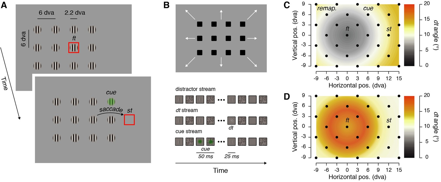
Stimulus displays and stimulus eccentricity effects.
(A) Participants fixated on the fixation target (ft) and prepared a saccade towards the saccade target (st) presented either to the right or to the left of the fixation between 700 and 900 ms after the trial onset. On each trial, 12 visual streams (40 Hz flickering vertical Gabors and masks) were shown and in two out of the three trials a cue was flashed (50 ms) either above or below a virtual line between the fixation and the saccade targets (note that the stimuli here are sketched to increase their visibility, actual stimuli match those shown in the visual stream depiction). (B) The arrangement of visual streams can take several positions (see Materials and methods), to cover the whole display across trials. Participants reported the orientation of a discrimination target (dt), a tilted Gabor, presented within all trials at a time maximizing the occurrence of its offset within the 150 ms preceding the saccade. (C–D) The discrimination target was shown across trials at 32 different positions (see black dots) covering 24 dva horizontally and 18 dva vertically and including four main positions of interest (the fixation target: ft; the saccade target: st, the cue: cue; and the remapped location of the cue: remap.). The tilt of the discrimination target was titrated to yield comparable performance at differently cued eccentricities from the fixation target. We adjusted these tilts in a preliminary task made either while participants kept their eyes steady at the fixation target (C) peripheral remapping threshold task (see Materials and methods), or prepared a saccade (D) foveal remapping threshold task (see Materials and methods). The maps show dt tilt angles averaged across participants in these two threshold tasks.
Figure 2
Stimulus timing and sensitivity maps.
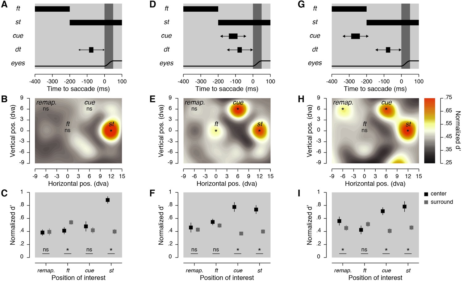
(A,D,G) Stimulus timing. Participants prepared a saccade at the offset of the fixation target (ft), which corresponded to the onset of the saccade target (st). In the 150 ms before the saccade, a discrimination target (dt) was briefly shown at one of the 32 possible positions. Then, no cue was shown (A), or a cue was shown 50 ms before the dt and about 100 ms before the saccade (D), or a cue was shown 200 ms before the dt and about 250 ms before the saccade (G). (B,E,H) Normalized sensitivity maps. Averaged normalized sensitivity (d') observed across participants and displayed using a color-coded linear scale going between 0.25 and 0.75 (see Materials and methods). Asterisks indicate significant differences (p < 0.05) in sensitivity found between a particular position of the dt and the average of all the other tested positions. (C,F,I) Averaged normalized d’ obtained at four positions of interest (black squares) and at their corresponding surrounding positions (dark gray squares). Error bars show SEM and asterisks indicate significant comparisons (p < 0.05).
Figure 3

Cue vs. no-cue subtraction maps.
Individual normalized sensitivity (d') is subtracted between conditions and the difference is normalized to obtain maps with convention as in Figure 2. Subtractions are made between trials in which the cue was shown ~100 ms before the saccade (A) or ~250 ms before the saccade (B) to trials in which it was not shown.
Figure 4
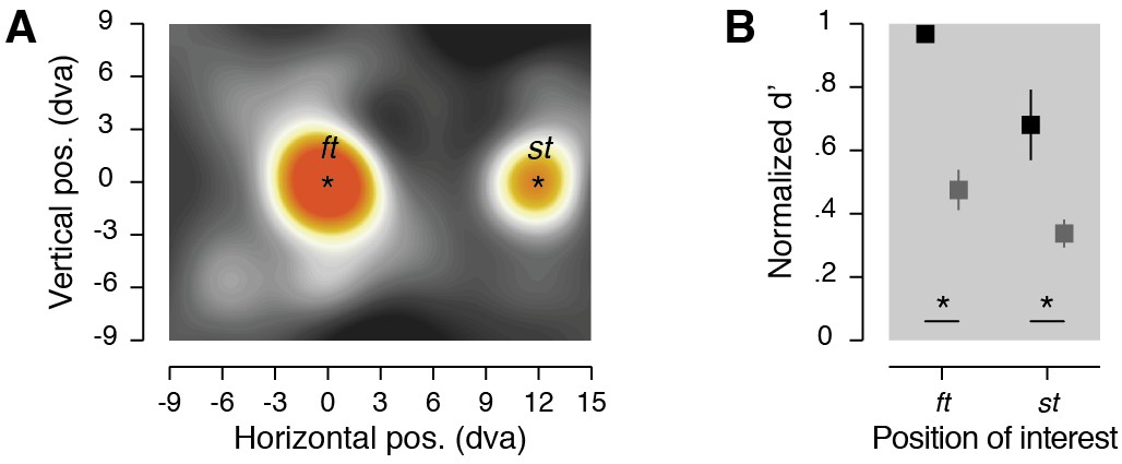
Foveal remapping task results.
(A) Normalized sensitivity maps. Averaged normalized sensitivity (d'). (B) Averaged normalized sensitivity (d')obtained at two positions of interest (see center in black) and at their corresponding surround positions (see surround in dark gray). Conventions and color scale are as in Figure 2.
Additional files
-
Transparent reporting form
- https://doi.org/10.7554/eLife.37598.006
Download links
A two-part list of links to download the article, or parts of the article, in various formats.
Downloads (link to download the article as PDF)
Open citations (links to open the citations from this article in various online reference manager services)
Cite this article (links to download the citations from this article in formats compatible with various reference manager tools)
Pre-saccadic remapping relies on dynamics of spatial attention
eLife 7:e37598.
https://doi.org/10.7554/eLife.37598


