Immune-mediated hookworm clearance and survival of a marine mammal decrease with warmer ocean temperatures

  1. Mauricio Seguel  Is a corresponding author
  2. Felipe Montalva
  3. Diego Perez-Venegas
  4. Josefina Gutiérrez
  5. Hector J Paves
  6. Ananda Müller
  7. Carola Valencia-Soto
  8. Elizabeth Howerth
  9. Victoria Mendiola
  10. Nicole Gottdenker
  1. University of Georgia, United States
  2. Pontificia Universidad Catolica de Chile, Chile
  3. Universidad Andrés Bello, Chile
  4. Universidad Austral de Chile, Chile
  5. Universidad Santo Tomas, Chile

Abstract

Increases in ocean temperature are associated with changes in the distribution of fish stocks, and the foraging regimes and maternal attendance patterns of marine mammals. However, it is not well understood how these changes affect offspring health and survival. The maternal attendance patterns and immunity of South American fur seals were assessed in a rookery where hookworm disease is the main cause of pup mortality. Pups receiving higher levels of maternal attendance had a positive energy balance and a more reactive immune system. These pups were able to expel hookworms through a specific immune mediated mechanism and survived the infection. Maternal attendance was higher in years with low sea surface temperature, therefore, the mean hookworm burden and mortality increased with sea surface temperature over a 10-year period. We provide a mechanistic explanation regarding how changes in ocean temperature and maternal care affect infectious diseases dynamics in a marine mammal.

https://doi.org/10.7554/eLife.38432.001

eLife digest

Every year off the coasts of Chile, Guafo Island becomes a nursery for South American fur seals pups. Mother fur seals leave their young on the beaches, going out at sea to hunt for fish before returning to the shore to nurse. These first few months are dangerous for young seals, with many dying because of hookworms, parasites that latch to the wall of the bowels to suck blood. However, the immune system of the pups is usually able to mount a response and fight off these parasites.

Even though the pups stay on land, their lives depend on the health of the ocean that feeds their nursing mothers. In recent years, sea temperature has been rising rapidly, which modifies winds and water currents. This can set off a chain of events that results in fewer fish being available for seals and other marine mammals to eat. Researchers know that years with warmer waters are associated with changes in the pattern of the mothers’ hunting trips, more pups’ deaths, and a weaker immune system in young fur seals. However, the mechanisms that connect these different factors are still unclear.

To explore this, Seguel et al. followed South American fur seals colonies on Guafo Island for several years, tracking the mothers’ trips and monitoring the health of the pups by looking at their levels of blood sugar, whether they carry hookworms, and certain elements of their immune system. Results showed that in years when the sea is warmer, fur seal mothers are gone hunting for longer: they spend less time nursing their young, which then grow slower. These young seals also have lower levels of blood sugar, and so they have less energy to create the immune response necessary to clear off parasitic worms. In fact, in years with warmer seas, almost half of the pups die from hookworm infections.

The work by Seguel et al. shows that warmer oceans directly weaken the immune defenses of certain marine mammals. If temperatures keep rising, infectious diseases may kill more of these animals. Further work is now needed to explore if strategies could be developed to help seal populations, for example by treating the pups with drugs that eliminate the parasites.

https://doi.org/10.7554/eLife.38432.002

Introduction

Marine mammals are a diverse group of top predators highly sensitive to changes in aquatic ecosystems (Constable et al., 2014). Within this group, fur seals and sea lions (otariids) breed and give birth on land but forage at sea, alternating periods of foraging in the ocean with periods of offspring attendance and nursing on land (income breeders) (Stephens et al., 2009). Therefore, otariids, like other marine mammals, are highly sensitive to local changes in prey distribution and abundance (Trillmich et al., 1991, Constable et al., 2014, Elorriaga-Verplancken et al., 2016). One of the most important indexes of the abundance of marine mammal prey is sea surface temperature (SST) (Soto et al., 2006, Elorriaga-Verplancken et al., 2016). Warmer SST indicates reduced nutrient upwelling, which is associated with reduced primary productivity and abundance of mesopelagic marine organisms (Lewandowska et al., 2014). This decrease in food resources forces otariid females to change their foraging strategies by increasing their foraging trip lengths, resulting in decreased time spent on land with their pup (maternal attendance) (Trillmich et al., 1991, Costa, 2008). These changes in patterns of maternal attendance have been associated with decreased pup growth and increased mortality (Soto et al., 2006, Jeanniard-du-Dot et al., 2017). Regardless, the mechanisms that drive decreased survival during years with low ocean productivity have not been intensely explored beyond assuming that this results from direct mortality because of starvation. However, in some otariid populations, in years with abnormal SST, the immune competence of pups decreases (Banuet-Martínez et al., 2017), suggesting that environmental variables can affect the health of marine mammals by impairing their immune function. If these immunological changes impact offspring survival, there could be additional negative consequences between a warmer ocean, health, and survival of marine vertebrates.

In some marine mammal populations, infectious diseases are one of the most significant causes of mortality among young individuals (Gulland and Hall, 2007; Spraker et al., 2007; Seguel et al., 2013). In otariids, hookworms (Uncinaria sp.) have been described in nearly all species, and while some populations suffer few adverse effects, others experience up to 70% of hookworm-related mortality being one of the most significant infectious diseases of young fur seals and sea lions (Spraker et al., 2007; Lyons et al., 2011a; Seguel et al., 2013; Seguel and Gottdenker, 2017). Fur seals are infected with hookworms (Uncinaria sp.) during their first 1–4 days of life through their mother’s colostrum (Lyons et al., 2011b; Seguel et al., 2018). These nematodes live in the small intestine where they bite the mucosa to feed on blood, causing substantial tissue damage, anemia, and death (Marcus et al., 2015, Seguel et al., 2017Seguel et al., 2018); however, it is unclear how the host responds to this infection. Long term studies in fur seal populations show that hookworm prevalence and mortality varies over time, but the mechanisms driving these patterns are unknown (Lyons et al., 2011a; Seguel et al., 2013). In this paper, we describe how oceanographic environmental variables, via the modification of maternal care, are associated with immune-mediated parasite clearance, and survival of a marine mammal, the South American fur seal (SAFS, Arctocephalus australis).

Results

Hookworm disease dynamics and mortality in fur seal pups

The hookworm (Uncinaria sp.) prepatent period varied from 14 to 18 days and based on the coprological analyses and necropsies of recaptured pups, the number of days a pup released hookworm eggs (infectious period) ranged from 5 to 55 days (2014–15 and 2017, mean = 25.7 ± 10.9, n = 146). Seven to 15 days before having a negative coprological test, fur seal pups experienced a decline of more than 50% in the number of eggs shed in previous exams. At this stage, pups were considered to be in a hookworm clearance state. When presenting the first negative coprological exam, they were considered to have cleared hookworm infection (Figure 1A). Between 81% to 100% of pups examined through necropsy between 2005–08 (n = 124) and 2012–17 (n = 154) had evidence of hookworm infection, and hookworm-related mortality corresponded to 13–50% of all pups found dead (n = 56, Figure 1B). Total hookworm mortality could be calculated in a subset of marked pups in 2014 (n = 38), 2015 (n = 53), and 2017 (n = 54) (Figure 1—source data 1). Hookworms killed 42.1% of pups born in 2014, 20.7% of pups born in 2015, and 24% of pups born in 2017 at Guafo Island (GLM, 2014 = 1.02 ± 0.47, Z = 2.16, p = 0.0304). Based on multimodel inference using generalized linear mixed models, pups that had higher hookworm burden, delayed hookworm clearance, and lower plasma concentration of parasite specific IgG, blood urea nitrogen, and glucose were more likely to die from hookworm disease (Figure 1C–F) (Supplementary file 1 and 2). Therefore, the most important host-related factors affecting hookworm mortality were energy balance and immune response against the parasite. The parasite-related factors affecting mortality suggested that hookworm clearance, by reducing infectious period and hookworm burden, enhanced host survival.

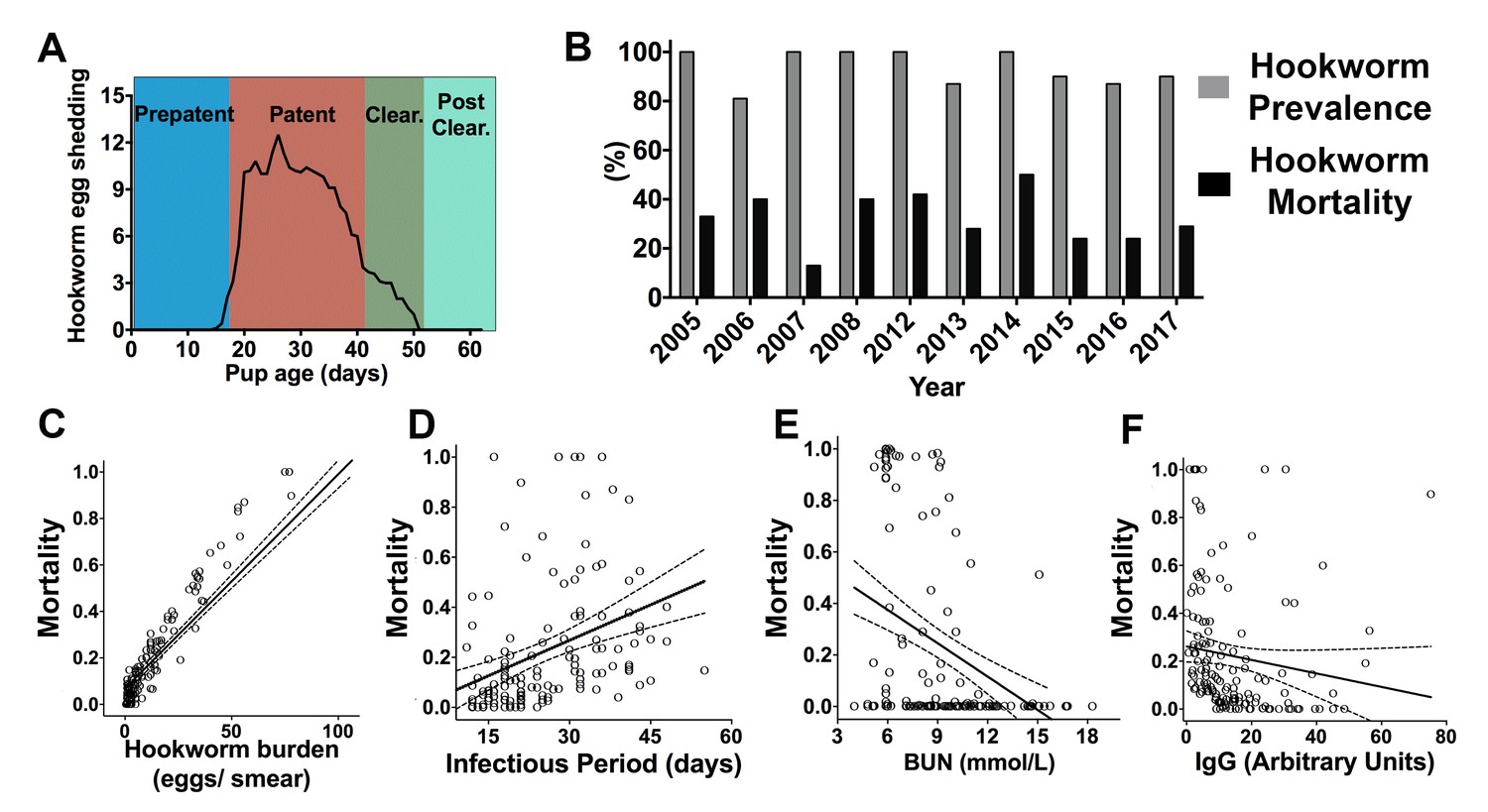
Hookworm infection dynamics and predictors of hookworm mortality in South American fur seal (Arctocephalus australis) pups at Guafo Island, southern Chile.

(A) Hookworm egg shedding patterns through infection stages. (B) Hookworm prevalence and mortality through different reproductive seasons (2005–08, 2012–17). (C–F) Predictors of hookworm mortality in generalized linear mixed models (GLMM) (2014, 2015, 2017) vs observed hookworm mortality. Higher hookworm burdens (GLMM, 1.47 ± 0.56, Z = 2.61, p = 0.009), longer infectious periods (GLMM, 0.17 ± 0.06, Z = 2.87, p = 0.004), and lower plasma concentrations of blood urea nitrogen (BUN) (GLMM, −0.45 ± 0.23, Z = −1.97, p = 0.049) and parasite-specific IgG (GLMM, −0.41 ± 0.17, Z = −2.45, p = 0.014) increased the probability of hookworm mortality. Raw data in: Figure 1—source data 1.

https://doi.org/10.7554/eLife.38432.003
Figure 1—source data 1

Predictors of hookworm-related mortality in South American fur seal pups.

https://doi.org/10.7554/eLife.38432.004

Hookworm clearance is immune-mediated

To determine the mechanisms that drive hookworm clearance and affect host mortality, the immune response to hookworms was investigated during 2017 at different infection stages in 54 fur seal pups, and compared to 24 hookworm-free (ivermectin-treated) age-matched controls (Figures 2, 4A and B, Figure 2—source data 1). The number of peripheral blood leukocytes (lymphocytes, macrophages, neutrophils, eosinophils, and basophils) was obtained as a basic tool to indirectly measure the level of proliferation of these different immune cell types in infected and control animals. During the patent and clearance period, fur seal pups that survived infection (n = 41) experienced a significant increase in the number of peripheral blood lymphocytes (GLMM, 0.9 ± 0.003, Z = 231, p = 2.0×10⁻¹⁶) and basophils (GLMM, 4.8 ± 0.08, Z = 56.7, p = 2.0×10⁻¹⁶), and had higher numbers of these cells when compared to age-matched controls and to the pups that died from hookworm infection (Figure 2A–B). The number of neutrophils in peripheral blood was similar between controls and pups that survived but slightly lower in pups that died (n = 13) from hookworm disease (GLMM, died = −0.53 ± 0.06, Z = −8.04, p = 9.1×10⁻¹⁶). During the patent period, lower numbers of monocytes were found in animals that died from hookworm disease compared to controls (GLMM, died = −0.88 ± 0.11, Z = −7.64, p = 1.54×10⁻¹⁴), and eosinophils were higher in animals that survived when compared to controls and animals that died (GLMM, survived = 0.86 ± 0.16, Z = 5.19, p = 2.0×10⁻⁷); however, during the clearance and post-clearance periods, eosinophils (GLMM, survived = 0.26 ± 0.14, Z = 1.88, p = 0.07) and macrophages (GLMM, survived = −0.03 ± 0.09, Z = 0.36, p = 0.71) were in similar numbers in pups that survived infection and controls. Pups that cleared the infection developed medium to high levels of parasite-specific IgG, whereas the level of these antibodies was significantly lower in pups that died from hookworm infection and almost non-existent in the control group (Figure 2J–M). There was moderate to marked immunolabelling of the hookworm intestinal brush border using serum from six pups with moderate to high levels of parasite-specific IgG (23–100 arbitrary units) (Figure 2I), suggesting that anti-hookworm antibodies bind proteins located in the hookworm intestine.

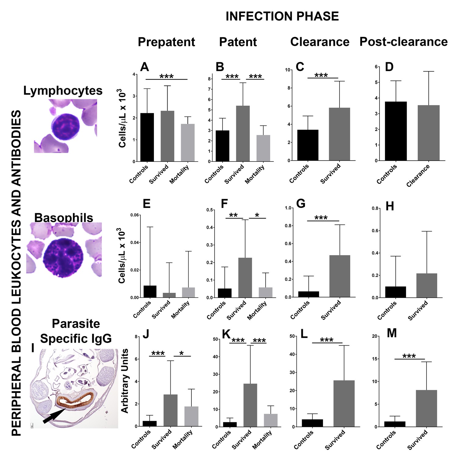
Changes in peripheral blood leukocytes and parasite-specific IgG antibodies during different phases of hookworm infection (Uncinaria sp.) in South American fur seal (Arctocephalus australis) pups (2017).

(A) Pups that die from hookworm disease have lower numbers of lymphocytes during the prepatent phase compared to controls and pups that survived (Generalized linear mixed model (GLMM), lymphocytes died = −0.52 ± 0.13, Z = −4.06, p = 4.92×10⁻⁵). Pups that survive hookworm infection have higher numbers of lymphocytes during the patent (B) (GLMM, lymphocytes survived = 0.80 ± 0.13, Z = 6.04, p = 1.54×10⁻⁹) and clearance (C) (GLMM, lymphocytes survived = 0.52 ± 0.12, Z = 4.30, p = 1.74×10⁻⁵) infection phases when compared to pups that died due to hookworm infection and/or age matched controls. (E–H) Pups that clear and survive hookworm infection have markedly higher numbers of basophils during the patent (GLMM, basophils survived = 7.46 ± 1.4, Z = 5.33, p = 1.0×10⁻⁷) and clearance (GLMM, basophils survived = 6.34 ± 0.9, Z = 7.07, p = 1.5×10⁻¹²) infection phases compared to controls and pups that died from hookworm infection. (I) Fur seal pups that clear hookworm infection produce parasite-specific IgG that binds the intestinal brush border of the fur seal hookworms (Uncinaria sp.) (arrow). (J–M) Fur seal pups that clear hookworm infection have higher levels of parasite-specific IgG during the prepatent (GLMM, IgG survived = 1.78 ± 0.36, Z = 4.86, p = 1.12×10⁻⁶), patent (GLMM, IgG survived = 2.27 ± 0.25, Z = 9.067, p = 2.0×10⁻¹⁶), clearance (GLMM, IgG survived = 1.80 ± 0.2, Z = 9.0, p = 2.0×10⁻¹⁶), and post-clearance (GLMM, IgG survived = 1.87 ± 0.25, Z = 7.3, p = 3.53×10⁻¹³) infection phases compared to controls and pups that died. Asterisk indicate groups are statistically different at alpha = 0.05. p-values code: *0.01 < 0.05, **0.001 < 0.01, *** < 0.001. Raw data in: Figure 2—source data 1.

https://doi.org/10.7554/eLife.38432.005
Figure 2—source data 1

Maternal attendance and health-related parameters in pups with different hookworm infection status.

https://doi.org/10.7554/eLife.38432.006

To determine the morphological and immune cell population changes in the anatomical site of hookworm infection, sections of small intestine and mesenteric lymph nodes were collected from pups that died from hookworm disease (n = 21), pups that were undergoing clearance (n = 18), and pups that were never infected with hookworms (controls, n = 6) (Figure 3—source data 1). The small intestine mucosa, submucosa, and the mesenteric lymph nodes of pups undergoing hookworm clearance contained larger numbers of T-lymphocytes when compared to pups that died from hookworm infection or pups never infected with adult Uncinaria sp (Generalized linear models with negative binomial distribution (GLM.NB), mucosa clearance = 0.86 ± 0.11, Z = 8.312, p = 2.0×10⁻¹⁶, submucosa clearance = 1.08 ± 0.16, Z = 6.86, p = 7.0×10⁻¹², mesenteric lymph node clearance = 0.78 ± 0.06, Z = 14.21, p = 2.0×10⁻¹⁶) (Figure 3). B-lymphocytes and plasma cells were more numerous in the mesenteric lymph node of pups clearing hookworm infection versus controls and pups dead from hookworm infection (GLM.NB, B-lymphocytes clearance = 0.29 ± 0.07, Z = 4.1, p = 4.1×10⁻⁵, plasma cell clearance = 0.59 ± 0.05, Z = 10.2, p = 2.0×10⁻¹⁶). Similarly, there were higher numbers of mast cells (GLM.NB, clearance = 1.14 ± 0.25, Z = 4.6, p = 4.2×10⁻⁶) and more mucus (GLM, clearance = 0.03 ± 0.003, Z = 7.84, p = 9.4×10⁻¹⁰) in the mucosa, and more leukocytes expressing IL-4 in the intestine (GLM.NB, clearance = 1.97 ± 0.15, Z = 12.72, p = 2.0×10⁻¹⁶) and mesenteric lymph node (GLM.NB, clearance = 1.57 ± 0.11, Z = 14.63, p = 2.0×10⁻¹⁶) of pups that cleared hookworm infection when compared to controls and pups with hookworm enteritis and bacteremia. Pups that died from hookworms, however, had larger numbers of macrophages in the intestinal submucosa (GLM.NB, mortality = 0.52 ± 0.09, Z = 5.79, p = 6.94×10⁻⁹) and mesenteric lymph nodes (GLM.NB, mortality = 0.63 ± 0.05, Z = 12.68, p = 2.0×10⁻¹⁶) compared to pups never infected with hookworms and pups clearing hookworm infection (Figure 3).

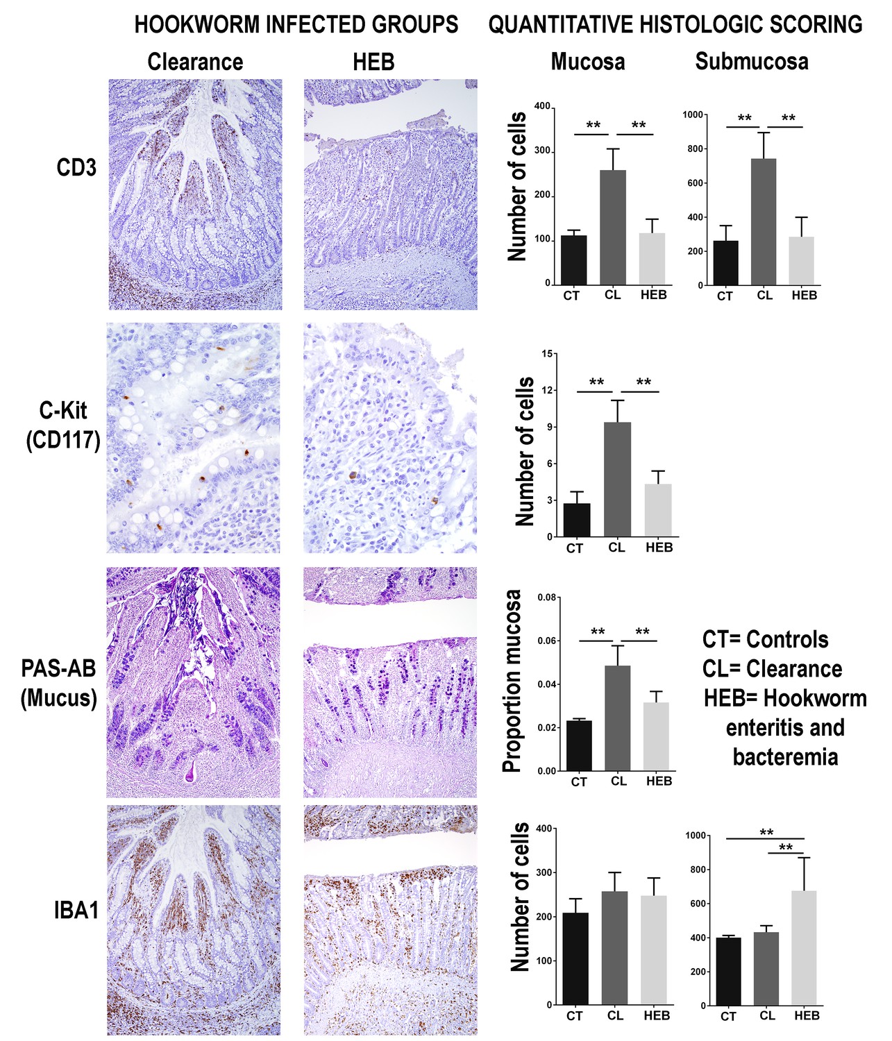
Intestinal immune response in different groups of South American fur seals (Arctocephalus australis) infected with hookworms (Uncinaria sp.) and controls.

During the clearance process, fur seal pups recruit numerous T-lymphocytes (CD3 stain) in the jejunum mucosa (Generalized linear models with negative binomial distribution (GLM.NB), clearance = 0.86 ± 0.11, Z = 8.312, p = 2.0×10⁻¹⁶) and submucosa (GLM.NB, clearance = 1.08 ± 0.16, Z = 6.86, p = 7.0×10⁻¹²). Mast cells (C-kit stain) are found in higher numbers in the intestinal mucosa of pups undergoing clearance (GLM.NB, clearance = 1.14 ± 0.25, Z = 4.6, p = 4.2×10⁻⁶). The intestinal mucosa of pups clearing hookworm infection contains a large amount of mucus (GLM, clearance = 0.03 ± 0.003, Z = 7.84, p = 9.4×10⁻¹⁰). Pups that die from hookworm enteritis and bacteremia (HEB) have lower numbers or proportions of these immune components but higher numbers of macrophages (IBA1 stain) in the jejunum submucosa (GLM.NB, mortality = 0.52 ± 0.09, Z = 5.79, p = 6.94×10⁻⁹). Asterisks indicate groups are statistically different at alpha = 0.05. p-values code: *0.01 < 0.05, ** <0.01. GLM.NB. Raw data: Figure 3—source data 1.

https://doi.org/10.7554/eLife.38432.007
Figure 3—source data 1

Leukocyte subsets and IL-4 immunohistochemical staining in intestine and mesenteric lymph node.

https://doi.org/10.7554/eLife.38432.008
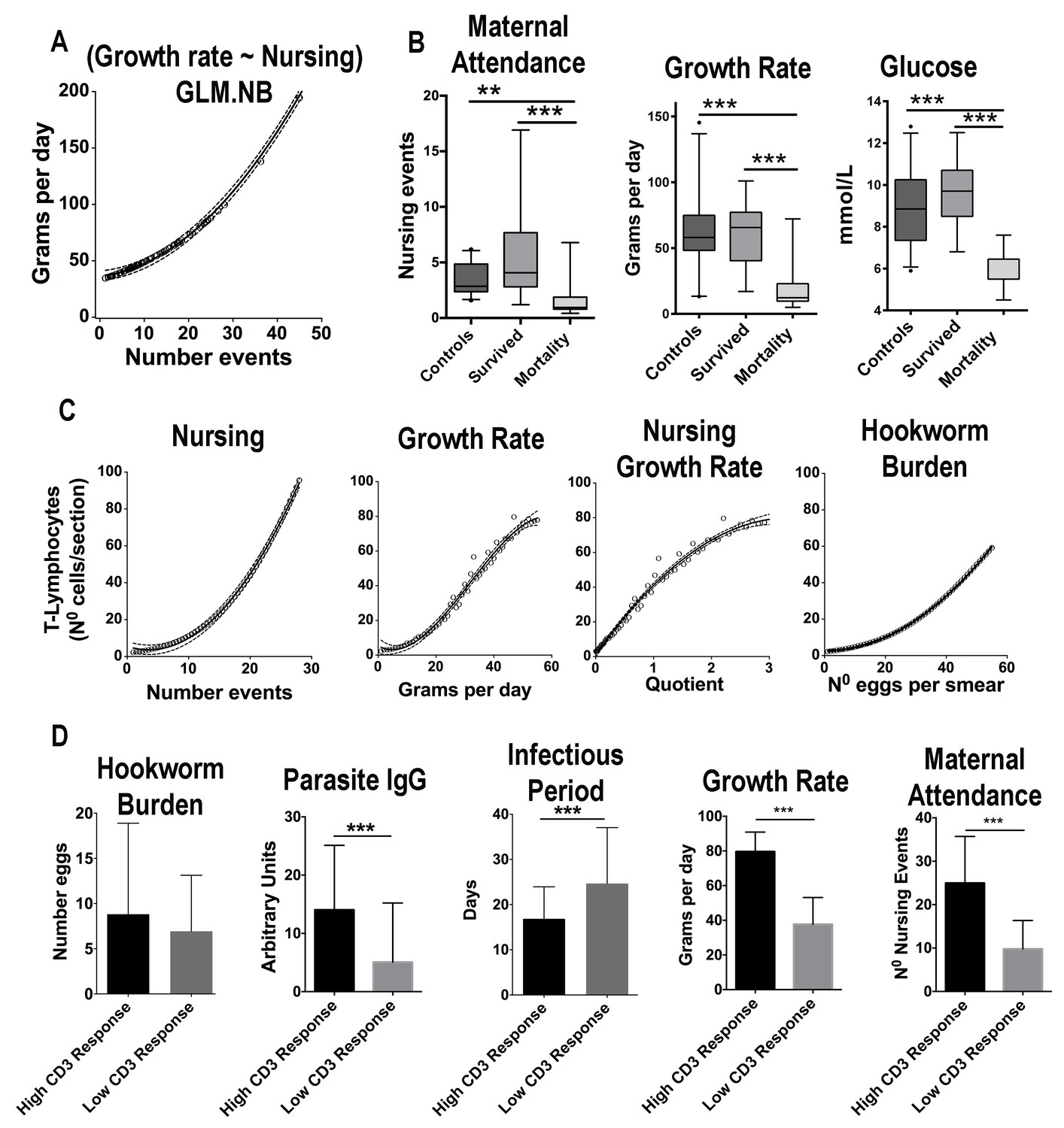
Maternal attendance affects South American fur seal pup’s growth rate, energy balance, and immune response against hookworms.

(A) The observed number of nursing events against predicted values of growth rate. With more nursing events pups grow faster (GLM.NB, 0.031 ± 0.006, Z = 5.53, p = 3.2×10⁻⁸). (B) Pups that survived hookworm infection had higher levels of maternal attendance (more nursing events) (GLM.NB, 0.78 ± 0.23, Z = 3.45, p = 5.5×10⁻⁴), faster growth rate (GLM.NB, 1.05 ± 0.16, Z = 6.6, p = 2.7×10⁻¹¹), and higher average levels of glucose (GLM, 3.0 ± 0.5, Z = 5.9, p = 1.02×10⁻⁷) compared to pups that died from hookworm disease; however, they had similar attendance and metabolic values compared to hookworm-free (control) pups (GLM.NB, 0.01 ± 0.13, Z = 0.13, p = 0.893, and GLM, 0.71 ± 0.38, t = 1.863, p = 0.066). (C) The observed values of number of nursing events, growth rate, interaction between nursing and growth rate and hookworm burden vs. the predicted values of CD3+ lymphocytes in section of skin in response to phytohemagglutinin (PHA) challenge. Pups with more nursing events (GLM.NB, 0.098 ± 0.02, Z = 4.39, p = 1.14×10⁻⁵), faster growth rate (GLM.NB, 0.04 ± 0.004, Z = 11.3, p = 2.0×10⁻¹⁶), and higher hookworm burden (GLM.NB, 0.009 ± 0.004, Z = 2.56, p = 0.01) had more recruitment of T-lymphocytes. (D) A subset of pups was divided into groups of low and high response to PHA challenge at 30 days old. Pups with higher CD3 lymphocyte response had higher average levels of parasite-specific IgG (GLM.NB, 1.11 ± 0.33, Z = 3.37, p = 7.5×10⁻⁴), shorter infectious period (GLM.NB, −0.38 ± 0.12, Z = −3.06, p = 2.1×10⁻³), faster growth rate (GLM.NB, 0.68 ± 0.06, Z = 10.9, p = 2.0×10⁻¹⁶), and higher levels of maternal attendance (GLM.NB, 0.94 ± 0.13, Z = 7.04, p = 1.92×10⁻¹). Hookworm burden was similar between the two groups (GLM.NB, low reactivity = −0.63 ± 0.37, Z = −1.67, p = 0.09). Raw data: Figure 4—source data 1 and Figure 4—source data 2.

https://doi.org/10.7554/eLife.38432.009
Figure 4—source data 1

Phytohemagglutinin immune challenge in 8-wk-old pups.

https://doi.org/10.7554/eLife.38432.010
Figure 4—source data 2

Phytohemagglutinin immune challenge in 4-wk-old pups.

https://doi.org/10.7554/eLife.38432.011

Maternal attendance affects fur seal pup hookworm clearance

Maternal attendance patterns and pup-related health parameters were assessed in the 2017 reproductive season (n = 78) (Figure 4A and B, Figure 4—source data 1). Among measured serum chemistry variables, the average level of blood glucose was the best predictor of growth rate (GLM.NB, 0.18 ± 0.03, Z = 5.9, p = 2×10⁻¹⁶). Among the considered external factors that could affect growth, the number of nursing events observed in a pup was the most significant predictor of growth rate, and although hookworm burden and hookworm infectious period were included in some top ranked models, their effect was not significant (Figure 4A, supplementary file 4 and 5). Additionally, there were no significant differences in growth rates between pups treated with ivermectin (n = 24) and non-treated (n = 54) (GLM.NB, 0.17 ± 0.15, Z = 1.16, p = 0.26). Nevertheless, when pups that died from hookworm disease were considered (n = 13), they had significantly slower growth rates (GLM.NB, −1.05 ± 0.16, Z = −6.6, p = 2.7×10⁻¹¹) compared to pups that survived (n = 41) and pups treated with ivermectin; however, the animals that died also had the lowest levels of maternal attendance (GLM.NB, −0.78 ± 0.23, Z = −3.45, p = 5.5×10⁻⁴) (Figure 4B). Regarding the factors that affected overall immune reactivity (Figure 4C, Figure 4—source data 1), pups with more nursing events, faster growth rate, and higher hookworm burden were more likely to recruit higher numbers of T-cells (CD3+ lymphocytes) in the skin in response to (Phytohemagglutinin) PHA challenge (Figure 4C, supplementary file 5 and 6).

Pups with lower parasite-specific IgG concentrations (GLMM.NB, coeff = −0.017 ± 0.002, Z = 6.54, p = 2×10⁻¹⁶, n = 146) and higher hookworm burden (GLMM.NB, coeff = 0.06 ± 0.022, Z = 2.78, p = 0.005, n = 146) had longer infectious periods (Supplementary file 7 and 8), suggesting that among measured immune parameters, parasite-specific IgG was the most significant factor affecting the permanence of hookworms in the intestine. Based on the PHA immune challenge performed when pups were 1-mo-old (Figure 4D, Figure 4—source data 1), animals with high T-cell response had higher levels of IgG, maternal attendance, glucose, growth rate, and shorter infectious periods at the end of the study when compared to the average levels in pups with low T-cell response (Figure 4D). However, hookworm burden was similar between the two groups (Figure 4D), suggesting, in conjunction with the previous analyses, that maternal attendance and growth rate accounted for most of the difference in T-cell reactivity between these groups.

In years with high sea surface temperature there is lower maternal attendance, immune response, and increased hookworm-induced mortality

SAFS females were observed more frequently arriving to the rookery from foraging trips early in the morning (2007 = 78/115, 67.8% returning events in the morning, 2017 = 87/135, 64% returning events in the morning). Foraging trip length was correlated with the number of nursing events, indicating that the more time females spend at sea makes it less likely to observe them nursing their pup (Figure 5A). In 2017, a year with SST above Guafo Island average, SAFS females (n = 21) spend more time foraging at sea compared to 2007 (n = 23), a year with SST below Guafo Island average, therefore in 2017 (n = 79) the level of maternal attendance and pup growth rate were lower than in 2007 (n = 128) (Figure 5B) (Figure 5A and B, Figure 5—source data 1). Between 2012 and 2017 (Figure 5C, Figure 5—source data 1), in years with high SST (e.g. 2014), the average concentrations of glucose, cholesterol, parasite-specific IgG, and peripheral blood lymphocytes and basophils were lower than in years with low SST (Figure 5C). Similarly, the average hookworm infectious period was shorter in years with low SST (GLM, X2 = 6.95, df = 1, p = 0.00036).

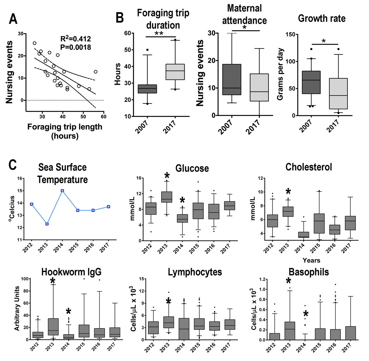
South American fur seals foraging behavior and maternal care patterns differ between seasons.

(A) The level of maternal attendance decreases as foraging trips become longer (linear regression, R2 = 0.412, p = 0.016; dashed lines represent 95% confidence intervals). (B) In a year with sea surface temperature (SST) below the historic Guafo Island average (2007), fur seal females foraging trips are shorter when compared to the mean foraging trip duration during a year with SST temperature above the historical average (2017) (unpaired T-test, t = 5.133, df = 42, p < 0.0001). Additionally, maternal attendance and pup growth rate in 2007 were higher than attendance and growth rates in 2017 (maternal attendance index: unpaired T-test, t = 2.060, df = 244, p = 0.04; growth rate: unpaired T-test, t = 2.85, df = 66, p = 0.0058). (C) Between 2012 and 2017 the mean values of glucose, cholesterol, parasite-specific IgG, peripheral blood lymphocytes and basophils followed an inverse pattern with mean SST at Guafo Island. In 2013, a year with low SST, pups had, on average, higher levels of glucose, cholesterol, parasite-specific IgG, lymphocytes and basophils when compared to the mean values of other reproductive seasons (Kruskal-Wallis with Dunn’s multiple comparison tests, Kruskal-Wallis statistic = 73.2–114.6, mean rank diff. = 63.98–203.8, p < 0.0001–0.023). In 2014, with the highest mean SST over the last 15 y at Guafo Island, fur seal pups had the lowest mean values of these metabolic and immune parameters (Kruskal-Wallis with Dunn’s multiple comparison tests, Kruskal-Wallis statistic = 73.2–114.6, mean rank diff. = −230.83,–83.4, p < 0.0001–0.017). (Asterisks indicate mean is significantly different from means of other seasons). Raw data in: Figure 5—source data 1 and Figure 5—source data 2.

https://doi.org/10.7554/eLife.38432.012
Figure 5—source data 1

Maternal attendance and growth rates in 2007 and 2017.

https://doi.org/10.7554/eLife.38432.013
Figure 5—source data 2

Immune and metabolic parameters in South American fur seal pups between 2012 and 2017.

https://doi.org/10.7554/eLife.38432.014

Over a 10-y period (2005–08, 2012–17) (Figure 6, Figure 6—source data 1 and 2), there was a significant positive correlation between mean hookworm burdens of necropsied pups and SST (Linear regression, Ad-R2 = 0.86, p < 0.001), and between hookworm mortality and SST (Ad-R2 = 0.56, p = 0.016); however, in the case of hookworm prevalence at necropsy the correlation with SST was not significant (Figure 6, supplementary file 911) (Ad-R2 = 0.29, p = 0.064). A similar but negative correlation existed between the same hookworm epidemiological parameters and average chlorophyll-a concentrations (Figure 6).

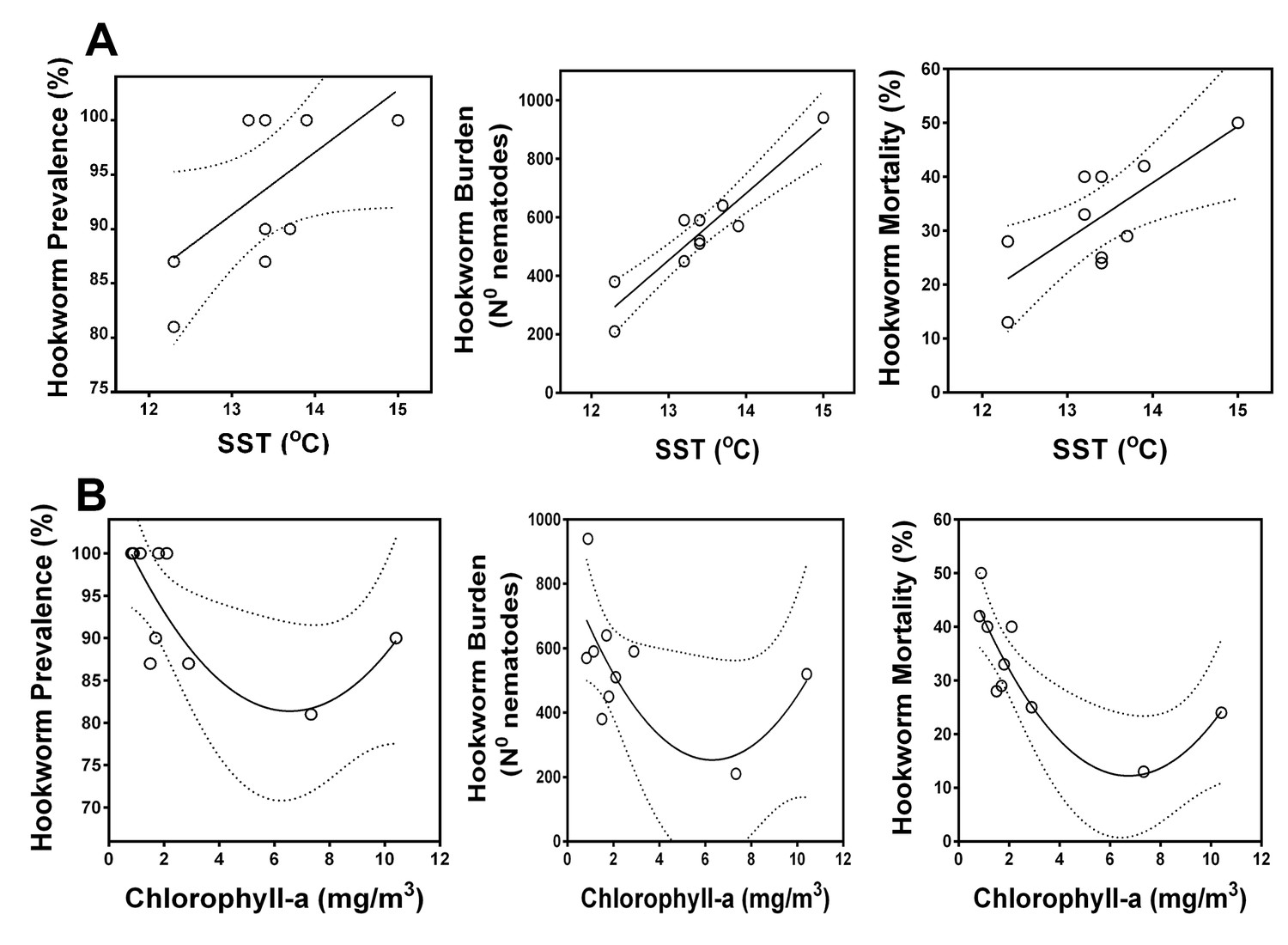
Correlation between oceanographic parameters (sea surface temperature and chlorophyll-a) and hookworm disease dynamics in South American fur seals (Arctocephalus australis) at the Chilean Patagonia (2005–08, 2012–17).

(A) Hookworm prevalence, burden, and mortality increase in years with warmer sea surface temperature (Linear regressions. Hookworm prevalence, Ad-R2 = 0.29, p = 0.064. Hookworm burden, Ad-R2 = 0.86, p < 0.001. Hookworm mortality, Ad-R2 = 0.56, p = 0.016). Hookworm prevalence, burden, and mortality decrease in some years with higher primary productivity (Second order polynomial regressions. Hookworm prevalence, Ad-R2 = 0.46, p = 0.046. Hookworm burden, Ad-R2 = 0.29, p < 0.123. Hookworm mortality, Ad-R2 = 0.70, p = 0.005). Dashed lines represent 95% confidence intervals. Raw data: Figure 6—source data 1 and Figure 6—source data 2.

https://doi.org/10.7554/eLife.38432.015
Figure 6—source data 1

Sea surface temperature data for Guafo Island.

https://doi.org/10.7554/eLife.38432.016
Figure 6—source data 2

Hookworm prevalence, burden, and mortality in South American fur seal pups at Guafo Island.

https://doi.org/10.7554/eLife.38432.017

Discussion

Hookworm disease causes significant mortality in SAFSs in the Chilean Patagonia and represents one of the most significant causes of death among pups (Seguel et al., 2013; Seguel et al., 2018). However, in some years, hookworm-induced mortality decreases significantly. We showed that variations in hookworm disease morbidity and mortality are associated with specific changes in the immune response against the parasite. Additionally, maternal care was the most important external factor affecting immune response and hookworm clearance. As otariid maternal care is influenced by ocean environmental conditions and prey availability (Trillmich et al., 1991; Soto et al., 2006; Jeanniard-du-Dot et al., 2017), indirect indicators of ocean productivity such as sea surface temperature are correlated with hookworm disease dynamics and overall fur seal pup survival. Changes in ocean productivity have been indicated as the main environmental factor driving marine mammal mortality (Trillmich et al., 1991; Soto et al., 2006), Costa, 2012, Elorriaga-Verplancken et al., 2016); however, the links between indexes of ocean productivity such as SST and mortality are based on circumstantial evidence, and the mechanisms that drive this correlation are unclear. Our study provides a mechanistic explanation regarding the specific pathophysiologic processes affected by changes in the marine environment, and explains how these environmental processes impact host physiology, immune response, and survival (Figure 7).

Proposed mechanism of South American fur seal response to environmental change in the context of endemic hookworm infection.

Changes in sea surface temperature (SST) are associated with changes in foraging trip length and patterns of maternal attendance in fur seals. In years with lower SST, fur seal pups receive more diurnal maternal attendance compared to years with high SST. Pups with more maternal attendance have better energy balance, higher T-lymphocyte reactivity, and produce higher levels of parasite-specific IgG. Pups with this immune profile eliminate hookworms from the intestine faster than pups with less reactive immune systems. Early hookworm clearance is one of the most important factors that drive hookworm-related mortality, therefore in years with higher SST fur seal pups die more often of hookworm disease.

https://doi.org/10.7554/eLife.38432.018

Based on the epidemiological data collected in the current study when fur seal pups are on average 2.5 mo old, adult hookworms cannot be found in the fur seal population because they have been cleared from the intestine or have died along with their host. This is consistent with what we have found in necropsies over more than 10 y at Guafo Island and with what has been suggested in other fur seal species (Lyons et al., 2011b; Seguel et al., 2018). Early hookworm clearance is a unique feature of otariids, as all studied natural hookworm infections of land mammals consist of long-term intestinal infections (Seguel and Gottdenker, 2017), suggesting that fur seals have developed efficient mechanisms to clear hookworms. Based on field experiments and the immune parameters measured in the present study, this parasite clearance process is mediated mostly by T-lymphocytes, basophils, Th2-type leukocytes, and production of parasite-specific IgG. These results are similar to changes in peripheral blood leukocytes in humans and rodents infected with hookworms (Loukas and Prociv, 2001; Cortés et al., 2017). In both systems, an increase in circulating T-lymphocytes and basophils is associated with hookworm clearance and resistance to re-infection (Cortés et al., 2017). Regarding parasite-specific IgG production, it is interesting that this antibody binds antigens located in a specific portion of the hookworm anatomy, the intestinal brush border. This anatomical location contains several digestive and heme-detoxifying enzymes that are crucial for the nematode blood digestion and survival (Williamson et al., 2003; Wei et al., 2016). In fur seals, it is possible that parasite-specific IgG reaches the nematode intestine with each blood meal, impairing the nematode blood digestion by blocking digestive enzymes, favoring clearance. A similar mechanism has been experimentally induced in dogs and humans through a hookworm vaccine using the digestive enzymes in the nematode intestinal brush border as antigens (Hotez et al., 2016; Diemert et al., 2017). These vaccines successfully avoid hookworm development and promote clearance (Hotez et al., 2016; Diemert et al., 2017). The morphologic and leukocyte population changes observed in the intestine of pups clearing hookworm infection are similar to those described in laboratory rodent models of hookworm infection. In these systems, T-lymphocytes, mast cells, and basophils are important players in the morphological changes of the intestine that facilitate nematode detachment and removal (Ohnmacht and Voehringer, 2010; Cortés et al., 2017). Therefore, as shown in laboratory animal models, it is likely that in fur seal pups, these changes in the intestinal mucosa and antibody production create a hostile environment for hookworm attachment and feeding, leading to clearance from the intestine.

Fur seal pups infected with hookworms for a shorter period of time were more likely to survive the infection. This suggests that there is a time-dependent effect of hookworms on the host, probably associated with host resource depletion, which could increase the risk of mortality from hookworm-induced anemia and peritonitis (Spraker et al., 2007; Seguel et al., 2017; Seguel et al., 2018). Additionally, pups with higher levels of parasite-specific IgG had shorter hookworm infection. This suggests that parasite-specific IgG is an immune element that contributes significantly to survival of fur seal pups. This is similar to what has been found in a wild population of Soay sheep (Ovis aries), where parasite-specific IgG is the best predictor of survival through the winter, precisely when there is scarcity of food resources and likely a more detrimental effect of parasites in the host (Watson et al., 2016). In most studied mammal species, IgG production is dependent on T-cell activity and caloric intake, particularly in neonates and children (Papier et al., 2014; Ibrahim et al., 2017). Similar processes could occur in fur seals given the differences observed in immune system reactivity and hookworm infection outcome in pups with different energy budget and maternal attendance patterns.

As expected, and as reported in other studies in pinnipeds, pups with higher levels of maternal attendance had higher growth rates (Francis et al., 1998; Georges and Guinet, 2000; Arnould and Hindell, 2001a). This could be related to pups spending more time with their mothers, shortening the fasting period. This is in line with the finding that in 2017 pups with more maternal attendance had higher glucose levels. Even though in some fur seal species pups can maintain relatively stable glucose levels during fasting (Arnould and Hindell, 2001a; Verrier et al., 2012), after a couple of days without nursing there is a significant decrease in blood glucose (Arnould et al., 2001b; Champagne et al., 2012). Therefore, it is not surprising that the average blood glucose levels and growth rate of pups were highly correlated in our study, suggesting that glucose could be a good marker of the energy balance in a pup, and an important factor to consider in the health assessment of wild pinnipeds given the connection between energy balance and immune system. For instance, recent studies have shown that Californian sea lion pups with better body condition and higher glucose levels have higher total IgG (Banuet-Martínez et al., 2017). In humans and laboratory animals, glucose metabolism is one of the most important factors driving T-cell activity and production of antigen-specific IgG (Mohammed et al., 2012; Palmer et al., 2015). In our study, glucose levels and maternal attendance were the most significant factors affecting the overall level of T-cell reactivity in fur seal pups, and pups with higher T-lymphocyte reactivity produced higher levels of parasite-specific IgG and expelled hookworms faster. This suggests that fur seal pups that spend more time with their mothers receive more and/or higher quality milk, favoring a positive energy balance, leaving more energy available for immune response, including proliferation of T-lymphocytes and production of anti-hookworm antibodies.

Fur seal and sea lion maternal attendance patterns can be affected by several factors, including prey availability, maternal experience, and body condition (Georges and Guinet, 2000; Arnould and Hindell, 2001a; Soto et al., 2006; McDonald et al., 2012a; McDonald et al., 2012b). Out of these variables, body condition and prey availability can be affected by changes in sea surface temperatures (SST) because of the role that ocean temperature plays in nutrient upwelling, primary productivity, and distribution of fish stocks (Soto et al., 2006, Costa, 2008). In the southern Pacific Ocean, warmer ocean temperature is associated with a marked decrease in primary productivity and fish stocks, these effects being particularly intense during El Nino Southern Oscillation (ENSO) events (Trillmich et al., 1991; Soto et al., 2006). Additionally, in ‘El Nino’ years, the isotopic signature of pup tissues changes, which suggest shifts in the foraging and attendance regimes of their mothers (Elorriaga-Verplancken et al., 2016). In the Northern Chilean Patagonia, in years with higher SST, there is an apparent increase in the foraging trip length and a decrease in the maternal attendance of SAFS females. Because of the nature of our data acquisition, we cannot discard changes in nocturnal maternal attendance patterns, although the similarity in the proportion of foraging events recorded in the morning in years with high and low SST suggests that this is a less likely possibility. The reported changes in attendance coincide with slower growth rates of fur seal pups in a year with high SST compared to a year with low SST, suggesting that there could be a decrease in prey availability for SAFSs in years with higher SST, forcing them to spend more time foraging in the ocean and less time nursing their pups. This interpretation of the results is in line with the proposed conceptual model on the effect of environmental fluctuation in parental attendance in sea lions and fur seals (Costa, 2008). This model suggests that when environmental variation affects prey resources, adult females will increase their foraging intensity effort and metabolic rate before increasing foraging trip length, because the latter almost always results in a decrease in the net energy delivered to the offspring (Costa, 2008; Costa, 2012). In this context, the changes in maternal attendance observed in our study could explain the decreased average levels of glucose and cholesterol in fur seal pups in years with higher SST. Given the results of field experiments, it could be that in years with pup lower energy balance, immune parameters could also be decreased in these animals. This was observed, particularly for immune parameters such as circulating lymphocytes, basophils, and parasite-specific IgG, which are key elements for prompt hookworm clearance. Additionally, as these immune elements drive hookworm permanence in the pups’ intestine (infectious period), in years with lower SST, hookworm infectious period was shorter, suggesting that environmental variables affect not only hookworm immune response but also transmission patterns.

The present findings suggest that indirectly, SST modifies hookworm infection dynamics in SAFSs by impacting on maternal attendance patterns, energy budget between dams and pups, and pup immune response. This observation is also supported by the correlation found between SST and hookworm burden and mortality over a 10-y period on Guafo Island. Similar long-term studies on hookworm infection of fur seals have found dramatic changes in hookworm prevalence and mortality over a 20–30-y period (Lyons et al., 2011b). It is possible that changes in prevalence are associated with modification in host population density, given the sensitivity of this parasite to host density constraint (Lyons et al., 2011a; Seguel et al., 2018). Our observations could explain why changes in prevalence and its correlation to environmental variables were not significant in our study, as the Guafo Island population has remained stable over the last decade (Pavés et al., 2016). However, the differences in immune response and hookworm dynamics in the studied population suggest that hookworm biomass and infection duration are the key parasite-specific components that drive mortality if prevalence is relatively stable. In line with these observations, the control of the immune system on the clearance process and the sensitivity of fur seal immune system to external factors such as maternal attendance, establish a pattern where parasite transmission is indirectly linked to environmental change in the ocean.

Global climate change is increasing ocean temperature, particularly in the Southern Hemisphere (Wijffels et al., 2016). Our findings highlight that under this scenario infectious diseases could have a more detrimental impact on populations of fur seals and sea lions in the future, but also provide a foundation for the study of climate change adaptation options for these species (Hobday et al., 2015). For instance, treatment of pups to eliminate or decrease parasite burden could be more productive in years with adverse environmental conditions or on otariids with less flexible foraging strategies (Costa, 2012). Considering the long-term impact of hookworm disease on the population fitness, host species extinction risk, and the importance of parasite biodiversity is critical before evaluating intervention strategies.

Conclusions

In Chilean Patagonia, during years with high SST, ocean productivity decreases, forcing adult female fur seals to increase their foraging trip length and decrease their levels of maternal attendance. Pups receiving less maternal care had reduced growth rates and decreased energy budget, impairing the ability of their immune system to mount an effective response against hookworms to expel the parasite from the intestine. These pups, with longer hookworm infection periods, usually die as a consequence of hookworm disease establishing a pattern in which hookworm disease severity and mortality are correlated to indexes of oceanographic environmental conditions such as sea surface temperature. The sensitivity of otariid hookworm disease to ocean temperature and marine productivity presents a scenario where global climate change could increase the extent and severity of a disease present in most fur seal and sea lion populations.

Materials and methods

Fur seal health assessments and mortality

Request a detailed protocol

From 2012 through 2017 South American fur seal pups were captured by hand every 7–15 d between December 15th and March 10th. At the first capture, pups were marked with a number in the fur using commercial hair decoloring solution. During each capture procedure, standard length, weight, sex, and body condition were recorded. The pup age was calculated based on the peak of parturition for Guafo Island rookery (December 15, Pavés et al., 2016) and based on the assessment of rest of placenta (1–3-d-old) and umbilical cord (2–7-d-old) during the first capture. For a subset of pups during 2014 (n = 10), 2015 (n = 20), and 2017 (n = 40), age was exactly known because their parturition was observed and they were marked 24 h later. Blood was drawn from the caudal gluteal vein of pups into EDTA, heparin, and plain (serum) vacutainer tubes. Plain blood tubes were centrifuged within 1–3 h post-collection in the field laboratory to obtain serum, which was preserved at −20°C until later long-term storage (−80°C) or analyses in the mainland laboratory. Plasma was obtained and stored following similar procedures with heparin non-coagulated blood. During each capture procedure, a rectal swab was collected and stored in Sheather’s sucrose for later semi-quantitative determination of hookworm egg burden according to standardized methods for this fur seal population (Seguel et al., 2018). Hookworm burden of pups found dead was determined by collection, sexing, and counting of all nematodes present in the small intestine and correlated with egg burden through a fecal swab collected during necropsy (Seguel et al., 2017; Seguel et al., 2018). Using non-coagulated blood, hematocrit, hemoglobin concentration, total red blood cell count (RBC), total white blood cell count (WBC), and differential leukocyte counting were determined for each pup as previously described (Seguel et al., 2016). The total serum concentrations of albumin, globulins, cholesterol, glucose, triglycerides, blood urea nitrogen, and creatinine were determined in the mainland laboratory using previously described methods for this population (Seguel et al., 2016). All blood and serum (or plasma) measured parameters were obtained for every capture procedure.

Because previous studies indicated a hookworm prevalence close to 100% in this population (Seguel et al., 2018), a ‘hookworm-free’ control group was created in 2017 by treating 60 pups with a subcutaneous injection of ivermectin (300 µg/kg) when they were between 1 and 7 d old. These pups were subjected to the same capture, handling, health assessments, and data acquisition procedures as indicated for the non-treated pups. These pups never presented hookworm eggs in their feces during the duration of the study.

In 2014 (n = 38), 2015 (n = 53), and 2017 (n = 54) marked pups were observed at least once a week during the study period. Pups observed dead were retrieved from the rookery to perform complete necropsies, collect tissue samples for histopathology, and determine the cause of death according to previously described diagnostic criteria (Seguel et al., 2011; Seguel and Gottdenker, 2017). The minimal number of pups to capture every year was calculated at the beginning of the reproductive season based on the known recapture rates at Guafo Island (60–80%) and sample size simulations to reach a power of at least 80% (R packages ‘pwr’ and ‘SIMR’). Therefore, hookworm disease outcome (dead vs. survived) was known in these pups and registered in the final data sheet to calculate total hookworm mortality and to fit models to identify the most significant health-related parameters that predicted hookworm mortality.

Immune challenge experiments

Request a detailed protocol

In 2017, a PHA immune challenge experiment was performed in a group of pups when they were approximately 8 wk old (n = 75). For these animals there were enough recaptures (at least four) to measure the average of all health-related parameters, hookworm infection history, and outcome (survival vs. death) at the end of the study period (February–March). The challenge consisted of injection of 0.1 ml of a 1.0 mg * ml−1 solution of phytohemagglutinin (PHA) into the interdigital skin of the right posterior flipper (Vera-Massieu et al., 2015). The same volume of a saline solution was injected in the same location of the left flipper (control). Swelling was measured in both injection sites 12 h after challenge and a 4-mm punch biopsy was collected from each site (PHA and control) following anesthesia with 5% isoflurane. Biopsy samples were stored in 10% buffered formalin and routinely processed for histopathology and immunohistochemistry for CD3. The number of CD3+ lymphocytes in control and treatment biopsies were counted and the difference between these two recorded and used in statistical analyses. In 2017, another subset of pups of known age (n = 55) was immune-challenged when they were approximately 30-d-old (during the acute hookworm infection phase). Sample size was calculated based on a minimum power of 80% using data collected in a preliminary study in 2016 (differences in skin swelling and T-cell recruitment). Pups were divided into two groups based on level of skin swelling and number of CD3+ lymphocytes detected during examination of biopsy samples. Animals with more than 20 CD3+ lymphocytes per section were considered high responders, whereas pups with less than 16 CD3+ lymphocytes were categorized as low responders. Only one pup was in the middle range (17 cells) and was not included in comparison analyses. At the end of the study period, data on growth rate, maternal attendance, and hookworm infection status were available for these pups.

Immune tests

Anti-hookworm ELISAs

Request a detailed protocol

A parasite-specific IgG ELISA was developed using whole worm extract as an antigen. Fresh hookworms were collected during necropsies at Guafo Island, washed in PBS, and frozen at −20°C in the field until transport to the mainland laboratory where they were stored at −80°C.

Thawed nematodes were macerated in phosphate buffered saline (PBS) using a glass homogenizer. The macerated nematodes were centrifuged at 15,000 RPM, 4°C for 1 h. Supernatant was collected, filtered, and total protein concentration determined using Bradford, bicinchoninic acid and ‘NanoDrop’ methods. Extracts were diluted in PBS for a final protein concentration of 1.6 µg/ml. High binding ELISA plates were coated overnight at 4°C using 100 µl per well of diluted (1:100) hookworm extract. Plates were washed with PBS/Tween and 100 µl of sample (fur seal serum) diluted in 5% dry milk/PBS were added to each well and incubated for 5 min at room temperature. A serial dilution of pooled fur seal serum from samples with high absorbance in previous experiments was used to construct a standard curve in each assay (positive controls). Serum from fur seal neonates and animals not exposed to hookworms (fur seal pups from populations without hookworms) were used as negative controls in each test and during standardization experiments. One hundred microliters of diluted (1:15,000) protein A (Vector laboratories, Burlingame, CA, USA) was added to each well and incubated for 5 min at 25°C. Plates were washed three times and 100 µl of TMB was added to each well and incubated for 30 min at room temperature. ELISA reaction was stopped using 100 µl per well of 1.0 N HCl and the plate was read at 450 nm wave length absorbance. The anti-hookworm IgG concentrations were calculated semi-quantitatively by comparing the optic density (OD) of the standard curve with the OD of the samples and reported as arbitrary units (AU). All these reactions were run in duplicate.

A similar procedure was used to standardize a parasite-specific IgE ELISA using a goat IgG anti-dog IgE antibody (Bethyl laboratories, Montgomery, TX, USA) as primary antibody and a rabbit anti-goat IgG as secondary antibody (Bethyl laboratories, Montgomery, TX, USA). However, we were unable to produce OD readings without a significant amount of noise (background staining) and data produced with this test were not further analyzed.

Special stains and immunohistochemistry

Request a detailed protocol

Sections of small intestine from necropsied pups and the skin sections of pups that underwent PHA immune challenges were routinely processed for histopathology and immunolabelled with antibodies against CD3, IBA1, CD79a, CD21, CD127 (c-kit), MUM1, and IL-4. The details of the antibodies used, retrieval, visualization methods, and dilutions are provided in supplementary file 12. The number of positive cells was recorded according to standard methods and used in data analyses.

General steps applied to all IHC protocols included deparaffinization of 4-µm tissue sections through immersion in xylene, and rehydration with graduated alcohols, antigen retrieval, quenching of endogenous peroxidase with hydrogen peroxide 3% for 15–20 min, incubation with primary antibody, blocking of nonspecific binding sites with a commercial blocking solution (Power Block, DAKO, Carpinteria, CA, USA), incubation with biotinylated secondary antibody (1:100 dilution, Vector Laboratories, Burlingame, CA) at room temperature for 20–30 min and with horseradish peroxidase-labeled streptavidin for 15 min (Biocare, Chicago, IL). Antigen-antibody complexes were visualized by incubation at room temperature for 5 min with diaminobenzidine (DAB) (Vector Laboratories, Burlingame, CA). Slides were counterstained with hematoxylin, dehydrated, and coverslipped. Tissue sections were observed in an optic microscope and representative sections photographed. The number of cells with positive imunolabelling was counted using the digital images with the use of the counting function of Adobe Photoshop. Sections from small intestine were stained through PAS-Alcian blue reaction to detect the number of goblet cells and amount of mucin produced. Slides were examined and standard sections photographed to calculate the amount of mucin present in the intestine. This number was calculated using Adobe Photoshop selection and calibration tools as the proportion of the total photographed area that stained positive with PAS-Alcian blue.

To detect the site of binding of anti-hookworm IgG in the body of nematodes, formalin-fixed hookworms obtained during necropsies of SAFS pups were routinely processed for histopathology (Seguel et al., 2017). Slides were cut at 4 µm, deparaffinized, and antigen retrieval was performed in citrate pH 6.0 for 10 min at 120°C. Blocking of nonspecific binding sites was done by incubation with 10% dry milk/PBS for 20 min and quenching of endogenous peroxidase by incubation with hydrogen peroxidase 3% for 30 min. After three washes with PBS, slides were incubated for 1 h at room temperature with SAFS pup serum that had the highest (strongly positive) or lowest (negative) absorbance during ELISA experiments, diluted (1:50) in 3% bovine serum albumin. After three washes with dilution buffer TWEEN (DBT), slides were incubated with biotinylated protein-A (1:1000 dilution) (Vector laboratories, Burlingame, CA) for 20 min at room temperature to detect IgG. Slides were washed three times with DBT and incubated with streptavidin horseradish peroxidase for 15 min at room temperature. Antigen-antibody complexes were visualized by incubation with DAB for 5 min at room temperature. Slides were counterstained with hematoxylin, dehydrated, and coverslipped.

Female foraging trip length, maternal attendance, and pup growth rates

Request a detailed protocol

In 2007 and 2017, observational studies were conducted in marked SAFS adult females and pup pairs (2007 n = 128, 2017 n = 78). Females were marked with paint spots delivered through paintball guns or by capture procedures using a net. In these procedures females were tagged in the front flippers (Allflex, Dallas, TX, USA) and marked with a number in the back using paint wax to facilitate their observation. Pups were captured, handled, and marked as previously described. Pups and females were observed daily for 1.5 h in the AM and 1.5 h in the PM. If a female was with her pup, this was characterized as a nursing event (regardless of whether the pup was suckling or not), and if a female was not present at the rookery and her pup was alone, the event was characterized as foraging (Francis et al., 1998; Kirkman et al., 2002; Trecu et al., 2010). All female-pup pairs showed high fidelity and allo-sucking events were not observed. The total number of nursing events observed for each pup was corrected by dividing them for the standard number of continuous observation events to avoid underestimation in pups with shorter observation periods (e.g. pups died). Therefore, for data analyses, the corrected number of observation events was used. Adult female foraging trip length was calculated by adding the number of observation periods (12-h intervals) when the female was not present at the rookery and her pup was alone (Francis et al., 1998; Kirkman et al., 2002; Trecu et al., 2010). Only animals with continuous observations were included in the data series (2007 n = 23, 2017 n = 21).

Pup growth rates were obtained through the following equation:

GR=(W2W1)/Δd,

where GR = growth rates (g/day),

W1= weight at first capture,

W2= weight at last capture,

Δd= d between first and last captures.

The minimal number of days used to obtain growth rates was 30 d (∆d ≥ 30) to assume a linear growth rate (Doidge et al., 1984).

Hookworm prevalence, ocean temperature, and primary productivity data

Request a detailed protocol

In Austral summers of 2004–2008 (n = 124) and 2012–2017 (n = 154), fresh fur seal pup carcasses were retrieved from the South American fur seal rookery at Guafo Island, Northern Chilean Patagonia (43° 35′ 34.9″ S, 74° 42′ 48.53″ W). Complete necropsies and histopathology were performed on these carcasses as previously described, to determine the cause of death of each pup (Seguel et al., 2011; Seguel and Gottdenker, 2017). During necropsies all parasites were collected and stored in 5% formalin for later counting. The median of the number of hookworms per pup in a given year was used as a measurement of hookworm burden for that particular season. The yearly prevalence of hookworm infection was calculated as the total number of pups with hookworms at necropsy divided by the total number of pups necropsied during that season.

Sea surface temperature and chlorophyll-a satellite data was retrieved from the NASA earth observation (NEO) website (https://neo.sci.gsfc.nasa.gov). The latitude and longitude to retrieve the chlorophyll-a and temperature data were selected to represent standard points 25 to 50 km west, south, north, and east of the South American fur seal rookery at Guafo Island. This approach was used to represent all the potential foraging areas of fur seals at Guafo Island (Data S8, sea surface temperature).

Data analyses

Hookworm mortality models

Request a detailed protocol

To identify factors that affected hookworm-induced pup mortality in 2014, 2015, and 2017, generalized linear mixed effect models (GLMM) were fitted using year as random effect (R package ‘glmmTMB’, Brooks et al., 2017). A multimodel selection approach and statistical inference was performed as recommended for data from natural systems (Burnham et al., 2011; Grueber et al., 2011). Predictors of mortality tested in different models included the average serum or plasmaconcentrations of albumin, globulins, cholesterol, glucose, and triglycerides, average blood hemoglobin concentration, pup growth rate, hookworm infectious period, the average and highest hookworm burden detected in the pup, and the average numbers of peripheral blood lymphocytes, neutrophils, eosinophils, basophils, and macrophages and their interactions. Growth rate and glucose were never fitted in the same models because of high correlation between these variables (r2 = 0.67). The output of each model and graphics of residuals were assessed to check model assumptions, overdispersion (residuals deviance), goodness of fit, and predictors coefficients and standard errors. Multiple models were constructed by adding and deleting predictors and their interactions based on biological predictions and model outputs. The selected fitted models that met quality assessment in terms of fulfillment of assumptions, overdispersion, and fit were later ranked based on second order Akaike’s information criteria (AICc). Additionally, Akaike weights and pseudo-R-squared for mixed models (Nakagawa and Schielzeth, 2013; Nakagawa et al., 2017) were obtained to compare models explanation of the data. Models with a delta AICc <2.0 were considered equally explanatory and were later averaged using the ‘model average’ function in the multimodel inference ‘R software’ package ‘MuMIn’ (Bartoń, 2017). Predictor coefficients, standard errors and p-values were assessed and reported in the text and supplementary tables.

Capture-recapture health assessments

Request a detailed protocol

Based on the recapture data, pups were assigned to different phases of the hookworm infection. These included the prepatent period, which corresponded to the phase when a pup had fresh or recently dried umbilical cord and was negative for hookworms at coprological examination; the patent period, which corresponded to the capture when pups had hookworm eggs in their feces; the clearance period, which corresponded to the capture when pups had a significant decline (at least 50%) in their hookworm burden when compared to their previous coprological exam; and the post-clearance period, which corresponded to the captures when pups with previously positive coprological exam had no hookworm eggs in their feces. Pups were also divided into animals that died from hookworm disease (mortality group), animals that cleared hookworms and survived (survived group), and age-matched control pups. These animals never presented patent hookworm infection because they were treated with ivermectin during their first 5 d of life. The immunological parameters obtained through complete blood cell counts (CBCs), serum chemistry, and ELISAs were compared between the different groups and different infection stages through GLMMs (‘glmmTMB’ R package) using pup group and infection stage as random effects and pup identification number as a nested random effect within a group or infection stage to account for repeated measures (Bolker et al., 2009). Additionally, the mean of metabolic parameters and maternal nursing events of pups that cleared and survived hookworm infection were compared with age-matched control pups and pups that died from hookworm disease through GLM with Gaussian or negative binomial distribution according to data distribution.

Pups found dead from non-hookworm related causes were assumed to be at hookworm clearance based on previous clinical data on that particular pup and findings at necropsy. These pups usually died from drowning or trauma. Pups were assumed to die because of hookworm enteritis and bacteremia according to previously established criteria to diagnose this condition (Seguel et al., 2017). The number of immune cells and amount of mucin in these two groups were compared through GLMs with Gaussian or negative binomial distribution.

Models for growth rate and PHA immune challenge response

Request a detailed protocol

To determine the best serum chemistry markers (BUN, cholesterol, glucose, albumin, globulins, hemoglobin, and triglycerides) to predict growth rate, several GLMs with negative binomial distribution were fitted, assessed, and reported as previously indicated for models of mortality. Additionally, similar models were fitted and assessed including only the external factors that could affect growth in the pups. The global model included hookworm burden, hookworm infectious period, number of nursing events, sex, and ivermectin treatment as predictors.

To determine the factors that affected the level of T-lymphocytes response in the pups challenged with PHA, GLMs with negative binomial distribution were fitted using hookworm burden, number of leukocytes in peripheral blood (basophils, lymphocytes, etc.), growth rate, scaled body mass, sex, and concentration of glucose, cholesterol, and hemoglobin as predictors of the number of CD3+ cells in skin biopsies in the global model. Model fitting and selection procedures were performed as described for the binomial models for hookworm mortality (multimodel inference approach). Models were ranked based on AICc, with top ranked models (delta AICc >2.0) averaged and the output of the averaged models reported. The differences in health parameters, maternal attendance, growth rate, and metabolic parameters between pups with high CD3 response and low CD3 response were assessed through Gaussian or negative binomial GLMs according to type and distribution of data points.

Temporal variation on health, maternal attendance, and environmental conditions.

Request a detailed protocol

The differences in the mean values of foraging trip length, maternal attendance, and pup growth rate between the 2007 and 2017 breeding seasons were assessed through student’s T-test. The differences in the mean values of glucose, cholesterol, basophils, lymphocytes, and parasite-specific IgG for the breeding seasons between 2012 and 2017 were assessed through Kruskal-Wallis tests with posterior Dunn’s multiple comparison test.

To assess a potential correlation between SST, chlorophyll-a, and hookworm-related variables in the fur seal population, the average of SST and chlorophyll-a measurements for the months of December through March (fur seal reproductive season) at the different geographic points assessed were correlated (through Spearman-rho) with hookworm prevalence, mortality, and burden. The month and geographic point with the highest r-square was selected to run linear and polynomial regression models to describe the correlation between these two variables. The best fit model was selected based on the lowest AICc and high R2 values for that particular relationship.

All statistical analyses were performed in ‘R’ statistical software version 3.3.2 (R Core Development Team, 2017) and statistical significance was set at alpha = 0.05 for all tests.

Data and material availability

Request a detailed protocol

All data are available in the manuscript or the supplementary materials.

Data availability

All data generated or analysed during this study are included in the manuscript and/or uploaded as supplementary materials.

References

    1. Arnould JPY
    2. Green JA
    3. Rawlins DR
    (2001b) Fasting metabolism in Antarctic fur seal (Arctocephalus gazella) pups
    Comparative Biochemistry and Physiology Part A: Molecular & Integrative Physiology 129:829–841.
    https://doi.org/10.1016/S1095-6433(01)00339-7
    1. Brooks M
    2. Kristensen K
    3. van Benthem KJ
    4. Magnusson A
    5. Berg CW
    6. Nielsen A
    7. Skaug HJ
    8. Maechler M
    9. Bolker BM
    (2017)
    glmmTMB Balances Speed and Flexibility Among Packages for Zero-inflated Generalized Linear Mixed Modeling
    The R Journal 9:378–400.
  1. Book
    1. Champagne D
    2. Crocker DE
    3. Fowler MA
    4. Houser DS
    (2012)
    Comparative Physiology of Fasting, Starvation, and Food Limitation
    McCue M. D, editors. Berlin Heidelberg: Springer.
    1. Costa DP
    (2012)
    The Effects of Noise on Aquatic Life
    A Bioenergetics Approach to Developing a Population Consequences of Acoustic Disturbance Model, The Effects of Noise on Aquatic Life, New York, Springer, 10.1007/978-1-4419-7311-5_96.
    1. García-aguilar MC
    2. Aurioles-gamboa D
    (2003)
    Cuidado materno en el lobo marino de California de Los Islotes, Golfo de California, México Maternal care in the California sea lion at Los Islotes, Gulf of California, Mexico.
    Ciencias 29:.
    1. R Core Development Team
    (2017)
    R: A language and environment for statistical computing
    R Foundation for Statistical Computing.
    1. Trecu VF
    2. Tassino B
    3. Páez E
    (2010)
    Comportamiento maternal de Arctocephalus Australis, en Isla de Lobos - Uruguay
    Revista De Etologia 9:29–40.

Decision letter

  1. Ian T Baldwin
    Senior Editor; Max Planck Institute for Chemical Ecology, Germany
  2. Christian Rutz
    Reviewing Editor; University of St Andrews, United Kingdom
  3. Urszula Krzych
    Reviewer; Walter Reed Army Institute of Research, United States
  4. Dan Costa
    Reviewer

In the interests of transparency, eLife includes the editorial decision letter and accompanying author responses. A lightly edited version of the letter sent to the authors after peer review is shown, indicating the most substantive concerns; minor comments are not usually included.

Thank you for submitting your article "Immune mediated hookworm clearance and survival of a marine mammal decreases with warmer ocean temperatures" for consideration by eLife. Your article has been reviewed by four peer reviewers, and the evaluation has been overseen by Christian Rutz as Reviewing Editor and Ian Baldwin as Senior Editor. The following individuals involved in the review of your submission have agreed to reveal their identity: Urszula Krzych (Reviewer #1); Dan Costa (Reviewer #4).

The reviewers have discussed the reviews with one another and the Reviewing Editor has drafted this decision to help you prepare a revised submission.

1) Collectively, the four reviewers have made a relatively large number of technical comments, and we have decided to append their full reports to this decision letter. During the consultation phase, there were no disagreements between reviewers on technical points, so we would like to ask you to carefully consider their combined feedback in your revision. Briefly, amongst other things, the reviewers: (a) pointed out that the relationship between foraging-trip duration and nursing events could be an artefact, if females shifted attendance ashore more into the night during periods of high temperatures; (b) noted that there is no direct evidence for a connection between attendance and pup glucose levels, since attendance was measured in 2007 and 2017, while glucose levels were only recorded in 2017; (c) cautioned that the fact that lymphocyte counts were higher in pups that survived could simply be a reflection of a developmental stage in immune maturity; (d) requested clarification regarding the PHA immune challenge results and some of the statistical analyses; (e) asked whether faecal counts were confirmed to be negative for 'control' animals treated with ivermectin, given the possibility of reinfection through the milk; (f) raised the possibility to enhance pup survival through targeted intervention; and (g) highlighted that the observed patterns fit models that assess disturbance effects on the foraging ability of marine predators.

Otherwise, there was broad consensus that the presentation of the work requires attention, specifically with regards to the following three points:

2) While there was agreement that the study's broad approach is a key strength, the reviewers felt that the different components could be integrated better.

3) The editors had noted at the initial assessment stage that the reporting of sample sizes should be improved and asked that this be addressed in the full submission. The reviewers have independently picked up on this point again, so clearly more work is required to achieve a satisfactory presentation. Please ensure, for the entire manuscript: (a) that every result stated in the main text, or shown in the figures, is accompanied by an unambiguous statement of the sample size and any other relevant information (such as sampling period and criteria for sub-sampling); and (b) that all results are clearly linked to detailed methodological descriptions in the Materials and methods section. To help with (a) and (b), we suggest you produce a summary data inventory in tabulated form that lists all the datasets collected, with information on methods and sample sizes. Where appropriate, table entries should refer explicitly to results reported in the main text, the figures, and the supplementary data files. Finally, since eLife does not impose page/word limits, supplementary text and methods should be integrated into the main body of the paper, for the benefit of the readers.

4) The work needs to be situated better in the existing literature on the topic. The reviewers highlighted the omission of two important edited volumes, by Gentry and Kooyman, (1986) and Trillmich and Ono, (1991), respectively, and suggested several other relevant studies that should be discussed. There was a feeling that claims should be toned down in places, and that earlier work that found similar (or contrasting) results needs to be reviewed more comprehensively. Finally, in some cases, citations seemed out of place, so the referencing should be checked carefully throughout.

Reviewer #1:

This paper by F. Montalva documents a very interesting set of events that span many years of studying the South American fur seals in Guafo Islands. The authors provide a correlation between rising sea surface temperature, which causes food shortage and in turn forces mothers into a longer foraging time resulting in a shorter maternal attendance to pups and higher incidence of hookworm infection owing to decreased immune function.

I will address my comments regarding only the immune response aspect of this study. The approaches that the authors are using to evaluate immune responses in fur seal pups with or without hookworm infection at prepatent, patent and cleared infection phases are rather very basic, e.g., enumerating immune cell types, measuring levels of specific IgGs and a limited number of cytokines. But this type of study with fur seals does not actually warrant a more sophisticated immune evaluation, nor are there appropriate reagents to utilize for state-of-the-art for evaluating immune responses in fur seals. The measurements are performed in peripheral blood, mesenteric lymph nodes. In addition, the authors include results from immuno-histopathology cross-section of the intestine, the site of the hookworm infection. The results from experimental probing touch upon innate, T cell and B cell responses and are sufficient to support their correlation (claim) that immune responses are responsible for eliminating hookworm infection and hence increase survival among the recovered pups. Enhanced levels of some of the immune cells were observed both in the peripheral blood as in mesenteric lymph nodes of pups that cleared the infection.

I am not particularly familiar with the PHA (a mitogen) challenge assay, but it seems reasonable and the results support the involvement of T cells during the infectious phase.

I would like to suggest that because this article spans so many different areas of biology, the authors include some explanation as to why they chose to examine these particular cell types, e.g., neutrophils, basophils, mast cells, T cell, B cells in seal pups +/- hookworm infection. For those less familiar with immunology, the function of these particular cell types may be unknown. I do find some explanation for measuring particular cell types in the Supplementary text. Perhaps a more expanded version of this explanation could be moved to the main text of the paper.

Reviewer #2:

The manuscript describes how hookworm infections affect the development of pups of the South American fur seal. This study nicely connects marine environmental conditions, maternal foraging behavior and pup hookworm infection, immune response, growth and survival. The authors provide data from the year 2017 that females that were observed nursing the pup more frequently increased pup growth rate. Pups that died were observed most rarely together with the mother. They conclude that under colder SST females spend less time at sea on foraging sojourns thereby providing more nutrition (or more frequently nutrition) to the pup which enables faster clearance of the hookworm infection due to stronger immune reaction thereby reducing pup mortality. The study compares a long series of observation (10 years) and comes to the conclusion that climate change through changes in the marine ecosystem will negatively affect pup survival in this species.

I am concerned that for most statements in the manuscript I could not find sample sizes for the described analyses, but only stats. One has to go to the Excel sheets provided as additional supplementary files to find that information. This I consider unacceptable.

References to the literature are quite unbalanced (many mistakes in the references, for example author lists inconsistent with respect to given names)

Georges and Guinet, 2000 missing "on Amsterdam Island".

I was surprised that the classic book by Gentry and Kooyman (eds) Maternal Strategies on land and at sea. Princeton (1986) was not cited as it is the classic on the effects of marine conditions on maternal attendance patterns.

Similarly, the book by Trillmich and Ono (eds) Pinnipeds and El Niño. Springer Verlag (1991) on the effects of El Niño on attendance behavior and survival of various species of pinnipeds would seem of central relevance to the issue.

Results:

Hookworm dynamics are apparently based on data for 146 pups, but it remains unclear whether these were samples from all study years or just a select period (or spread over all years 2005-2008 and 2012-2017). Hookworm prevalence and mortality in Figure 1B is given on a percent basis, but again no sample sizes are provided. In subsection “Hookworm disease dynamics and mortality in fur seal pups” we hear about a subset of marked pups. So, how can you be sure that the hookworm infections calculated for the other years did not count the same pups multiple times?

The immune characteristics of control, surviving and dying pups are based on pups sampled in 2017. We are not told how "controls" are defined (one can find it in subsection “Data Analyses”). I suppose you mean uninfected pups? Again, sample sizes (n=55? As in the Materials and methods section)?

The same applies to the analysis of the relations between maternal attendance and pup growth and survival in the 2017 cohort.

Subsection “In years with high sea surface temperature there is lower maternal attendance, immunity, and increased hookworm induced mortality”. The relationship between foraging trip duration (how was it measured? On how many females?) and "nursing events" (these should better be called attendances as nursing was not necessarily observed) is based on the years 2007 and 2017. If females shifted attendance ashore more into the night during periods of high temperatures, this relationship could be an artifact.

Subsection “Female foraging trip length, maternal attendance, and pup growth rates”: How many female-pup pairs were observed in 2007 and 2017?

Subsection “Female foraging trip length, maternal attendance, and pup growth rates”: How can you estimate within a time period of 30 days what is the minimal number of days to derive a linear estimate of growth rate? Try to explain more clearly what you did.

Subsection “Fur seal population, ocean temperature and primary productivity data”. Give the number of pups necropsied per year.

Discussion

You have no direct evidence of the connection between attendance and pup glucose levels, since attendance differences were measured for 2007 and 2017, but glucose levels only for 2017. Though your speculation here seems reasonable you should be wording it more careful given the lack of direct evidence.

Francis, Boness and Ochoa-Acuña, (1998); Georges and Guinet, (2000); Arnould, (2001) do not even mention glucose. These papers only communicate data on attendance pattern.

Methods

Subsection “Fur seals health assessments and mortality” If the Seguel et al., standard method for determining hookworm burden is still not published, briefly explain how it works.

Subsection “Fur seals health assessments and mortality”: How many marked pups were observed in 2014, 2015 and 2017?

Subsection “Fur seals health assessments and mortality”: "The number of pups captured each year was calculated based on the known recapture rates at Guafo Island (60% to 80%) and sample size simulations to reach a power of at least 80% (R packages "pwr" and "SIMR")".

I do not understand what you want to say here. Why do you have to calculate the number of pups caught? You should know your sample sizes?

Reviewer #3:

This manuscript describes an interesting and timely study. My concerns are three-fold: one, the authors greatly 'oversell' their results and largely ignore other studies that have shown similar (or contrasting) results. Two, some of the key concepts relevant to the study appear to be either treated superficially or not well understood. Three, in some parts, I found the methods used not clear. For instance, it was very difficult to follow whether the same animals used for the PHA challenges were the ones used to quantify IgG, and whether the samples were collected during the challenges. This is important in terms of the discussion of the results. In regards to the statistical analyses, some key results are omitted, and it is my opinion that they could have used a more robust statistical framework to analyze their data and avoid type I errors. Furthermore, I often sensed that this paper is really two studies that they combined in one manuscript but that were not initially related. In particular, the hookworm analysis seems to be a study on its own, and it was difficult to link it to the main story of environmental-related immune alterations.

The major concerns are listed below:

Abstract: The sentence 'Our results provide a mechanistic explanation of how changes in ocean temperature affect immunity and survival of marine mammals' is misleading. If anything, the authors provided evidence that a common and virulent pathogen plays a role in pup mortality when climatic conditions are less than ideal. Extrapolation of this interesting result is unlikely to hold across other species affected by different pathogens. Please rephrase.

Introduction. The sentence 'Regardless, the mechanisms that drive decreased survival during years with low ocean productivity have not been explored beyond assuming that is due to direct mortality because of starvation' oversees previous studies that have looked at this link. For instance, the recent work by Banuet-Martines and others examined immune competence during years with low ocean productivity and reported a glucose-limited mechanism that correlated with lower immune responses and mortality.

Introduction and Discussion section. The use of the word 'reactivity' when speaking of the immune system appears to be misused in the context of the sentence. Immune reactivity relates to specific cellular activities, which are not related to environmental variables but rather to the presence or absence of specific receptors.

Introduction. The statement that among marine mammals infectious diseases are one of the most significant causes of disease of young individuals is erroneous and misleading. Of course, it holds true for some species that have been studied, but there is certainly no evidence to support this statement.

Introduction. The sentence 'These nematodes live in the small intestine where they bite the mucosa to feed on blood, causing substantial tissue damage, anemia and death (10-12), however it is unclear how the host responds to this infection' largely ignores the various studies published on hookworm-related mortality in phylogenetically related species, such as the Northern fur seal and the California sea lion. Please review the literature and rephrase.

subsection “Hookworm disease dynamics and mortality in fur seal pups”. Please be specific (one or two weeks later is very wide and could be relevant, particularly at the age of the pups they studied).

Subsection “Hookworm disease dynamics and mortality in fur seal pups”. Please provide numbers. A 'subset' of pups does not allow a reader to understand the relevance of their findings.

Subsection “Hookworm disease dynamics and mortality in fur seal pups”. The presentation of the statistics is uncommon. I would like to see the percentage of variance explained, and at least some information on homoscedasticity (perhaps as a supplementary table). Additionally, it appears that the authors' grasp on the statistical analyses selected for the study is not very strong (see some of my other comments). For instance, in subsection “Data analyses”, they state that 'the mean of metabolic parameters and maternal nursing events of pups that cleared and survived hookworm infection were compared to the mean of these parameters in age matched control pups and pups that died due to hookworm disease through GLM with Gaussian or negative binomial distribution according to data distribution'. Generalized linear models do not 'compare the means'. Please rephrase or select a proper analysis to challenge the working hypotheses.

Figure 1. The writing is very confusing. The authors graphed 'predicted mortality', but the models appear to use observed mortality as a response variable, not predicted mortality. Please rewrite to ensure clarity.

Subsection “Hookworm clearance is immune-mediated”. The statement that pups that survived experienced an increase in the number of blood lymphocytes is misleading. Lymphocyte counts undergo ontogenetic variations, and their finding does not necessarily imply causation, as the authors appear to intend. Nematode parasites rarely activate lymphocytes, and the fact that lymphocyte counts were higher in the pups that survived could simply be a reflection of a developmental stage in immune maturity. If they had pups of the same age that were not infected, and these pups had lower lymphocyte counts, then the authors' statement would be valid. Furthermore, the authors appear to contradict themselves (see subsection “Maternal attendance affects fur seal pup hookworm clearance”).

Subsection “Hookworm clearance is immune-mediated”. This section appears somewhat unlinked to the study's goals. As it reads, it would seem that this is a separate story. The same thing happens in the Discussion section.

Subsection “Maternal attendance affects fur seal pup hookworm clearance”. This section was somewhat cryptic to me. PHA challenges in pinnipeds induce an initial innate response, with limited infiltration of T cells, and is mostly explained by neutrophil infiltration. According to the authors, they measured swelling and obtained the biopsies 12 hours post challenge, which means that the majority of the response would not be driven by lymphocytes. Furthermore, as written, it appears that the authors propose that having higher T-cell counts have higher maternal attendance, blood glucose, and growth rates, which in any case would be the other way around (higher attendance, blood glucose and growth rates leading to better responses). It is essential that this point is clear, as it is framed within the ecological immunology framework, and is in line with previous findings in both pinnipeds (e.g. Banuet-Martines et al., 2017) and birds (Martin et al., 2004).

Figure 3 legend. Are these really 'predicted values of growth rate'? Also, was the relationship only observed for CD3+ lymphocytes? How about other CD subsets?

Subsection “In years with high sea surface temperature there is lower maternal attendance, immunity, and increased hookworm induced mortality”. 'SAFS females foraging trip length was correlated with the number of nursing events, indicating that the more time females spend at sea less likely is to observe them nursing their pup'. This statement is obvious. It is as saying that 'the less time a mother spends with her pup, the less the pup is seen to be with the mother'.

Subsection “In years with high sea surface temperature there is lower maternal attendance, immunity, and increased hookworm induced mortality”. 'Similarly, the average hookworm infectious period was shorter in years with low SST (GLM, Χ2=6.95, df=1, P=0.00036).' This is an important finding that was barely discussed in the appropriate section.

Discussion section. The authors state that they showed that the variations in hookworm disease dynamics are associated with specific changes in the immune response against hookworms. Although I do believe that they showed a mechanistic explanation for how hookworm-related mortality (and morbidity) can vary, they certainly did not study hookworm 'disease dynamics'. This would be a very different study, one that would need a much more thorough ecological framework, which was not carried out here.

Discussion section. As before (see my comments above), the authors are underscoring previous studies to highlight their own results. This is not very professional, and suggests, at the very least, a lack of knowledge on the literature surrounding their study.

Discussion section. I do not understand what the authors wish to communicate with the phrase: 'This suggest that there is a time dependent effect of hookworms on the host, probably associated with host resources depletion, which increases the risk of mortality due to hookworm induced peritonitis'. What evidence of host-resource depletion do the authors have? Also, what evidence is there that (if any) resource depletion leads to hookworm induced peritonitis?

In the same sentence, please include a reference to talk about hookworm induced peritonitis which was first described in another otariid species (Spraker et al.).

Discussion section. Please discuss the results in the context of what has already been published in this regard. A clear link between IgG production and glucose levels in otariid pups during high SST events has already been reported (Martines Banuet et al., 2017), and in the context of the various studies on isotopic signatures of maternal feeding habits during oceanographic alterations.

The title of the subsection reads 'Fur seal population, ocean temperature and primary productivity data', but there is no mention on population censuses.

Subsection “Data analyses”. The authors state that the control animals were those treated with ivermectin during the first five days of life. However, based on what is known of the infectious cycle of hookworms in pinnipeds, pups are re-infected constantly via maternal transmission (in the milk). I would like to know if they did any tests to ascertain that the fecal counts were indeed negative. Otherwise, considering the pups as 'controls' is inadequate.

Subsection “Data analyses”. I am curious to why the authors selected modest t-test or Kruskal-Wallis analysis here. This did not allow for them to consider co-variates that they had already identified.

Subsection “Data analyses”. Model selection based purely on AICc is incomplete. Did the authors compare models statistically? Please provide more information or update the models. I suggest the authors familiarize themselves with model selection criteria. For instance, Kullback's symmetric divergence or deviance based criteria.

Reviewer #4:

This is an interesting and important manuscipt. This paper is in the area of disease ecology and is a great example of how disease processes are affected by environmental processes. The manuscript was well written.

My major concern is that the use of SST be clearly defined as an index and not as a causative agent. I think the authors understand that but in a few places, the written is a little imprecise.

https://doi.org/10.7554/eLife.38432.034

Author response

Collectively, the four reviewers have made a relatively large number of technical comments, and we have decided to append their full reports to this decision letter. During the consultation phase, there were no disagreements between reviewers on technical points, so we would like to ask you to carefully consider their combined feedback in your revision. Briefly, amongst other things, the reviewers: (a) pointed out that the relationship between foraging-trip duration and nursing events could be an artefact, if females shifted attendance ashore more into the night during periods of high temperatures; (b) noted that there is no direct evidence for a connection between attendance and pup glucose levels, since attendance was measured in 2007 and 2017, while glucose levels were only recorded in 2017; (c) cautioned that the fact that lymphocyte counts were higher in pups that survived could simply be a reflection of a developmental stage in immune maturity; (d) requested clarification regarding the PHA immune challenge results and some of the statistical analyses; (e) asked whether faecal counts were confirmed to be negative for 'control' animals treated with ivermectin, given the possibility of reinfection through the milk; (f) raised the possibility to enhance pup survival through targeted intervention; and (g) highlighted that the observed patterns fit models that assess disturbance effects on the foraging ability of marine predators.

We appreciate the thoughtful and constructive criticism of all reviewers. Since the editor highlighted the key parts of the study that required revision and/or clarification we reply to these comments now and we will proceed with more specific comments highlighted by each reviewer later.

(a) Potential artifact of the relationship between foraging trips and nursing events.

The reviewers point that in years with higher sea surface temperature (SST), females could shift their attendance more during night, producing an artifact in our data. We cannot completely rule out this possibility, however, given the nature of our data collection we think this is unlikely. As indicated in the Materials and methods section, the calculation of foraging trip length was performed in a 12-hour interval based on the observation hours. Therefore, the only chance to cause such artifact for the calculation of foraging trip length would be that females in years with higher SST arrived to the rookery more often just after the last observation time (after 9:00-10:00 PM). These females would be observed the next morning (7:00-8:00 AM) and therefore their foraging trip length could be increased in up to 11 hours. If this artifact occurred, we would expect to see a higher number of females recorded coming back from their foraging trip in the morning during 2017 compared to 2007. Traditionally, we see more females coming back from foraging trips very early in the morning (one of the reasons for the selection of our observation times, besides making observations easier), however we detected no significant differences in the proportion of observations in the morning in 2007 (78/115, 67.8%) vs 2017 (87/135, 64%).

In the case of nursing events, these could have been more frequent at night hours during 2017, causing fewer observations of this behavior during 2017. However, if pups during 2017 had a higher proportion of nursing events that we did not record compared to 2007 we could expect to see similar or higher growth rate in pups during 2017. According to some of the literature cited in the article and suggested by the reviewers, maternal attendance is one of the most significant factors affecting otariid pups’ growth rate (Costa, 2008, McDonald et al., 2012a), however other factors such as milk quality and energy expenditure due to environmental constraints can play a significant role (McDonald et al. 2012b). Therefore, although we cannot completely assume a direct relationship between maternal attendance and growth, if the other external factors affecting growth remained equal (e.g. pups movements, weather) we could infer that the differences are most likely due to differences in the levels of maternal attendance.

Finally, the main point of this part of the study is that SST is associated with changes in the attendance patterns of SAFS. That is the main point of the comparison between 2007 and 2017 and although it would have been ideal to have attendance data for more years this was not possible due to logistical reasons. Regardless, if the changes observed have the artifact of more nursing events at night, still means that there are differences in the attendance patterns in years with low and high SST. The difference in growth rate of pups also suggest that these changes in attendance could have resulted in differences in growth rates between these years.

These thoughts were incorporated in the results and discussion as follow:

Results section: “SAFS females were observed more frequently arriving to the rookery from foraging trips early in the morning (2007= 78/115, 67.8% returning events in the morning, 2017= 87/135, 64% returning events in the morning).”

Discussion section: “Although, due to the nature of our data acquisition we cannot discard changes in nocturnal maternal attendance patters, the similarity in the proportion of foraging events recorded in the morning in years with high and low SST suggest this is a less likely possibility. Furthermore, the reported changes in attendance coincide with slower growth rates of fur seal pups in a year with high SST compared to a year with low SST, suggesting that there could be a decrease in prey availability for SAFSs in years with higher SST, forcing them to spend more time foraging in the ocean and less time nursing their pups. This interpretation of the results is in line with the proposed conceptual model on the effect of environmental fluctuation in parental attendance in sea lions and fur seals (Costa, 2008). This model suggests that when environmental variation affect prey resources, adult females will increase their foraging intensity effort and metabolic rate before increasing foraging trip length, because the latter almost always result in a decrease in the net energy delivered to the offspring (Costa 2008, 2012).”

(b) No direct evidence of connection between maternal attendance and glucose levels.

Blood glucose levels depend on several factors in mammals, however, the most important in free-ranging animals are food intake and energy expenditure. The argument of the reviewers is that there is no evidence for a link between glucose levels and maternal attendance because attendance was measured only during 2017 and 2007, whereas glucose only in 2017. Unfortunately, we did not collect serum samples in 2007, therefore glucose levels and other metabolites or immune parameters could not be measured during that season. However, we had serum collected from 2012 through 2017, allowing us to measure metabolic and immune parameters these years in order to test the prediction that the mean values of these parameters in the population would change along with the SST. The data showed in Figure 3A and table S2 (current Supplementary Files 3 and 4), suggest that maternal attendance is the most important external factor affecting growth rate of pups in a regular season (2017). Therefore, since data on maternal attendance was not available for the years 2012 through 2016 we tried to provide the closest indicator of energy input in the pups. Growth rate would have been ideal however for 2012 and 2016 we do not have growth rates because there was not enough number of recaptures with the required time span (at least 30 days). Therefore, the idea was try to measure or compare the best “internal” proxy for the pup’s growth. Based on GLMs, the best pup related or metabolic predictors of growth rate was the mean glucose levels and to a lesser extent cholesterol. This is the reason why these values are presented in figure 4C instead of nursing events or growth rate.

An additional reason on why we preferred to report glucose levels instead of growth rates is because in similar studies in otariids, glucose and body mass have been used as a proxy for the energy balance of the animal (Banuet-Martinez et al., 2017). Therefore, to make our results more comparable with these studies we selected this metabolite.

Regarding the link between glucose and maternal attendance, at least during 2017 maternal attendance was the most important external factor that predicted glucose levels. Although these results do not imply cause-effect but association if we assume the current models of energy flow in otariid pups we could infer that pups with more maternal attendance receive more energy and have higher glucose levels. This paragraph of the Discussion section was edited as follow to incorporate the reviewers and editor comments:

“As expected, and as reported in other studies in pinnipeds, pups with higher levels of maternal attendance had higher growth rates (Francis et al., 1998, Georges and Guinet, 2000, Arnould and Hindell, 2001). […] In humans and laboratory animals, glucose metabolism is one of the most important factors driving T-cell activity and production of antigen specific IgG (Mohammed et al., 2012, Palmer et al., 2015).”

(c) Lymphocytes counts higher in pups that survived could reflect immune system maturity.

We think this is very unlikely because the comparisons were performed against age matched controls. In all immunological test the animals age was taken into account because we have previously reported in this population that mean values of leukocytes in blood change as pups get older (Seguel et al., 2016). Therefore, controls were selected based on their estimated age and animals that died were also compared against controls and the values of animals that survived at the same age. We have included additional statements in the Materials and methods section to highlight how age was determined and how comparisons between groups were performed on animals of the same age.

Materials and methods section: “The pup age was calculated based on the peak of parturition for Guafo Island rookery (Paves et al.2016) and based on the assessment of rest of placenta (1-3 days old) and umbilical cord (2-7 days old) during the first capture. For a subset of pups during 2014 (n=10), 2015 (n=20) and 2017 (n=40), age was exactly known because their parturition was observed and they were marked 24 hours later”.

“Pups were also divided in animals that died due to hookworm disease (mortality group), animals that cleared hookworms and survived (survived group) and age-matched control pups”.

(d) Clarification regarding PHA immune challenge and some of the statistical analyses.

We have addressed specific comments regarding PHA immune challenge in the responses to the different reviewers. Here we summarize some of the points explained and modification incorporated in the manuscript.

Phytohemagglutinin (PHA) is a lectin protein that once injected in the skin induces a local inflammatory reaction that includes T-lymphocyte recruitment and proliferation. Other cell types such as macrophages, neutrophils and even eosinophils are also observed in the inflammation site (personal observations and Vera-Massieu et al. 2015 “Induction of an Inflammatory response is context dependent in the California sea lion”). In our study, we used several modifications of the approach described in Banuet-Martinez et al. 2017 in order to measure T-cell response instead of non-specific inflammation or skin swelling. First, based on previous experiments, we took biopsies 12 hours after PHA injection because this was the minimum time on which we detected no differences in the number of T-lymphocytes recruited (we compared samples taken 4, 6, 12 and 24 hours after injection). In the study by Banuet-Martinez et al. and in most of the wildlife literature, researchers measure skin swelling after PHA challenge. By measuring only swelling, is impossible to dissect which particular cellular response was stronger in particular animals. In our study, after collection of skin biopsies we performed histopathology and immunohistochemistry for CD3 in order to label T-cells in the skin biopsies. This procedure was also repeated in control skin biopsies from the same animals where only saline instead of PHA was injected. Later, we counted the number of cells in a previously determined number of fields in samples and controls and the difference between these two was the number recorded for that animal and used in the statistical models. Therefore, we feel this procedure gives a better measurement of T-lymphocyte recruitment and proliferation in response to an inflammatory stimulus than what has been described previously in the wildlife and ecology literature.

The Materials and methods sections has been edited as follows “In 2017, a PHA immune challenge experiment was performed in a group of pups when they were approximately 8-weeks-old (n=75). […] Only one pup was in the middle range (17 cells) and was not included in comparison analyses. At the end of the study period data on growth rate, maternal attendance and hookworm infection status was available for these pups”.

Regarding the statistical analyses, the presentation of models in the supplementary files has been changed to make their interpretation clearer. Additionally, in the Materials and methods section the analyses were separated and restructured to follow the same structure as the presentation of the results. The methods used to construct and select models were also expanded as follows:

“Hookworm mortality models

To identify factors that affected hookworm induced pup mortality in 2014,2015 and 2017, generalized linear mixed effect models (GLMM) were fitted using year as random effect (R package “glmmTMB”, Brooks et al., 2017). […] Models with a delta AICc <2.0 were considered equally explanatory and were later averaged using the “model average” function in the multimodel inference “R software” package “MuMIn” (Barton, 2017). Predictors coefficients, standard errors and p-values were assessed and reported in the text and supplementary tables.”

(e) Were fecal counts confirmed to be negative in control pups given the possibility of reinfection with hookworms through the milk?

Since hookworm prevalence is approximately 100% among pups in this population we created a control, hookworm-free pups by treating them with ivermectin when they were 1-7 days-old. These pups were subjected to the same capture and handling procedures as hookworm-infected pups, therefore fecal sampling and parasitological exams were always performed. None of the treated pups shed hookworm eggs during the study. In the text, Materials and methods section, we have expanded the details on the control groups:

“Because previous studies indicated a hookworm prevalence close to 100% in this population (Seguel et al., 2018), a “hookworm-free” control group was created in 2017 by treating 60 pups with a subcutaneous injection of ivermectin (300 µg/Kg) when they were between 1 and 7 days old. These pups were subjected to the same capture, handling, health assessments and data acquisition procedures as indicated for the non-treated pups. These pups never presented hookworm eggs in their feces during the duration of the study.”

(f) Raised the possibility of enhanced pup survival through targeted intervention

We have incorporated these thoughts in the context of current literature in the last paragraph of the Discussion section:

“Global climate change is increasing ocean temperature, particularly in the Southern Hemisphere (Wijffels et al., 2016). Our findings highlight that under this scenario infectious diseases could have a more detrimental impact on populations of fur seals and sea lions in the future, but also provide the foundation for the study of climate change adaptation options for these species (Hobday et al., 2015). For instance, treatment of pups to eliminate or decrease parasite burden could be more productive in years with adverse environmental conditions or on otariids with less flexible foraging strategies (Costa, 2012). Considering the long term impact of hookworm disease on the population fitness, the host species extinction risk and the importance of parasite biodiversity is critical before evaluating intervention strategies.”

(g) Highlighted that the observed patterns fit models that assess disturbance effects on the foraging ability of marine predators.

We have incorporated these comments in several parts of the Discussion including the following:

“This interpretation of the results is in line with the proposed conceptual model on the effect of environmental fluctuation in parental attendance in sea lions and fur seals (Costa, 2008). This model suggests that when environmental variation affect prey resources, adult females will increase their foraging intensity effort and metabolic rate before increasing foraging trip length, because the latter almost always result in a decrease in the net energy delivered to the offspring (Costa, 2008, 2012).”

Otherwise, there was broad consensus that the presentation of the work requires attention, specifically with regards to the following three points:

2) While there was agreement that the study's broad approach is a key strength, the reviewers felt that the different components could be integrated better.

We have edited the first and second part of the result section to make a better link between these sections:

“Therefore, the most important host related factors affecting hookworm mortality were energy balance and immune response against the parasite. The parasite related factors affecting mortality suggested that hookworm clearance, by reducing infectious period and hookworm burden, enhanced host survival.

Hookworm clearance is immune-mediated

In order to know the mechanisms that drive hookworm clearance and affect host mortality, the immune response to hookworms was investigated during 2017 at the different infection stages in 54 fur seal pups and compared to 24 hookworm-free (ivermectin-treated) age matched controls. During the patent [...]”

Additionally, we have incorporated Figure 7 that summarizes the major findings and interpretation of the study. We have included this figure at the end of the summary paragraph of the discussion, however we leave to the editorial team if the figure is better situated at the end of the results, as Supplementary file or as graphical abstract.

3) The editors had noted at the initial assessment stage that the reporting of sample sizes should be improved and asked that this be addressed in the full submission. The reviewers have independently picked up on this point again, so clearly more work is required to achieve a satisfactory presentation. Please ensure, for the entire manuscript: (a) that every result stated in the main text, or shown in the figures, is accompanied by an unambiguous statement of the sample size and any other relevant information (such as sampling period and criteria for sub-sampling); and (b) that all results are clearly linked to detailed methodological descriptions in the Materials and methods section. To help with (a) and (b), we suggest you produce a summary data inventory in tabulated form that lists all the datasets collected, with information on methods and sample sizes. Where appropriate, table entries should refer explicitly to results reported in the main text, the figures, and the supplementary data files. Finally, since eLife does not impose page/word limits, supplementary text and methods should be integrated into the main body of the paper, for the benefit of the readers.

Sample sizes have been incorporated through the text, paying attention to the sampling period and group sizes. Additionally, we constructed the data inventory proposed by the reviewer and all data sets and the inventory are uploaded with this submission.

Supplementary text and Supplementary Materials and methods have been moved to the main manuscript.

4) The work needs to be situated better in the existing literature on the topic. The reviewers highlighted the omission of two important edited volumes, by Gentry and Kooyman, (1986) and Trillmich and Ono, (1991), respectively, and suggested several other relevant studies that should be discussed. There was a feeling that claims should be toned down in places, and that earlier work that found similar (or contrasting) results needs to be reviewed more comprehensively. Finally, in some cases, citations seemed out of place, so the referencing should be checked carefully throughout.

All citations have been reviewed and put in the journal format. The following new references (many of them suggested or highlighted by the reviewers) have been included in the Introduction, Discussion section and Materials and methods sections.

Constable et al., 2014; Costa, 2007; Costa, 2012; Grueber et al., 2011; Hobday, Chambers and Arnould, 2015; McDonald et al., 2012a; McDonald et al., 2012b; Mohammed et al., 2012; Nakagawa and Schielzeth, 2013; Nakagawa, Schielzeth and Johnson, 2017; Stephens et al., 2009; Watson et al., 2016; Wijffels et al., 2016

Reviewer #1:

[…] I would like to suggest that because this article spans so many different areas of biology, the authors include some explanation as to why they chose to examine these particular cell types, e.g., neutrophils, basophils, mast cells, T cell, B cells in seal pups +/- hookworm infection. For those less familiar with immunology, the function of these particular cell types may be unknown. I do find some explanation for measuring particular cell types in the Supplementary text. Perhaps a more expanded version of this explanation could be moved to the main text of the paper.

As stayed by the reviewer we used the immunological tools that we were able to validate in this species, using most of the time other animal’s species (e.g. dog) reagents.

We have incorporated the comments regarding rationale for the use of different cells and immunological measurements in the Results section as follows:

“In order to know the mechanisms that drive hookworm clearance and affect host mortality, the immune response to hookworms was investigated during 2017 at different infection stages in 54 fur seal pups, and compared to 24 hookworm-free (ivermectin-treated) age matched controls.”

“The number of peripheral blood leukocytes (lymphocytes, macrophages, neutrophils, eosinophils and basophils) were obtained as a basic tool to indirectly measure the level of proliferation of these different immune cell types in infected and control animals.”

“In order to determine the morphological and immune cell population changes in the anatomical site of hookworm infection, sections of small intestine and mesenteric lymph nodes were collected from pups that died due to hookworm disease (n=21), pups that were undergoing clearance (n=18) and pups that were never infected with hookworms (controls, n=6).”

Reviewer #2:

[…] I am concerned that for most statements in the manuscript I could not find sample sizes for the described analyses, but only stats. One has to go to the Excel sheets provided as additional supplementary files to find that information. This I consider unacceptable.

References to the literature are quite unbalanced (many mistakes in the references, for example author lists inconsistent with respect to given names)

Georges and Guinet, 2000 missing "on Amsterdam Island".

I was surprised that the classic book by Gentry and Kooyman (eds) Maternal Strategies on land and at sea. Princeton (1986) was not cited as it is the classic on the effects of marine conditions on maternal attendance patterns.

Similarly, the book by Trillmich and Ono (eds) Pinnipeds and El Niño. Springer Verlag (1991) on the effects of El Niño on attendance behavior and survival of various species of pinnipeds would seem of central relevance to the issue.

The mentioned references were not added in the first version to privilege the inclusion of more current literature in the subject. However, the same principles discussed in the literature mentioned by the reviewer, particularly Trillmich et al. are valid and current. We have included Trillmich et al. in the current version of the manuscript.

Regarding sample sizes, we have included the “n=” or “d.f” in each test reported in the text and figure legends.

Results:

Hookworm dynamics are apparently based on data for 146 pups, but it remains unclear whether these were samples from all study years or just a select period (or spread over all years 2005-2008 and 2012-2017). Hookworm prevalence and mortality in Figure 1B is given on a percent basis, but again no sample sizes are provided. In subsection “Hookworm disease dynamics and mortality in fur seal pups” we hear about a subset of marked pups. So, how can you be sure that the hookworm infections calculated for the other years did not count the same pups multiple times?

The immune characteristics of control, surviving and dying pups are based on pups sampled in 2017. We are not told how "controls" are defined (one can find it on line 539-540). I suppose you mean uninfected pups? Again, sample sizes (n=55? As in the Materials and methods section)?

The same applies to the analysis of the relations between maternal attendance and pup growth and survival in the 2017 cohort.

Hookworm infectious period and clearance patterns were measured in 2014, 2015 and 2017. We have included the following numbers in the report of the results “(2014-15 and 2017, mean=25.7 ± 10.9, n=146)”.

Hookworm prevalence was calculated based on animals necropsies from 2004-08 and from 2012-2017 as indicated in Figure 1B. Sample sizes were incorporated in the text of this part of the Results: “Between 81% to 100% of pups examined through necropsy between 2005-2008 (n=124) and 2012-2017 (n=154) had evidence of hookworm infection, and hookworm-related mortality corresponded to 13%-50% of all pups found dead (n=56, Figure 1B”.

Regarding the possibility of counting a pup more than one time for prevalence estimates, this would be very unlikely given that all pups were marked with a number in the fur. In the next season, pups from the previous season are not present in the rookery given the natural history of the studied species. Even if a few pups could remain, these are attaining foraging independence and are morphologically very different from pups of the new season (a difference of more than 15 Kg plus change in fur color).

Regarding control pups we now report the numbers and characteristics of infected and control groups in the Results: “In order to know the mechanisms that drive hookworm clearance and affect host mortality, the immune response to hookworms was investigated during 2017 at the different infection stages in 54 fur seal pups and compared to 24 hookworm-free (ivermectin-treated) age matched controls.”

The sample sizes were also incorporated in the result section regarding maternal attendance and pup growth.

Subsection “In years with high sea surface temperature there is lower maternal attendance, immunity, and increased hookworm induced mortality”. The relationship between foraging trip duration (how was it measured? On how many females?) and "nursing events" (these should better be called attendances as nursing was not necessarily observed) is based on the years 2007 and 2017. If females shifted attendance ashore more into the night during periods of high temperatures, this relationship could be an artifact.

This point was clarified in the response to the main points highlighted by the editor and all reviewers.

Subsection “Female foraging trip length, maternal attendance, and pup growth rates”: How many female-pup pairs were observed in 2007 and 2017?

The following information was added to the sentence “In 2007 and 2017, observational studies were conducted in marked SAFS adult females and pup pairs (2007 n=128, 2017 n=78)”

Subsection “Female foraging trip length, maternal attendance, and pup growth rates”: How can you estimate within a time period of 30 days what is the minimal number of days to derive a linear estimate of growth rate? Try to explain more clearly what you did.

Since growth rate is not linear, we used a minimum number of days between captures (n=30) to calculate the growth rate as a linear function. This is based on similar studies in pinnipeds (see Doidge et al., 1984).

The text was edited as follow to clarify this point “The minimal number of days used to obtain growth rates was 30 days (∆d≥30) in order to assume a linear growth rate (Doidge et al., 1984). “

Subsection “Fur seal population, ocean temperature and primary productivity data”. Give the number of pups necropsied per year.

The sentence was edited as follow “In Austral summers of 2004-2008 (n=124) and 2012-2017 (n=154)”.

I preferred to give the number by period since reporting the total number each year will make the reading more confusing.

Discussion

You have no direct evidence of the connection between attendance and pup glucose levels, since attendance differences were measured for 2007 and 2017, but glucose levels only for 2017. Though your speculation here seems reasonable you should be wording it more careful given the lack of direct evidence.

This paragraph was rewritten as follow to incorporate the comments from this and other reviewers:

“As expected, and as reported in other studies in pinnipeds, pups with higher levels of maternal attendance had higher growth rates (Francis et al., 1998, Georges and Guinet, 2000, Arnould and Hindell, 2001). […] For instance, recent studies have shown that California sea lion pups with better body condition and higher glucose levels have higher total IgG (Banuet-Martinez et al., 2017).”

Francis, Boness and Ochoa-Acuña, (1998); Georges and Guinet, (2000); Arnould, (2001) do not even mention glucose. These papers only communicate data on attendance pattern.

The references have been changed next to the proper statement as indicated in the response to the previous comment.

Methods

Subsection “Fur seals health assessments and mortality” If the Seguel et al., standard method for determining hookworm burden is still not published, briefly explain how it works.

The reference has been changed to “Segue et el., 2018”. This manuscript is now published in an open access journal.

Subsection “Fur seals health assessments and mortality”: How many marked pups were observed in 2014, 2015 and 2017?

The sentence was edited to read “In 2014 (n=38), 2015 (n=53) and 2017 (n=54) marked pups were observed at least once a week during the study period.”

Subsection “Fur seals health assessments and mortality”: "The number of pups captured each year was calculated based on the known recapture rates at Guafo Island (60% to 80%) and sample size simulations to reach a power of at least 80% (R packages "pwr" and "SIMR")".

I do not understand what you want to say here. Why do you have to calculate the number of pups caught? You should know your sample sizes?

The sentence refers to the minimum number of animals to capture every year. The sentence was edited to read “The minimal number of pups to capture every year was calculated at the beginning of the reproductive season based on the known recapture rates at Guafo Island (60% to 80%) and sample size simulations to reach a power of at least 80% (R packages “pwr” and “SIMR”)”.

Reviewer #3:

[…] The major concerns are listed below:

Abstract: The sentence 'Our results provide a mechanistic explanation of how changes in ocean temperature affect immunity and survival of marine mammals' is misleading. If anything, the authors provided evidence that a common and virulent pathogen plays a role in pup mortality when climatic conditions are less than ideal. Extrapolation of this interesting result is unlikely to hold across other species affected by different pathogens. Please rephrase.

The sentence was changed to “We provide a mechanistic explanation regarding how changes in ocean temperature and maternal care affect infectious diseases dynamics in a marine mammal.”

Introduction. The sentence 'Regardless, the mechanisms that drive decreased survival during years with low ocean productivity have not been explored beyond assuming that is due to direct mortality because of starvation' oversees previous studies that have looked at this link. For instance, the recent work by Banuet-Martines and others examined immune competence during years with low ocean productivity and reported a glucose-limited mechanism that correlated with lower immune responses and mortality.

The sentence was changed to “have not been intensely explored beyond assuming that is due to direct mortality because of starvation”.

We do not intent to disregard similar studies, in fact in the following sentences we discuss in detail the findings on the study mentioned by the reviewer (Banuet-Martinez et al., 2017). However, this study does not provide information regarding mortality or survival of the studied species, only provides data related to energy balance and immune competence in years with different SST.

Introduction and Discussion section. The use of the word 'reactivity' when speaking of the immune system appears to be misused in the context of the sentence. Immune reactivity relates to specific cellular activities, which are not related to environmental variables but rather to the presence or absence of specific receptors.

The word “reactivity” was changed for “immune function”.

Introduction. The statement that among marine mammals infectious diseases are one of the most significant causes of disease of young individuals is erroneous and misleading. Of course, it holds true for some species that have been studied, but there is certainly no evidence to support this statement.

The sentence was reworded as “In some marine mammal populations”.

Additionally, the references provided are just a few examples of studies on marine mammals populations where infectious diseases are among the most significant causes of mortality. As in many other animal groups (and humans), particularly those dependent on parental care, infectious diseases are usually more prevalent among young individuals.

Introduction. The sentence 'These nematodes live in the small intestine where they bite the mucosa to feed on blood, causing substantial tissue damage, anemia and death (10-12), however it is unclear how the host responds to this infection' largely ignores the various studies published on hookworm-related mortality in phylogenetically related species, such as the Northern fur seal and the California sea lion. Please review the literature and rephrase.

To the best of our knowledge there are no studies on the immune response or overall host reaction to hookworm infection in otariids. The closest study dealing with these variables is the work by Marcus et al., 2015. However, in the mentioned study, due to the particular reproductive ecology of the studied host species, it was not possible to completely isolate the effect of the parasite. Most of the studies that have measured the effect of hookworms on otariids have focused on the epidemiology and effect on growth rate. Because of the space limitations within the paragraphs and particular sections is not possible to cite all otariid hookworm literature but now we cite the mentioned study by Marcus et al., and two recent reviews (Lyons et al., 2011 and Seguel and Gottdenker, 2017) that summarize the major aspects of hookworm life history traits, diversity and effects on populations of northern fur seals and also in all wildlife species. The latter review (written by the first and last authors of this manuscript) contains an updated list of the literature on otariids hookworms and their major findings.

Subsection “Hookworm disease dynamics and mortality in fur seal pups”. Please be specific (one or two weeks later is very wide and could be relevant, particularly at the age of the pups they studied).

The sentence was changed to Seven to 15 days before […]”

Subsection “Hookworm disease dynamics and mortality in fur seal pups”. Please provide numbers. A 'subset' of pups does not allow a reader to understand the relevance of their findings.

The number of pups included in the analysis in each year was incorporated.

Subsection “Hookworm disease dynamics and mortality in fur seal pups”. The presentation of the statistics is uncommon. I would like to see the percentage of variance explained, and at least some information on homoscedasticity (perhaps as a supplementary table). Additionally, it appears that the authors' grasp on the statistical analyses selected for the study is not very strong (see some of my other comments). For instance, in subsection “Data analyses”, they state that 'the mean of metabolic parameters and maternal nursing events of pups that cleared and survived hookworm infection were compared to the mean of these parameters in age matched control pups and pups that died due to hookworm disease through GLM with Gaussian or negative binomial distribution according to data distribution'. Generalized linear models do not 'compare the means'. Please rephrase or select a proper analysis to challenge the working hypotheses.

The explanation on the statistical tests used, their reasoning, assessment of homogeneity of variance and proper references has been expanded in the Materials and methods sections. We have incorporated the comments from this and other reviewers. One of the reviewers’ suggestions, regarding percentage of variance explained by the models, was incorporated as pseudo-R-squared measurements from GLMM as recommended by Nakagawa and Schielzeth, 2013.

Additionally, we have changed the format of presentation of models output and now each model is divided in 2 tables. One reporting the predictors included in top ranked models along with basic information for interpretation (delta AIC, pseudo R-squares) and a second table reporting the multimodel (averaged) coefficients, SE and P values of predictors included in the top ranked models. This approach will increase the number of Supplementary file but it will make interpretation and assessment of models easier and less confusing.

Regarding the comment on the “mean of metabolic parameters” does not imply that we compared “means” through GLMs but rather that to obtain the value of each parameter of a pup we used the average metabolic values for that individual (each pup had 4 to 8 measurements throughout the study) since many of these are quite variable depending on fasting state (e.g. BUN). However, this last part of the paragraph is redundant since we do not include the results of those analyses and the GLMM approach with nested random effects was preferred and it is what is reported and explained earlier in the same paragraph. Therefore, we have deleted the section highlighted by the reviewer and edited the “Data Analyses” subsection to make easier to relate the description of methods with results presented in the manuscript.

Figure 1. The writing is very confusing. The authors graphed 'predicted mortality', but the models appear to use observed mortality as a response variable, not predicted mortality. Please rewrite to ensure clarity.

The label on Figure 1 was corrected (“Predicted mortality” was changed for “Mortality”). The figures legend was modified as “Predictors of hookworm mortality in generalized linear mixed models (GLMM) (2014, 2015, 2017) vs observed hookworm mortality.”

Subsection “Hookworm clearance is immune-mediated”. The statement that pups that survived experienced an increase in the number of blood lymphocytes is misleading. Lymphocyte counts undergo ontogenetic variations, and their finding does not necessarily imply causation, as the authors appear to intend. Nematode parasites rarely activate lymphocytes, and the fact that lymphocyte counts were higher in the pups that survived could simply be a reflection of a developmental stage in immune maturity. If they had pups of the same age that were not infected, and these pups had lower lymphocyte counts, then the authors' statement would be valid. Furthermore, the authors appear to contradict themselves (see subsection “Maternal attendance affects fur seal pup hookworm clearance”).

We clarified this point previously in the response to the general comments highlighted by the editor as critical. In summary, as stated in the Material and methods and later in the Results section these pups were compared to age matched controls. I think there was some confusion related to the reporting of results in this section. First, we clarify that pups that survive increase the number of lymphocytes. As written in the results we do not interpret the finding but just mention it. This is the expected result given the known ontogenic changes observed in the peripheral blood leukocytes of this species (see for instance Seguel et al., 2016). These changes were observed only in pups that survived and controls, but not in pups that died due to hookworm disease. In other words, pups that did not survive infection failed to follow the expected curve of increase in lymphocytes. Later we report the comparison with age matched controls and pups that died due to hookworm infection. Based on these findings we interpret later in the Discussion section. The fact that pups infected had more lymphocytes than age matches controls suggest an effect of hookworm infection or hookworm treatment in the peripheral blood lymphocytes.

We do not think the results are contradictory, but complementary. In this analysis we compare hookworm burden between infected individuals with high and low CD3 response. First, the analysis is not the same as presented in Figure 2. In this case we are measuring a specific lymphocyte subset in tissues. In Figure 2 we measure all lymphocytes in peripheral blood. Additionally, this assay was performed when pups were 30 days old, therefore a subset of pups were already dead or dying due to hookworm disease and could not be included. Other factor affecting the responses is that many animals with high burden maintain a high CD3 lymphocyte and antibody response and survive hookworm infection. In the design of this experiment the main objective was to determine the main factors associated with high and low CD3 lymphocytes response in the pups, including hookworm infection as one of those factors. Therefore, is not surprising that hookworm burden was not significantly different between pups with high and low CD3 response.

Subsection “Hookworm clearance is immune-mediated”. This section appears somewhat unlinked to the study's goals. As it reads, it would seem that this is a separate story. The same thing happens in the Discussion section.

Based on the results of the first section, our main conclusions were that hookworm related variables, pup’s energy balance, and immune response against hookworm impacted mortality due to hookworms across different seasons. The hookworm related factors affecting mortality were burden and infectious period, which are impacted (Figure 1A) by the clearance process. Therefore, in order to understand how mortality is driven we needed to understand the mechanisms behind hookworm clearance.

We have added the following sentences at the end of the first section to make emphasis in this link: “Therefore, the most important host related factors affecting hookworm mortality were energy balance and immune response against the parasite. The parasite related factors affecting mortality suggested that hookworm clearance, through reduced infectious period and hookworm burden enhanced host survival.”

Later, in the beginning of the second section we have added the following sentences to make the necessary link between these two sections:

“In order to know the mechanisms that drive hookworm clearance and affect host mortality, the immune response of fur seal pups to hookworms was investigated at the different infection stages and in hookworm free (ivermectin treated) age matched controls.”

Subsection “Maternal attendance affects fur seal pup hookworm clearance”. This section was somewhat cryptic to me. PHA challenges in pinnipeds induce an initial innate response, with limited infiltration of T cells, and is mostly explained by neutrophil infiltration. According to the authors, they measured swelling and obtained the biopsies 12 hours post challenge, which means that the majority of the response would not be driven by lymphocytes. Furthermore, as written, it appears that the authors propose that having higher T-cell counts have higher maternal attendance, blood glucose, and growth rates, which in any case would be the other way around (higher attendance, blood glucose and growth rates leading to better responses). It is essential that this point is clear, as it is framed within the ecological immunology framework, and is in line with previous findings in both pinnipeds (e.g. Banuet-Martines et al., 2017) and birds (Martin et al., 2004).

Phytohemagglutinin (PHA) is a lectin protein that once injected in the skin induces a local inflammatory reaction that includes T-lymphocyte recruitment and proliferation. Other cell types such as macrophages, neutrophils and even eosinophils are also observed in the inflammation site (personal observations and Vera-Massieu et al., 2015 “Induction of an Inflammatory response is context dependent in the California sea lion”). In our study, we used several modifications of the approach described in Banuet-Martinez et al., 2017 in order to measure T-cell response instead of non-specific inflammation or skin swelling. First, based on previous experiments, we took biopsies 12 hours after PHA injection because this was the minimum time on which we detected no differences in the number of T-lymphocytes recruited (we compared samples taken 4, 6, 12 and 24 hours after infection). In the study by Banuet-Martinez et al. and in most of the wildlife literature, researchers measure skin swelling after PHA challenge. By measuring only swelling, is impossible to dissect which particular cellular response was stronger in particular animals. In our study, after collection of skin biopsies we performed histopathology and immunohistochemistry for CD3 in order to label T-cells in the skin biopsies. This procedure was also repeated in control skin biopsies from the same animals where only saline instead of PHA was injected. Later, we counted the number of cells in a previously determined number of fields in samples and controls and the difference between these two was the number recorded for that animal and used in the statistical models. Therefore, we feel this procedure, gives a better measurement of T-lymphocyte recruitment and proliferation in response to an inflammatory stimulus than what has been described previously in the wildlife and ecology literature.

We have edited the Materials and methods section accordingly to make these points clearer and also we have modified the redaction of the results and discussion to explain better the suggested link between higher maternal attendance -> blood glucose -> growth rates -> immune response -> hookworm clearance, according to the reviewer’ suggestions.

Figure 3 legend. Are these really 'predicted values of growth rate'? Also, was the relationship only observed for CD3+ lymphocytes? How about other CD subsets?

In Figure 3A we show predicted values of Growth Rate as a function of the number of nursing events. In Figure 3C we show the predicted number of CD3 lymphocytes in response to changes in Nursing Growth rate, and hookworm burden.

Regarding the other CD subsets, unfortunately we were not able to perform CD4 and CD8 to differentiate T cells subpopulations, because the antibodies available for the closely related host species (dog) only worked on frozen tissue sections (of dogs) and due to logistical reasons we were not able to preserve fresh tissues from the fur seal pups (because of the isolated area where we worked and limited freezer space). However, we measured other leukocytes including macrophages (with Iba1) and B-lymphocytes (CD21) however we detected no significant differences in the number of these cells and in the case of B-lymphocytes these were very low numbers in the control and PHA treated biopsies.

Subsection “In years with high sea surface temperature there is lower maternal attendance, immunity, and increased hookworm induced mortality”. 'SAFS females foraging trip length was correlated with the number of nursing events, indicating that the more time females spend at sea less likely is to observe them nursing their pup'. This statement is obvious. It is as saying that 'the less time a mother spends with her pup, the less the pup is seen to be with the mother'.

We agree that this statement seems obvious, however we think is important to include it because rule out some potential bias in the recording of the number of nursing events. If the number of nursing events recorded were not actually linked to female presence or absence from the rookery but to observer ability to find the pups or movements of a female within the rookery we would expect to see a milder correlation and lower R2. Additionally, given the comment of other reviewers regarding the potential changes in the nocturnal attendance patterns in years with higher SST, the fact that in 2007 and 2017 the relationship between nursing events and foraging trip remained similar suggest that such effect is less likely. This was explained in more detail in the response to the general comments highlighted by the editor.

Subsection “In years with high sea surface temperature there is lower maternal attendance, immunity, and increased hookworm induced mortality”. 'Similarly, the average hookworm infectious period was shorter in years with low SST (GLM, Χ2=6.95, df=1, P=0.00036).' This is an important finding that was barely discussed in the appropriate section.

The following sentences have been added to the Discussion section:

“Additionally, since these immune elements drive hookworm permanence in the pups’ intestine (infectious period), in years with lower SST, hookworm infectious period was shorter, suggesting that environmental variables affect not only hookworm immune response but also transmission patterns.”

Discussion section. The authors state that they showed that the variations in hookworm disease dynamics are associated with specific changes in the immune response against hookworms. Although I do believe that they showed a mechanistic explanation for how hookworm-related mortality (and morbidity) can vary, they certainly did not study hookworm 'disease dynamics'. This would be a very different study, one that would need a much more thorough ecological framework, which was not carried out here.

This manuscript is a continuation or complementary to a work recently published (Seguel et al., 2018) and provided to the reviewers as suggestion from the editorial team. In that study hookworm disease dynamics or ecological aspects are investigated in terms of the natural history of the parasite. Regardless of having investigated these aspects in other studies we have changed the wording of the paragraph to better reflect what is shown in this study (“dynamics” was replaced by “morbidity and mortality”).

Discussion section. As before (see my comments above), the authors are underscoring previous studies to highlight their own results. This is not very professional, and suggests, at the very least, a lack of knowledge on the literature surrounding their study.

We do not think we are underscoring previous studies. To the best of our knowledge there are not other studies that have linked environmental variables with specific immune response to a particular disease and mortality in a marine mammal system. In this paragraph we only highlight the most significant findings of the study and we support some of our statements with some of the most classical studies related to those statements (e.g. ocean conditions affect otariid foraging regimes). It is not our intention in this summary paragraph to make an in-depth discussion of the literature. Additionally, in later sections, we cannot cite all the literature related to otariid foraging ecology and we had to select those studies that resembled our approach more closely to make comparisons. If studies use different methodologies or measure different traits is hard to compare results or put them in the context of our study. Something similar occur with the literature related to hookworm disease, immune response and fur seal reproductive ecology. We have even left some of our previous studies out of the discussion because they do not add significant information in the context of this study. We think that try to synthesize information and discuss the most relevant studies is not “unprofessional” or denotes “lack of knowledge” of the literature, all the contrary, this approach requires an in deep analysis of all studies to select those that are technically sound and more correct to compare with our results, especially if the results in the previous literature or the overall predominant information in the literature is different from what we found in our system.

Regardless of this discussion, we have made an effort to provide additional references particularly to recent studies and some of the “classical” studies suggested by reviewers #3 and #4.

Discussion section. I do not understand what the authors wish to communicate with the phrase: 'This suggest that there is a time dependent effect of hookworms on the host, probably associated with host resources depletion, which increases the risk of mortality due to hookworm induced peritonitis'. What evidence of host-resource depletion do the authors have? Also, what evidence is there that (if any) resource depletion leads to hookworm induced peritonitis?

In the same sentence, please include a reference to talk about hookworm induced peritonitis which was first described in another otariid species (Spraker et al.).

In the study cited in this paragraph (Seguel et al., 2017) we have previously explored the potential causes of hookworm peritonitis. We observed that along with hookworm burden, the number of red blood cells in the hookworm intestine was a significant predictor of hookworm peritoneal penetration and peritonitis. Therefore, in that manuscript we propose that the availability of resources for the hookworm could be one of the factors that lead hookworms to dig deeper in the intestinal mucosa and penetrate the intestinal wall. Although additional studies are necessary to test this hypothesis, our other study (Seguel et al., 2018) also shows that hookworms in fur seal pups do not experience density dependent restriction in blood feeding in the intestine, supporting the hypothesis that fur seal hookworms extract host resources in a high rate.

We have edited this sentence as follow to clarify this point:

“This suggest that there is a time dependent effect of hookworms on the host, probably associated with host resources depletion, which could increase the risk of mortality due to hookworm induced anemia and peritonitis (Lyons et al., 2011b, Seguel et al., 2017, 2018).”

Regarding the comment about the first description of hookworm peritoneal penetration in otariids, this was provided by Dr Terry Spraker in a report published in 2004. That study is a case series report on a few individuals. Later in 2007, Dr Spraker et al. published a classical study on the pathology of hookworm infection in sea lions, including description of the prevalence of peritoneal penetration. Since this finding has always been considered relevant in the context of hookworm infection in other species, Dr Lyons et al. published the following study Lyons et al., 2011. In this study, the authors test the hypothesis that infection with the “wrong” parasite species lead to peritoneal penetration. The authors finally reject this hypothesis in the light of their findings, but interestingly found that hookworms in the peritoneal cavity were smaller than those in the intestine, leading to the test in our mentioned study (Seguel et al., 2017) of the “worm starvation” hypothesis. Therefore, I have included Lyons et al. reference instead of Spraker et al. However, I have to caution that the authors of that study (Lyons et al.) do not interpret their data in the context of availability of host resources for the parasite.

Discussion section. Please discuss the results in the context of what has already been published in this regard. A clear link between IgG production and glucose levels in otariid pups during high SST events has already been reported (Martines Banuet et al., 2017), and in the context of the various studies on isotopic signatures of maternal feeding habits during oceanographic alterations.

The following sentences were added to the discussion:

“More recent studies have shown that California sea lion pups with better body condition and higher glucose levels have higher total IgG (Banuet-Martinez et al., 2017). In humans and laboratory animals, glucose metabolism is one of the most important factors driving T-cell activity and production of antigen specific IgG (Mohammed et al., 2012, Palmer et al., 2015).”

The studies on changes in isotopic signatures are discussed later in the section on effects of SST on otariids foraging patterns and health.

The title of the subsection reads 'Fur seal population, ocean temperature and primary productivity data', but there is no mention on population censuses.

The title was changed to “Hookworm prevalence, ocean temperature and primary productivity data”.

Subsection “Data analyses”. The authors state that the control animals were those treated with ivermectin during the first five days of life. However, based on what is known of the infectious cycle of hookworms in pinnipeds, pups are re-infected constantly via maternal transmission (in the milk). I would like to know if they did any tests to ascertain that the fecal counts were indeed negative. Otherwise, considering the pups as 'controls' is inadequate.

There are few studies on the epidemiology and life cycle of hookworms in pinnipeds (see the review by Lyons et al., 2011 and our review on hookworms of wildlife, Seguel and Gottdenker, 2017. Additionally, studies by Castinel et al., 2017 and Marcus et al., 2015). However, all these studies suggest that if reinfection occurs this is not common or extended through time. Additionally, our description of the life cycle of hookworms in South American fur seals suggest that patent infection only occurs through colostrum during the first 1-7 days of the pups life (Seguel et al., 2018).

Regarding the management of control pups, these animals were subjected to the same clinical procedures as infected pups, which included coprological analyses to detect hookworm eggs. All ivermectin-treated pups (controls) never shed hookworm eggs during the duration of the study.

The following paragraph was added to the Materials and methods section.

“Because previous studies indicated a hookworm prevalence close to 100% in this population (Seguel et al., 2018), a “hookworm-free” control group was created in 2017 by treating 60 pups with a subcutaneous injection of ivermectin (300 µg/Kg) when they were between 1 and 7 days old. These pups were subjected to the same capture, handling, health assessments and data acquisition procedures as indicated for the non-treated pups. These pups never presented hookworm eggs in their feces during the duration of the study.”

Subsection “Data analyses”. I am curious to why the authors selected modest t-test or Kruskal-Wallis analysis here. This did not allow for them to consider co-variates that they had already identified.

The purpose of this analysis was only to compare the means of several health and attendance parameters during different years. We did not attempt to examine co-variates since this was performed in earlier sections of the manuscript. Based on the type of data and the distribution and dispersion of the data we considered that these conservative tests were enough to test significant differences between years.

Subsection “Data analyses”. Model selection based purely on AICc is incomplete. Did the authors compare models statistically? Please provide more information or update the models. I suggest the authors familiarize themselves with model selection criteria. For instance, Kullback's symmetric divergence or deviance based criteria.

A multimodel inference approach was used and model selection was based on information criteria but also hypothesis testing throughout the model fitting process as described in Burnham et al., 2011. The following sentences have been added to the methods section to clarify the model fitting and selection procedures employed.

“A multimodel selection approach and statistical inference was performed as recommended for ecological data (Burnham et al., 2011, Grueber et al., 2011).”

“The output of each model and graphics of residuals were assessed to check models assumptions, overdispersion (residuals deviance), goodness of fit and predictors coefficients and standard errors. Multiple models were constructed by adding and deleting predictors and their interactions based on biological predictions and models outputs. The selected fitted models that met quality assessment in terms of fulfillment of assumptions, overdispersion and fit were later ranked based on second order Akaike’s information criteria (AICc). Additionally, Akaike weights and pseudo-R-squared for mixed models (Nakagawa and Schielzeth 2013, Nakagawa et al. 2017) were obtained to compare models explanation of the data. Models with a delta AICc <2.0 were considered equally explanatory and were later averaged using the “model average” function in the multimodel inference “R software” package “MuMIn” (Barton 2017). Predictors coefficients, standard errors and p-values were assessed and reported in the text and supplementary tables.”

https://doi.org/10.7554/eLife.38432.035

Article and author information

Author details

  1. Mauricio Seguel

    Department of Pathology, College of Veterinary Medicine, University of Georgia, Athens, United States
    Present address
    Odum School of Ecology, University of Georgia, Georgia, United States
    Contribution
    Conceptualization, Data curation, Formal analysis, Supervision, Funding acquisition, Validation, Investigation, Visualization, Methodology, Writing—original draft, Project administration, Writing—review and editing
    For correspondence
    mseguel@uga.edu
    Competing interests
    No competing interests declared
    ORCID icon "This ORCID iD identifies the author of this article:" 0000-0002-0465-236X
  2. Felipe Montalva

    Facultad de Ciencias Biologicas, Pontificia Universidad Catolica de Chile, Santiago, Chile
    Contribution
    Data curation, Investigation, Methodology, Writing—review and editing
    Competing interests
    No competing interests declared
  3. Diego Perez-Venegas

    PhD Program in Conservation Medicine, Facultad de Ecología y Recursos Naturales, Universidad Andrés Bello, Santiago, Chile
    Contribution
    Data curation, Formal analysis, Funding acquisition, Investigation, Methodology, Project administration
    Competing interests
    No competing interests declared
  4. Josefina Gutiérrez

    1. Programa de Investigación Aplicada en Fauna Silvestre, Facultad de Ciencias Veterinarias, Universidad Austral de Chile, Valdivia, Chile
    2. Facultad de Ciencias Veterinarias, Instituto de Patología Animal, Universidad Austral de Chile, Valdivia, Chile
    Contribution
    Data curation, Investigation, Methodology, Project administration
    Competing interests
    No competing interests declared
  5. Hector J Paves

    Departamento de Ciencias Básicas, Universidad Santo Tomas, Osorno, Chile
    Contribution
    Conceptualization, Data curation, Supervision, Funding acquisition, Investigation, Methodology, Project administration
    Competing interests
    No competing interests declared
  6. Ananda Müller

    Instituto de Ciencias Clínicas Veterinarias, Universidad Austral de Chile, Valdivia, Chile
    Contribution
    Data curation, Formal analysis, Investigation, Methodology
    Competing interests
    No competing interests declared
  7. Carola Valencia-Soto

    Programa de Investigación Aplicada en Fauna Silvestre, Facultad de Ciencias Veterinarias, Universidad Austral de Chile, Valdivia, Chile
    Present address
    Facultad de Medicina Veterinaria, Universidad San Sebastián, Santiago, Chile
    Contribution
    Data curation, Formal analysis, Investigation, Methodology
    Competing interests
    No competing interests declared
  8. Elizabeth Howerth

    Department of Pathology, College of Veterinary Medicine, University of Georgia, Athens, United States
    Contribution
    Data curation, Formal analysis, Investigation, Methodology
    Competing interests
    No competing interests declared
  9. Victoria Mendiola

    Department of Pathology, College of Veterinary Medicine, University of Georgia, Athens, United States
    Contribution
    Data curation, Formal analysis, Methodology
    Competing interests
    No competing interests declared
  10. Nicole Gottdenker

    Department of Pathology, College of Veterinary Medicine, University of Georgia, Athens, United States
    Contribution
    Conceptualization, Formal analysis, Funding acquisition, Methodology, Writing—original draft, Project administration, Writing—review and editing
    Competing interests
    No competing interests declared

Funding

Morris Animal Foundation (D16ZO-413)

  • Mauricio Seguel

Society for Marine Mammalogy (Small grants in aid)

  • Mauricio Seguel

Rufford Foundation (N 18815–1)

  • Mauricio Seguel

The funders had no role in study design, data collection and interpretation, or the decision to submit the work for publication.

Acknowledgements

We appreciate the logistical support of the Chilean Navy, Artisanal fishermen of Quellon (Vessel crews Marimar II and Nautylus V), and the crews of the Chilean Navy lighthouse. We thank Amanda Hooper, Eugene DeRango, Elvira Vergara, Ignacio Silva, Dr. Lorraine Barbosa, Emma Milner, Sian Tarrant, Emily Morris, Suzette Miller, and Piero Becker for dedicated field assistance. We thank Dr. Vanesa Ezenwa for comments and insights in earlier versions of the manuscript.

This work was supported by The Rufford Small Grant Foundation (Grant N 18815–1), Morris Animal Foundation (Grant N D16ZO-413), and the Society for Marine Mammalogy Small Grants in aid awards 2015 and 2016.

Ethics

Animal experimentation: The experiments described in this manuscript were conducted with approval of the Chilean fisheries service and the University of Georgia animal use committee (IACUC #A2013 11-004-Y3-A0).

Senior Editor

  1. Ian T Baldwin, Max Planck Institute for Chemical Ecology, Germany

Reviewing Editor

  1. Christian Rutz, University of St Andrews, United Kingdom

Reviewers

  1. Urszula Krzych, Walter Reed Army Institute of Research, United States
  2. Dan Costa

Publication history

  1. Received: May 16, 2018
  2. Accepted: October 26, 2018
  3. Accepted Manuscript published: November 6, 2018 (version 1)
  4. Version of Record published: November 20, 2018 (version 2)

Copyright

© 2018, Seguel et al.

This article is distributed under the terms of the Creative Commons Attribution License, which permits unrestricted use and redistribution provided that the original author and source are credited.

Metrics

  • 1,782
    Page views
  • 193
    Downloads
  • 12
    Citations

Article citation count generated by polling the highest count across the following sources: Crossref, PubMed Central, Scopus.

Download links

A two-part list of links to download the article, or parts of the article, in various formats.

Downloads (link to download the article as PDF)

Open citations (links to open the citations from this article in various online reference manager services)

Cite this article (links to download the citations from this article in formats compatible with various reference manager tools)

  1. Mauricio Seguel
  2. Felipe Montalva
  3. Diego Perez-Venegas
  4. Josefina Gutiérrez
  5. Hector J Paves
  6. Ananda Müller
  7. Carola Valencia-Soto
  8. Elizabeth Howerth
  9. Victoria Mendiola
  10. Nicole Gottdenker
(2018)
Immune-mediated hookworm clearance and survival of a marine mammal decrease with warmer ocean temperatures
eLife 7:e38432.
https://doi.org/10.7554/eLife.38432

Further reading

    1. Ecology
    2. Evolutionary Biology
    Jason P Dinh, SN Patek
    Research Article Updated

    Evolutionary theory suggests that individuals should express costly traits at a magnitude that optimizes the trait bearer’s cost-benefit difference. Trait expression varies across a species because costs and benefits vary among individuals. For example, if large individuals pay lower costs than small individuals, then larger individuals should reach optimal cost-benefit differences at greater trait magnitudes. Using the cavitation-shooting weapons found in the big claws of male and female snapping shrimp, we test whether size- and sex-dependent expenditures explain scaling and sex differences in weapon size. We found that males and females from three snapping shrimp species (Alpheus heterochaelis, Alpheus angulosus, and Alpheus estuariensis) show patterns consistent with tradeoffs between weapon and abdomen size. For male A. heterochaelis, the species for which we had the greatest statistical power, smaller individuals showed steeper tradeoffs. Our extensive dataset in A. heterochaelis also included data about pairing, breeding season, and egg clutch size. Therefore, we could test for reproductive tradeoffs and benefits in this species. Female A. heterochaelis exhibited tradeoffs between weapon size and egg count, average egg volume, and total egg mass volume. For average egg volume, smaller females exhibited steeper tradeoffs. Furthermore, in males but not females, large weapons were positively correlated with the probability of being paired and the relative size of their pair mates. In conclusion, we identified size-dependent tradeoffs that could underlie reliable scaling of costly traits. Furthermore, weapons are especially beneficial to males and burdensome to females, which could explain why males have larger weapons than females.

    1. Ecology
    2. Evolutionary Biology
    Claudia Londoño-Nieto, Roberto García-Roa ... Pau Carazo
    Research Article Updated

    Strong sexual selection frequently leads to sexual conflict and ensuing male harm, whereby males increase their reproductive success at the expense of harming females. Male harm is a widespread evolutionary phenomenon with a strong bearing on population viability. Thus, understanding how it unfolds in the wild is a current priority. Here, we sampled a wild Drosophila melanogaster population and studied male harm across the normal range of temperatures under which it reproduces optimally in nature by comparing female lifetime reproductive success and underlying male harm mechanisms under monogamy (i.e. low male competition/harm) vs. polyandry (i.e. high male competition/harm). While females had equal lifetime reproductive success across temperatures under monogamy, polyandry resulted in a maximum decrease of female fitness at 24°C (35%), reducing its impact at both 20°C (22%), and 28°C (10%). Furthermore, female fitness components and pre- (i.e. harassment) and post-copulatory (i.e. ejaculate toxicity) mechanisms of male harm were asymmetrically affected by temperature. At 20°C, male harassment of females was reduced, and polyandry accelerated female actuarial aging. In contrast, the effect of mating on female receptivity (a component of ejaculate toxicity) was affected at 28°C, where the mating costs for females decreased and polyandry mostly resulted in accelerated reproductive aging. We thus show that, across a natural thermal range, sexual conflict processes and their effects on female fitness components are plastic and complex. As a result, the net effect of male harm on overall population viability is likely to be lower than previously surmised. We discuss how such plasticity may affect selection, adaptation and, ultimately, evolutionary rescue under a warming climate.