An unexpected noncarpellate epigynous flower from the Jurassic of China
- Jose Bienvenido Diez

- Manuel García Ávila
- Zhong-Jian Liu
- Hang Chu
- Yemao Hou
- Pengfei Yin
- Kaihe Du
- Xin Wang
- Chinese Academy of Sciences, China
- Universidad de Vigo, Spain
- Brisbane Botanical Gardens Mt Coot-tha, Australia
- Universidade de Vigo, Spain
- Fujian Agriculture and Forestry University, China
- China Geological Survey, China
- Nanjing Normal University, China
Figures
Figure 1 with 1 supplement
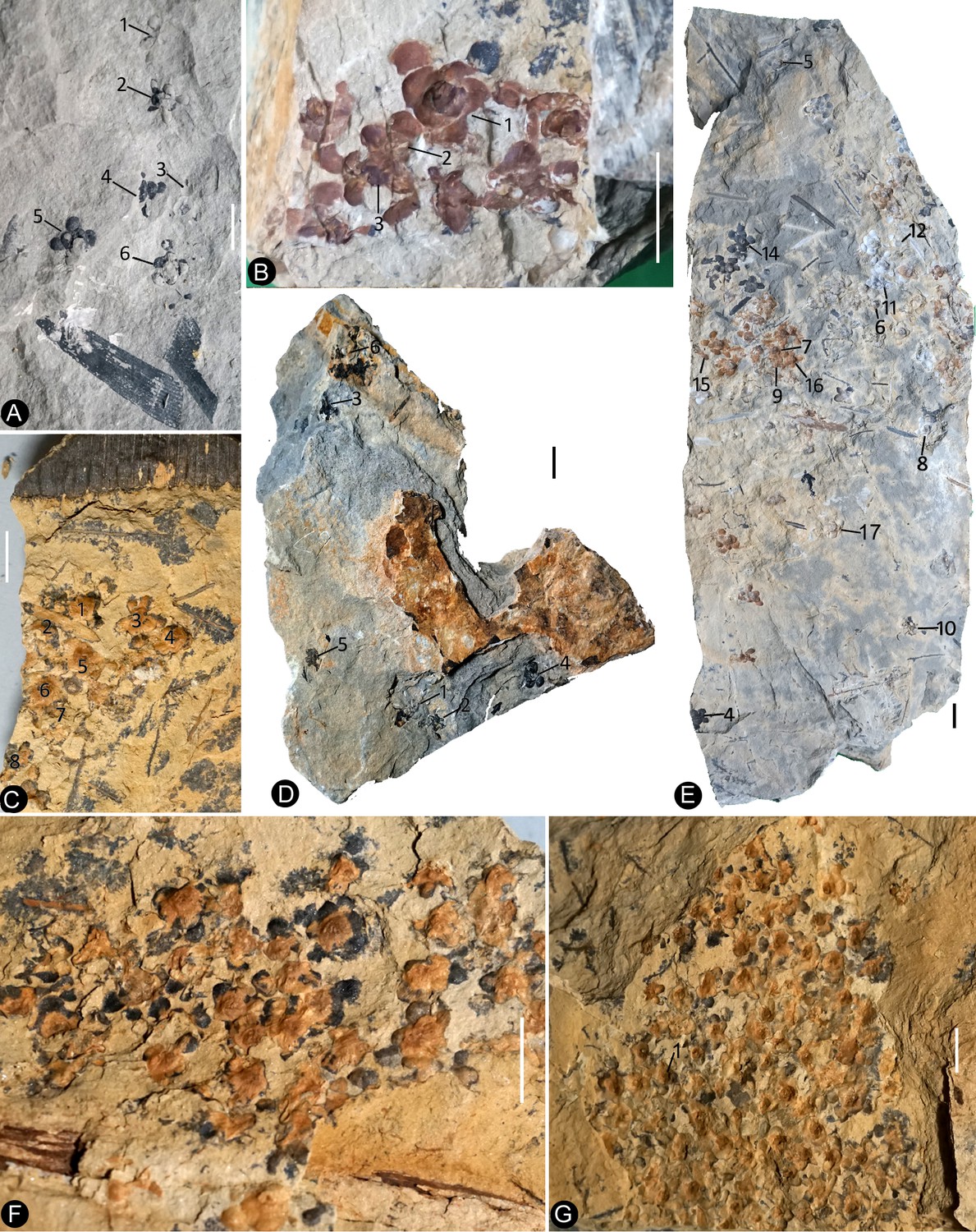
Siltstone slabs bearing Nanjinganthus.
All bars are 1 cm long. (A) Six flowers (1-6) on the same slab, and an associated triangular leaflet with parallel venation. PB22227. (B) Several flowers on the same slab. 1–3 are shown in detail in Figures 2f and 6d,e. PB22226. (C) Several flowers (1-8) on the same slab and the associated Nilssonia parabrevis (top). PB22220. (D) Several flowers (1-6) on the same slab. 1–3 are shown in detail in Figures 2h and 3a–c. PB22224. (E) Many flowers on the same slab. Some of the numbered ones are shown in detail in later figures. PB22222a. (F) A slab with numerous flowers. PB22221. (G) A slab almost fully covered with flowers. PB22228.
Figure 1—figure supplement 1
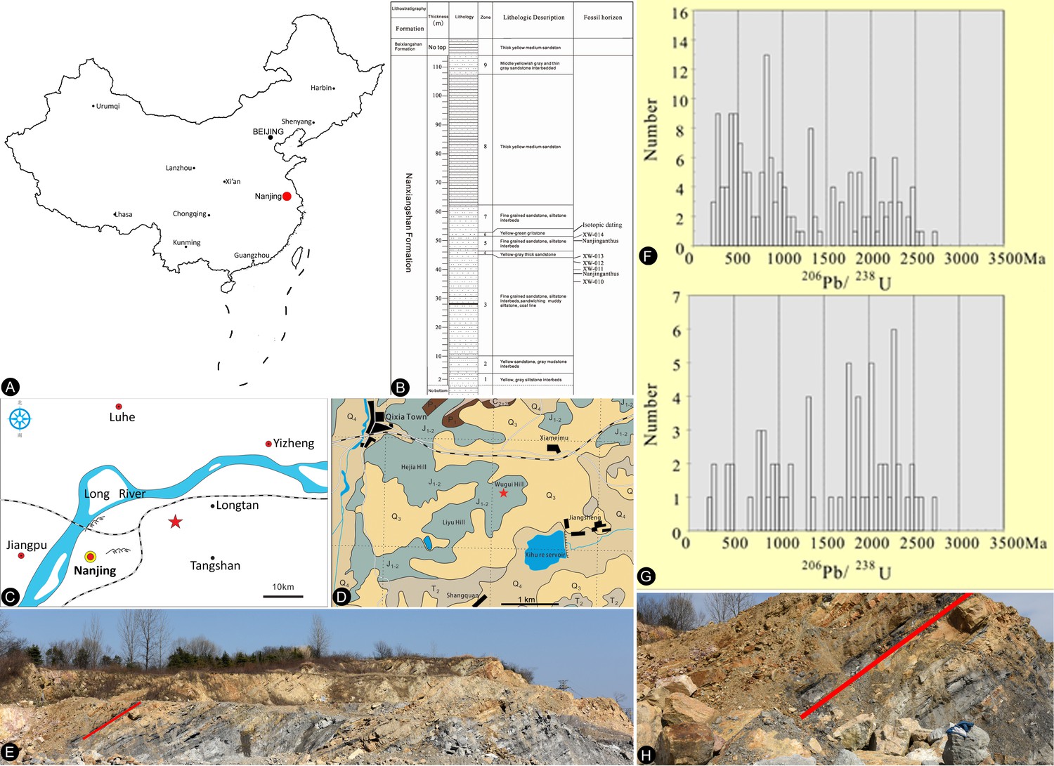
The type fossil locality of Nanjinganthus, Nanjing in China and isotopic dating.
(A) Fossil locality (red dot) in Nanjing, eastern China. (B) Stratigraphic column showing the fossiliferous strata, strata for isotopic dating, and layers (XW010 to XW014) sampled for palynological dating. (C) Type locality (red asterisk, N32˚08′ 19″, E118˚58′ 20″) of Nanjinganthus in the suburbs of Nanjing, China. (D) Geological map of the region near the type locality, dark green represents the outcrop of the Xiangshan Group. (E) Fossiliferous strata (red line) of the South Xiangshan Formation exposed in the quarry of Xiaoyetian Cement Company Ltd. Note one of the strata (red line) yielding many flowers of Nanjinganthus. (F–G) Comparison of relative probability plot of zircon data (F: all data; G: concordance >90%). (H) Close-up of one of the Nanjinganthus-yielding layers (red line).
Figure 2 with 1 supplement
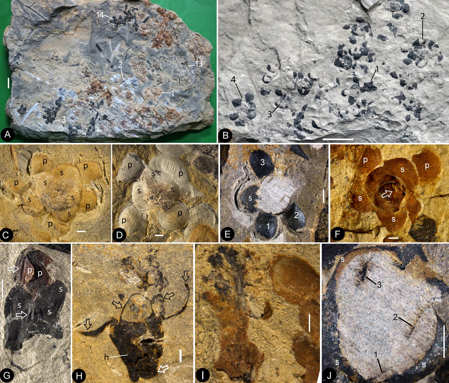
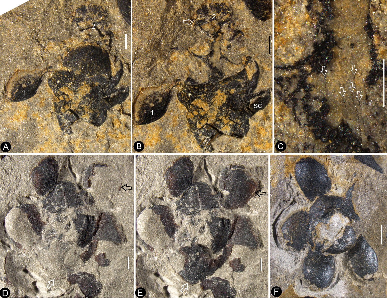
Flowers of Nanjinganthus preserved in different states and their details.
Bar = 1 mm except otherwise annotated. (A) Numerous flowers preserved on a single slab. Some of the numbered ones are detailed in later figures. PB22222B. Bar = 1 cm. (B) Numerous coalified flowers on the same slab. Some of the numbered ones are detailed in Figure 3d–e. PB22223. Bar = 1 cm. (C) Bottom view of Flower 1 in Figure 2a, showing five sepals (s) and five petals (p) with longitudinal ribs. PB22222B. (D) Bottom view of Flower 2 in Figure 2a, showing four sepals (s) and four petals (p) with longitudinal ribs. PB22222B. (E) Bottom view of the flower in Figure 3f, showing a sepal (s) and three petals (p) radiating from the center, which is obliquely broken to show the relationship among the sepals and petals as in Figure 2j. PB22278. (F) Top view of Flower 1 in Figure 1b with sepals (s), petals (p), and seeds (arrow, enlarged in Figure 6h) inside the receptacle. PB22226. (G) Side view of a flower bud (Flower 1 in Figure 2b) with longitudinal ribs (arrows) on the sepals (s) and petals (p). PB22223. (H) Side view of Flower 1 in Figure 1d, showing a receptacle (h), perianth (black arrows), and a dendroid style (white arrow). PB22224. (I) Side view of Flower 15 in Figure 1e, without sepals or petals. PB22222a. Bar = 1 mm. (J) Detailed view of the flower shown in Figure 2e, showing the arrangement of three petal bases (1-3) inside the sepals (s). These petals bases correspond to the three petals (1-3) in Figure 2e. PB22278.
Figure 2—figure supplement 1
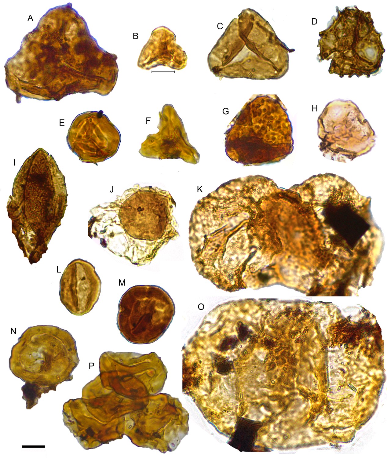
Frequently observed palynomorphs associated with Nanjinganthus.
Bar = 10 μm. (A) Cyathidites australis Couper. (B) C. minor Couper. (C) Deltoidospora sp. (D) Retitriletes clavatoides Döring. (E) Striatella seebergensis Madler. (F) Gleicheniidites senonicus Ross. (G) Manumia delcourtii (Pocock) Dybkjær. (H) Anulispora sp. (I) Cycadopites follicularis Wilson and Webster. (J) Perinopollenites elatoides Couper. (K) Platysaccus sp. (L) Monosulcites sp. (M, N) Classopollis spp. (O) Alisporites robustus Nilsson. (P) Tetrad Classopollis sp.
Figure 3 with 1 supplement
Individuals of Nanjinganthus.
Bar = 1 mm except otherwise annotated. (A–C, PB22224) (A) Flower 2 in Figure 1d (before the dégagement), showing the petal (1) and style (2) still embedded in the sediments. (B The same flower as in Figure 3a, after dégagement, showing the exposed dendroid style (white arrow) and petal (1), and the scale (sc) on the side of receptacle. (C) Detailed view of the style shown in Figure 3b with faint striations (arrows). Bar = 0.5 mm. (D–E) Flower 2 in Figure 2b after and before the organic material of the sepals (white arrows) and petals (black arrows) were removed for cuticle analysis. PB22223. (F) Bottom view of a flower before processing. Internal details are shown in Figure 2e,j. PB22278.
Figure 3—figure supplement 1
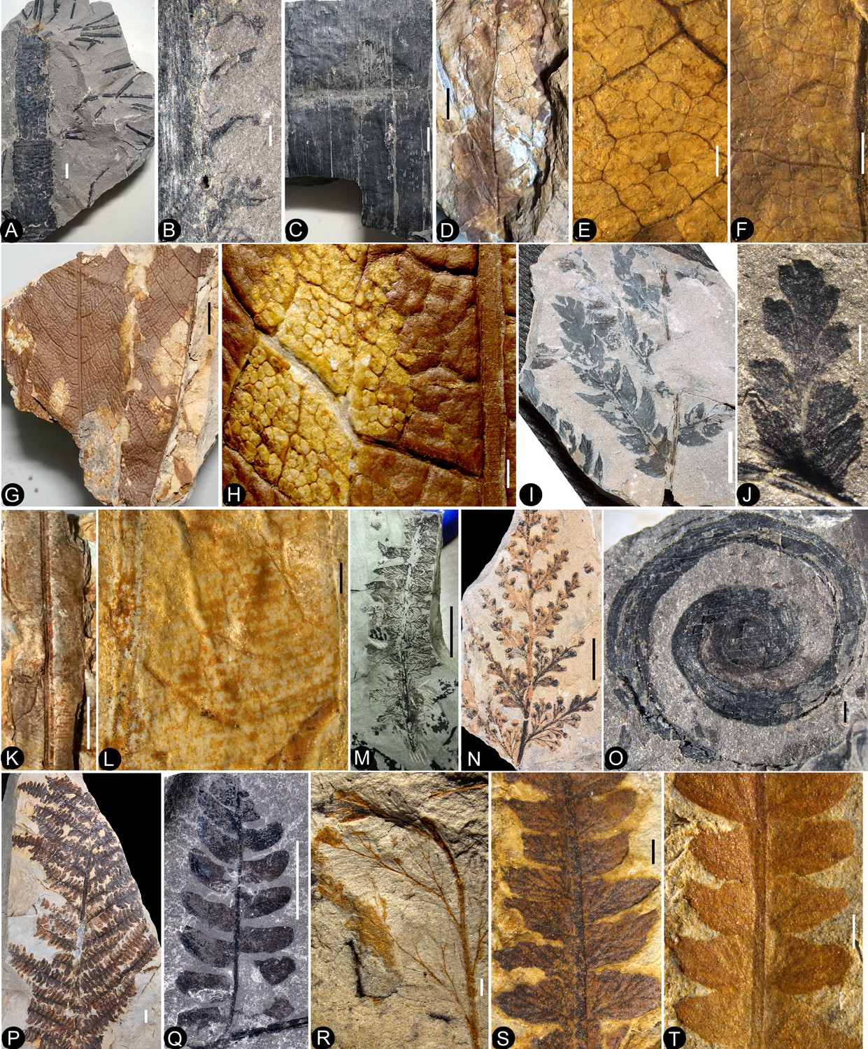
Fossil plants associated with Nanjinganthus.
Bar = 1 mm except otherwise annotated. (A) Neocalamites horridus. PB22234. Bar = 1 cm. (B). Detailed view of the prickles on the stem surface of Neocalamites horridus. (C). Neocalamites. PB22235. Bar = 1 cm. (D). Clathropteris meniscioides. PB22245. Bar = 1 cm. (E). Detailed view of the 4-order reticulate venation of Clathropteris meniscioides, enlarged from Fig. D. PB22245. (F). Reticulate venation of Clathropteris platyphylla. PB22232. (G). Two leaves of Clathropteris meniscioides. PB22233. Bar = 1 cm. (H). Detailed view of the 4-order reticulate venation, enlarged from Fig. G. (I). Cladophlebis. PB22214. Bar = 1 cm. (J). Coniopteris. PB22244. (K). Nilssoniopteris vittata with a robust midrib. PB22238. Bar = 1 cm. (L). Detailed view of the smooth margin and lateral veins branching off from the midrib, enlarged from Fig. K. PB22238. (M). Cladophlebis. PB22251. Bar = 1 cm. (N). ?Hymenophyllaceae. PB22213. Bar = 5 mm. (O). Spiropteris. PB22239. (P). Cladophlebis. PB22219. Bar = 1 cm. (Q). Cladophlebis. PB22217. Bar = 1 cm. (R). Raphaelia. PB22233. (S-T). Otozamites. PB22248B.
Figure 4 with 1 supplement
Nanjinganthus flowers preserved in various orientations and states.
Bar = 1 mm except otherwise annotated. (A) An oblique longitudinally split flower (Flower 11 in Figure 1e) with scales (sc), sepals (s), and petals (p). PB22222a. (B) A longitudinally split flower (Flower 12 in Figure 1e) with sepals (s) and petals (p). PB22222a. (C) Integral surface of an ovarian roof with a scar (arrow) left by a broken off style, from the flower shown in Figure 5h. PB22279. Bar = 0.5 mm. (D) Bottom view of a flower (Flower 14 in Figure 1e) with three sepals (s) and five petals (p) visible. PB22222a. (E) One of the sepals in Figure 4d, showing longitudinal ribs forking (arrow). PB22222a. (F) One of the petals in Figure 4d, showing longitudinal ribs. PB22222a. (G) Side view of a flower, showing scales (sc) on the ovary side and connate bracts (b) at the bottom. PB22229. (H) Detailed view of the connate bracts (b) and scales (sc) in Figure 4g. Note the outline (white line) of the fused bracts. PB22229. (I) The locule surrounded by the ovary wall (arrows) of the flower shown in Figure 4d. PB22222a.
Figure 4—figure supplement 1
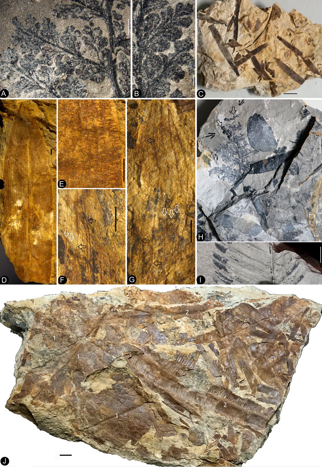
Fossil plants associated with Nanjinganthus.
Bar = 1 mm except otherwise annotated. (A) Pinnae of Coniopteris szeiana, enlarged from Fig. H. PB22243. Bar = 2 mm. (B) Detailed view of the pinnae of Coniopteris szeiana, enlarged from Fig. A. (C) Leaves of Desmiophyllum on a slab. PB22231. Bar = 1 cm. (D) A leaf of Ferganiella with midrib and parallel veins. PB22245. (E) Detailed view of the parallel veins of the leaf in Fig. D. (F) One of the leaves with at least two orders of longitudinal veins (white arrows) and occasional branching (black arrows), enlarged from Fig. G. (G) A leaf closely associated with Nanjinganthus with parallel veins (white arrows) and occasional branching (black arrows). PB22241. (H) Frond of Coniopteris szeiana associated with two Nanjinganthus (arrows). PB22243. Bar = 1 cm. (I) Ptilophyllum showing pinnae with parallel venation. Bar = 1 cm. (J) A slab loaded with fossils of Clathropteris meniscioides (1), Nilssonia parabrevis (2), and Ptilophyllum contiguum (3). PB22237. Bar = 1 cm.
Figure 5 with 1 supplement
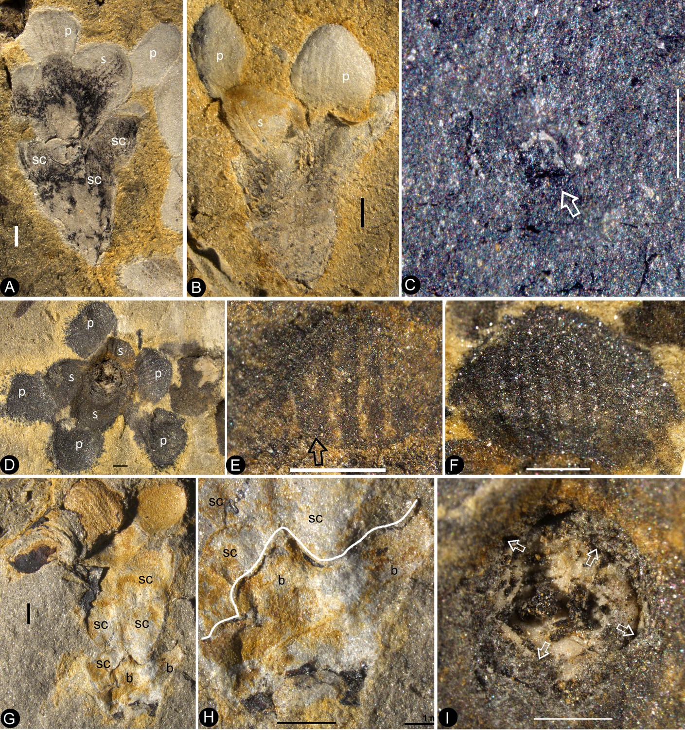
In situ seeds and flowers.
Bar = 1 mm except otherwise annotated. (A) A seed (outlined) inside the ovary of Flower 16 in Figure 1e. Note the oboval micropyle (black arrow) and funiculus (white arrow). PB22222a. Bar = 0.2 mm. (B) Detailed view of the funiculus (between the arrows) of the seed in Figure 5a. PB22222a. Bar = 0.1 mm. (C) A seed (detailed in Figure 6i) inside the ovary of Flower 7 in Figure 1e. PB22222a. (D) Detailed view of the oval micropyle (arrows) of the seed in Figure 5a. PB22222a. Bar = 0.1 mm. (E) A seed (arrow, detailed in Figure 6d–e) inside the receptacle in Flower two in Figure 1b. PB22226. (F, G) Two facing parts of the same flower (Flower 10 in Figure 1e). PB22222a. (H) Top view of a flower with organically-preserved sepals (s), petals (p) and integral ovarian roof (fr), which is detailed in Figure 4c. PB22279. (I) Side view of a longitudinally split flower with scales (sc) on ovary side, sepals (s), petals (p) and partially preserved style (arrow). PB22489. (J) Detailed view of basal portion of the style (between arrows) arrowed in Figure 5i, with faint longitudinal striations. PB22489. Bar = 0.2 mm. (K) Detailed view of the narrowing base (between arrows) of the right petal in Figure 5i. PB22489. Bar = 0.5 mm.( L) Detailed view of a sepal in Figure 5i. PB22489. Bar = 0.2 mm.
Figure 5—figure supplement 1
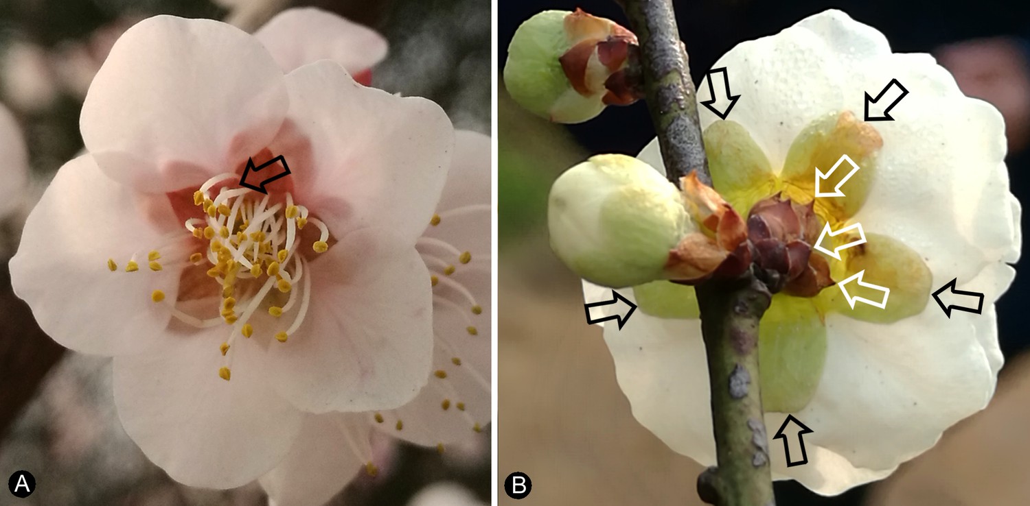
Flowers of a living angiosperm and its details.
(A) Top view showing the white petals with narrowing bases (arrow) and green sepals in the background. (B) Scales (white arrows), sepals (black arrows), and white petals.
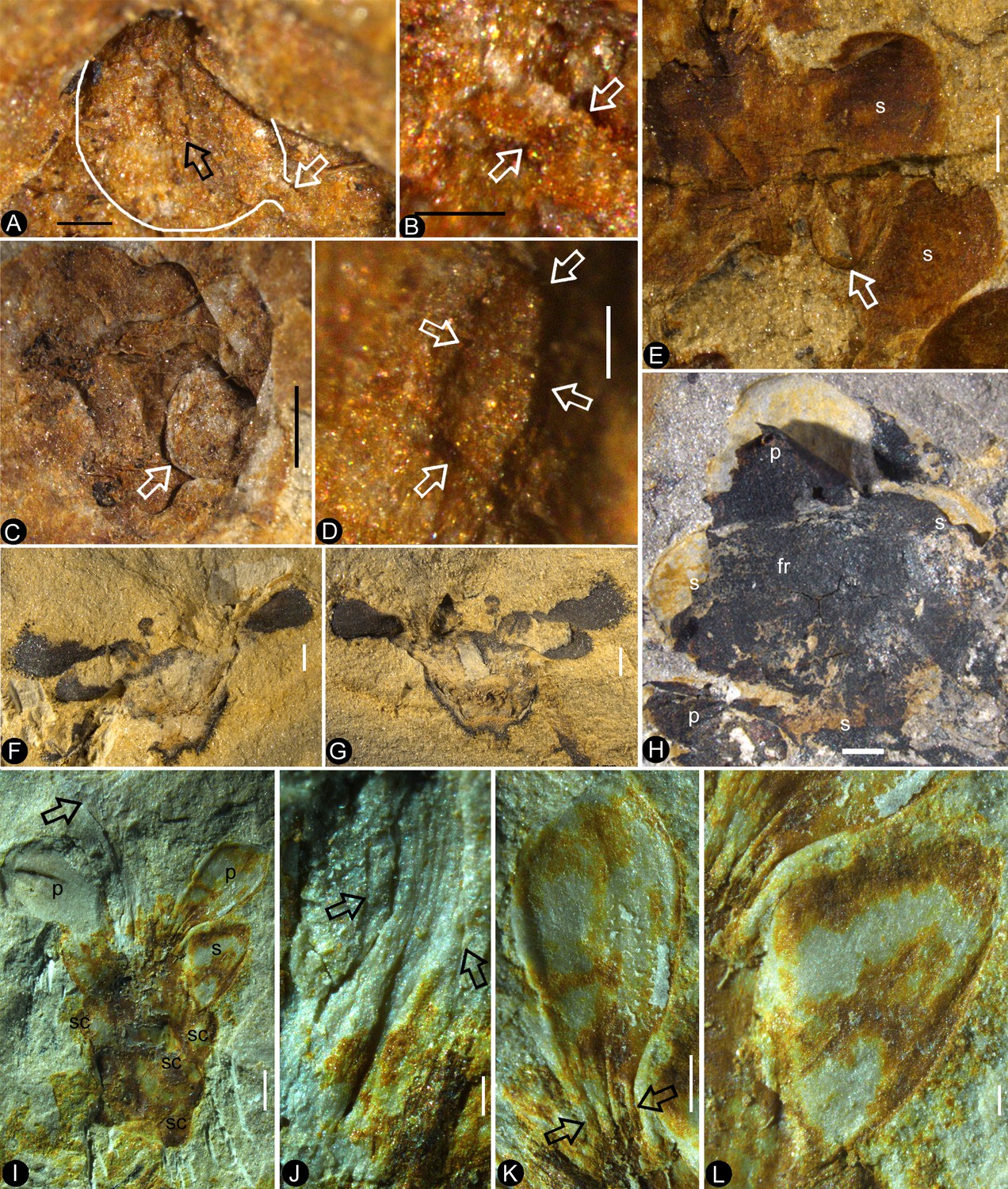
Figure 6
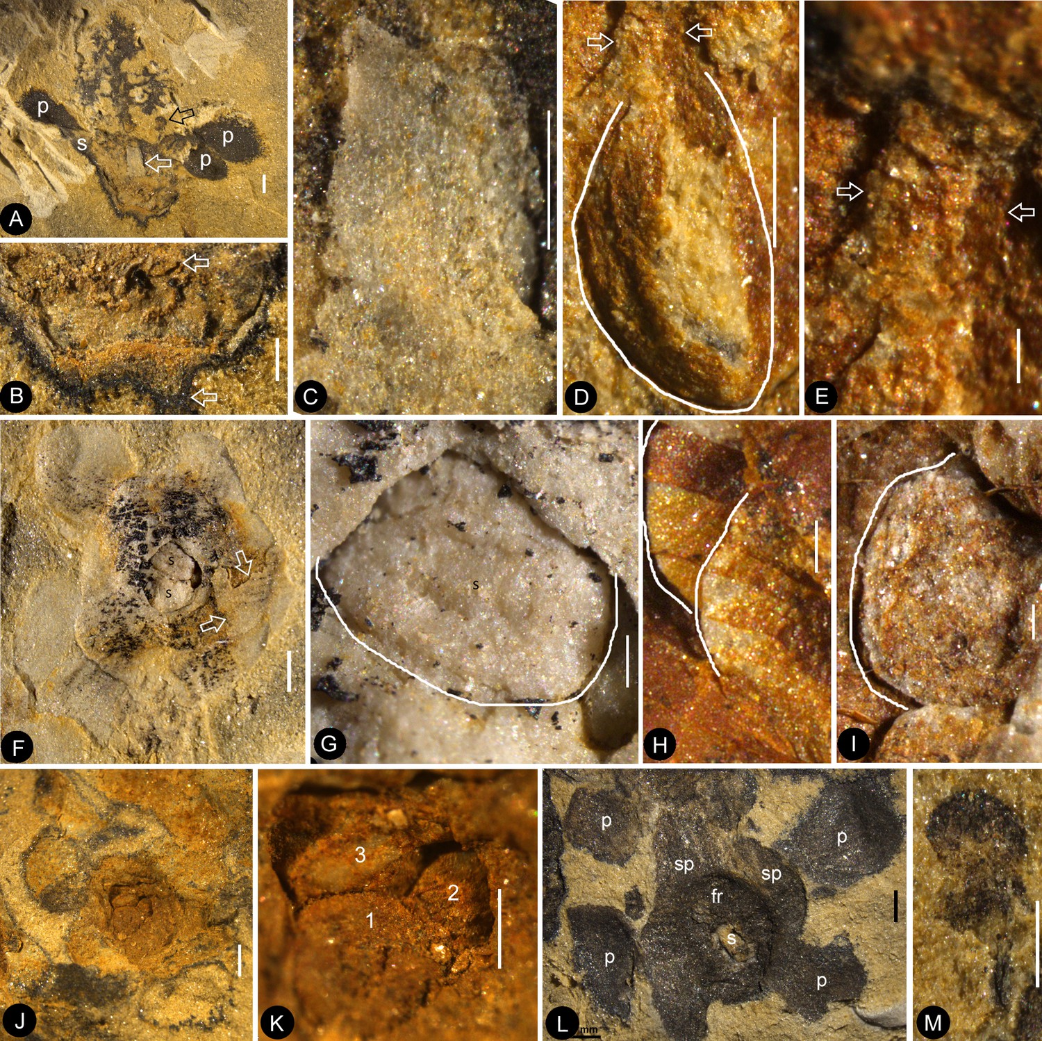
Dendroid style, in situ seeds, and details of flowers.
PB22222a, Bar = 1 mm except otherwise annotated. (A) A longitudinally split flower (counterpart of Flower 10 in Figure 1e, the same as in Figure 5f–g) showing the sepal (s) and petals (p), style base (white arrow), and an unknown organ (black arrow). (B) Detailed view showing the pedicel (lower arrow) terminating at the bottom of the ovary in Figure 6a. Note the level of ovarian roof (upper arrow). Bar = 0.5 mm. (C) Detailed view of the basal portion of the style marked by white arrow in Figure 6a. Bar = 0.5 mm. (D) A seed (white line) hanging by its funiculus (between arrows) on the ovarian wall of the Flower 2 in Figure 1b. PB22226. Bar = 0.5 mm. (E) Detailed view of the funiculus (between arrows) of the seed in Figure 6d. PB22226. Bar = 0.1 mm.( F) Top view of Flower 8 in Figure 1e with sepals and petals surrounding the ovary containing two seeds (s). Note the residue (arrows) of the ovarian roof. (G) Detailed view of one of the oval seeds (s) inside the ovary in Figure 6f. Bar = 0.2 mm. (H) Two seeds (white line), one overlapping the other, inside the ovary shown in Figure 2f. PB22226. Bar = 0.2 mm. (I) An oval seed (white line) inside the ovary of Flower 7 in Figure 1e. Bar = 0.2 mm. (J) Detailed view of Flower 1 in Figure 1g, showing seeds within ovary. PB22228. (K) Detailed view of three seeds (1-3) inside the ovary of the flower shown in Figure 6j. PB22228. Bar = 0.5 mm. (L) Top view of a flower showing petals (p), sepal (sp), seed (s) visible under the ovarian roof (fr). PB22222d. (M) Detailed view of the unknown organ (staminode?) marked by the black arrow in Figure 6a. Bar = 0.5 mm.
Figure 7
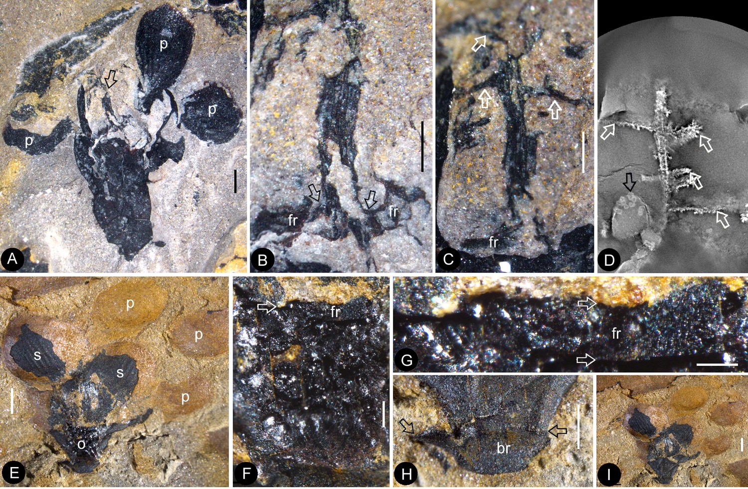
The flowers and their internal details.
(A-C) (E-I) stereomicroscopy; (D), micro-CL. Bar = 1 mm except otherwise annotated. (A) A flower carefully dégaged to expose the details of the gynoecium. Note the petals (p) and a style (arrow) in the center. PB22282. (B) Detailed view of the style in Figure 7a, showing its connection (arrows) to the ovarian roof (fr). PB22282. Bar = 0.5 mm. (C) Distal portion of the same style as in Figure 7b, showing its connection with the ovarian roof (fr) and dendroid form with lateral branches (arrows). PB22282. Bar = 0.5 mm. (D) Micro-CL slice 1169 showing a perianth element (black arrow) and branches (white arrows) of the style, embedded in sediments and thus invisible to naked eyes, of Flower 4 in Figure 1e. PB22222a. (E–I) PB22281. (E) Side view of an organically-preserved flower with sepals (s) and petals (p). Note the dark organic material in the ovary (o) and some sepals. The foreground portion of the receptacle has been removed (compare with Figure 7i), to show the details in Figure 7f–h. (F) Detailed view of the receptacle/ovary in Figure 7e. Note the ovarian roof (fr) preventing the outside (above) sediment (yellow color) from entering the ovarian locule. Bar = 0.2 mm. (G) Detailed view of the solid organically-preserved ovarian roof (fr) with integral outer (upper arrow) and inner (lower arrow) surfaces. Bar = 0.1 mm. (H) Bottom portion of the flower in Figure 7i, showing subtending bracts (br, arrows). Bar = 0.5 mm. (I) The flower in Figure 7e, before removing the foreground portion of the ovary.
Figure 8

Details of the sepal and petal.
(A-B) stereomicroscopy; (C) (E-G) fluorescence light microscopy; (D) TEM; (H) SEM. Bar = 1 mm except otherwise annotated. (A) A petal with a narrowing base. PB22280. (B) A partial petal from the Flower in Figure 3d–e, with the longitudinal rib (to the left) forking at the base (arrow) and the rib-free laminar area to the right. PB22223. (C) Elongated epidermal cells of the petal in Figure 8b . PB22223. Bar = 0.1 mm. (D) Transmission electron microscope view showing the cuticle (left, light color) of a petal. PB22223. Bar = 2 μm. (E) Elongated epidermal cells not in strict longitudinal files in the laminar portion of the petal in Figure 8b . Note the two newly formed epidermal cells (arrow). PB22223. Bar = 0.1 mm. (F) Ribs with elongated epidermal cells (left and right) alternating the between region with less elongated cells (middle) of the petal in Figure 8b . PB22223. Bar = 0.2 mm. (G Elongated (above) and isodiametric (below) epidermal cells on the sepal of Flower in Figure 3d–e. PB22223. Bar = 0.1 mm. (H) A stoma on the bract of the flower (marked by white arrow in Figure 2h). PB22224. Bar = 5 μm.
Figure 9
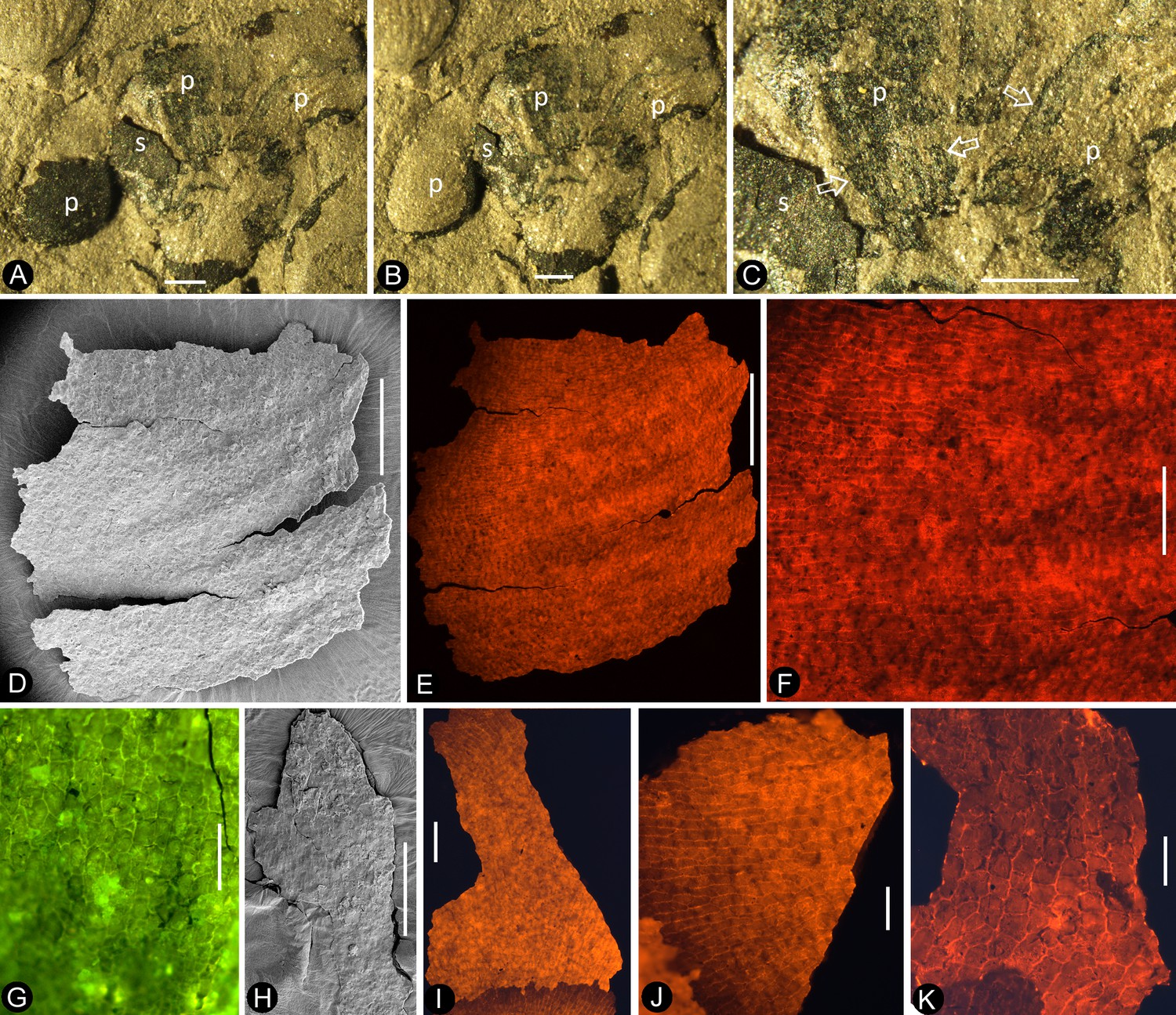
Petal and details of Nanjinganthus.
(A-C) light stereomicroscopy; (D) (H) SEM; (E-G) (I-K) fluorescence light microscopy. PB22223. Bar = 1 mm except otherwise annotated. (A) Side view of Flower 3 in Figure 2b, showing the arrangement of the petals (p) and sepal (s). (B) The same flower as in Figure 9a. Note that some organic material of the petal has been removed for detailed observation. (C). Margins (arrows) of the petal (p) with cuneate base and their relationship to the sepal (s). (D) The petal removed from Figure 9a. SEM. Bar = 0.5 mm. (E) Cellular details of the petal in Figure 9d Bar = 0.5 mm. (F) Elongated epidermal cells arranged in files, enlarged from Figure 9e. Bar = 0.2 mm. (G) Isodiametric epidermal cells in the laminar area portion of the petal in Figure 9e. Bar = 0.1 mm. (H) A fragment of the sepal seen in Figure 9a. Bar = 0.5 mm. (I) Cellular details of the sepal in Figure 9h. Bar = 0.2 mm. (J) Elongated epidermal cells arranged in files on the sepal in Figure 9i. Bar = 0.1 mm. (K) Isodiametric epidermal cells on the laminar area of the sepal in Figure 9a. Bar = 0.1 mm.
Figure 10
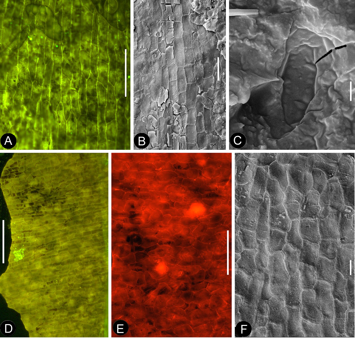
Cuticular details of Nanjinganthus.
A, D-E, Fluorescence light microscopy; B-C, F, SEM. PB22223. (A) Elongated epidermal cells in longitudinal files in the middle portion of the petal in Figure 8b. Bar = 0.1 mm. (B) Elongated epidermal cells on the rib of the petal in Figure 8b. Bar = 50 μm. (C) A possible stoma on the petal shown in Figure 8b. Bar = 2 μm. (D) Elongated epidermal cells in files on the sepal of flower in Figure 3d–e. Bar = 0.2 mm. (E) Isodiametric epidermal cells on the sepal of flower in Figure 3d–e. Bar = 0.1 mm. (F) Isodiametric epidermal cells on the sepal of flower in Figure 3d–e. Bar = 20 μm.
Figure 11
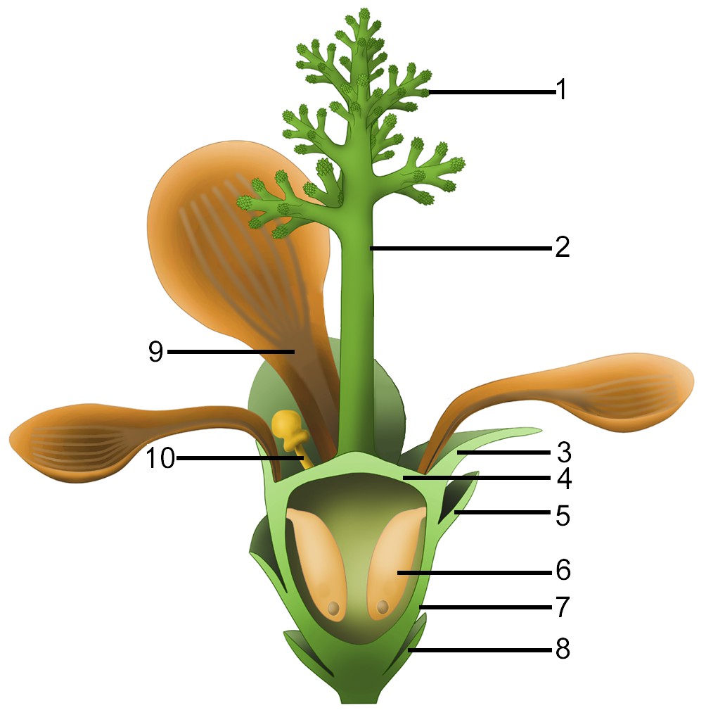
Idealized reconstruction of Nanjinganthus.
1, branches of dendroid style; 2, dendroid style; 3, sepal; 4, ovarian roof; 5, scale; 6, seed; 7, cup-form receptacle/ovary; 8, bract; 9, petal; 10, unknown organ (staminode?).
Tables
Table 1
Comparison between Nanjinganthus and Mesozoic gymnosperms.
https://doi.org/10.7554/eLife.38827.018| Nanjinganthus | Caytonia | Bennettitales | Corystospermales | Ginkgoales | Coniferales | Iraniales | Czekanowskiales | Pentoxylales | |
|---|---|---|---|---|---|---|---|---|---|
| Symmetry | Radial | Bilateral | Radial | Bilateral | Radial | Radial | Radial? | Bilateral | Radial? |
| With foliar appendages | Yes | No | No | No | No | ? | No | No | No |
| Enclosed seed | Yes | No | No | No | No | No? | ? | No | No |
| Opening in female part | No | Adaxial basal | Terminal? | Adaxial basal | N/A | N/A | ? | Distal slit | ? |
| Penetrating cone axis | No | Yes | Yes | Yes | ? | Yes | ? | Yes | Yes |
Additional files
-
Supplementary file 1
Number of flowers on each Nanjinganthus specimen.
- https://doi.org/10.7554/eLife.38827.020
-
Supplementary file 2
Summary of zircon LA–ICP–MS U–Pb data for fossiliferous layers samples of the South Xiangshan Formation.
Online Information 3D virtual image of the holotype of Nanjinganthus. Click on the link, using ctrl/shift and mouse, you can manipulate the image for your observation.
- https://doi.org/10.7554/eLife.38827.021
-
Transparent reporting form
- https://doi.org/10.7554/eLife.38827.022
Download links
A two-part list of links to download the article, or parts of the article, in various formats.
Downloads (link to download the article as PDF)
Open citations (links to open the citations from this article in various online reference manager services)
Cite this article (links to download the citations from this article in formats compatible with various reference manager tools)
An unexpected noncarpellate epigynous flower from the Jurassic of China
eLife 7:e38827.
https://doi.org/10.7554/eLife.38827


