Central Dicer-miR-103/107 controls developmental switch of POMC progenitors into NPY neurons and impacts glucose homeostasis
- Children’s Hospital Los Angeles, United States
- University of Southern California, California
- Inserm U1172, Lille 2 University of Health and Law, France
Figures
Figure 1 with 1 supplement
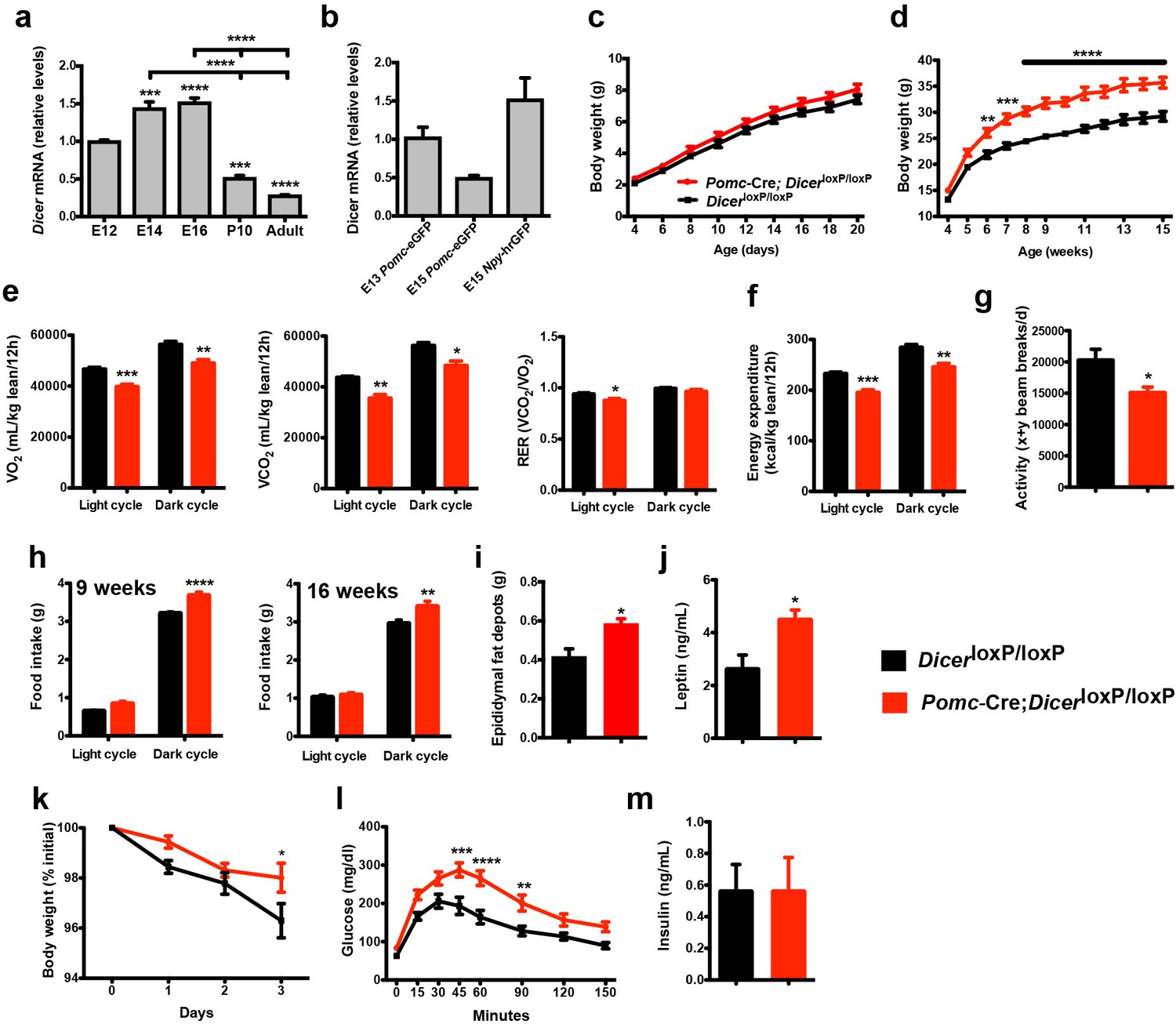
Loss of Dicer in Pomc expressing neurons causes metabolic dysregulation.
(a) Relative expression of Dicer mRNA in the hypothalamus of E12, E14, E16 embryos and in the mediobasal hypothalamus of P10, and adult mice (n = 3 – 5 per group). (b) Relative expression of Dicer mRNA in sorted Pomc-eGFP cells at E13 and E15 and Npy-hrGFP cells at E15 (n = 2 – 4 per group). (c) Pre- and (d) post-weaning growth curves (body weights) of DicerloxP/loxP and Pomc-Cre; DicerloxP/loxP male mice (n ≥ 8 per group). (e) VO2, VCO2 and respiratory exchange ratio (RER) of 15-week-old DicerloxP/loxP and Pomc-Cre; DicerloxP/loxP male mice (n = 4 – 6 per group). (f) Energy expenditure of 15-week-old DicerloxP/loxP and Pomc-Cre; DicerloxP/loxP male mice (n = 4 – 6 per group). (g) Locomotor activity of 15-week-old DicerloxP/loxP and Pomc-Cre; DicerloxP/loxP male mice (n = 4 – 6 per group). (h) Cumulative food intake of 9- and 16-week-old DicerloxP/loxP and Pomc-Cre; DicerloxP/loxP male mice (n = 5 – 6 per group). (i) Epididymal fat mass of 20- to 23-week-old male mice (n = 4 – 5 per group). (j) Plasma leptin levels in 20- to 23-week-old DicerloxP/loxP and Pomc-Cre; DicerloxP/loxP male mice (n = 4 – 5 per group). (k) Leptin-induced weight loss in 14-week-old DicerloxP/loxP and Pomc-Cre; DicerloxP/loxP male (n = 5 per group). (l) Glucose tolerance test of 10- to 11-week-old male mice (n ≥ 7 per group). (m) Plasma insulin levels in 20- to 23-week-old DicerloxP/loxP and Pomc-Cre; DicerloxP/loxP male mice (n = 6 per group). Values are shown as mean ±SEM. *p≤0.05 versus DicerloxP/loxP (e, g, i, j); **p≤0.01 versus DicerloxP/loxP (d, e, f, h, l); ***p≤0.001 versus E12 WT (a), versus DicerloxP/loxP (d, e, f, l); ****p≤0.0001 versus E12 WT (a), versus E14 WT (a), versus E16 WT (a), versus DicerloxP/loxP (d, l). Statistical significance was determined using 2-tailed Student’s t test (e, f, g, i, j, m), 1-way ANOVA followed by Turkey’s post hoc test (a, b) and 2-way ANOVA followed by Bonferroni’s post hoc test (c, d, h, k, l).
Figure 1—figure supplement 1
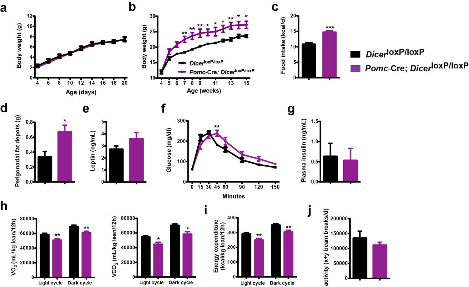
Altered metabolism in female mice lacking Dicer in Pomc-expressing neurons.
(a) Pre- and (b) post-weaning growth curves (body weights) of DicerloxP/loxP and Pomc-Cre; DicerloxP/loxP female mice (n = 7–8 per group). (c) Cumulative food intake of 15-week-old DicerloxP/loxP and Pomc-Cre; DicerloxP/loxP female mice (n = 4 per group). (d) Perigonadal fat mass of 15-week-old DicerloxP/loxP and Pomc-Cre; DicerloxP/loxP female mice (n ≥ 6 per group). (e) Plasma leptin levels in 15-week-old DicerloxP/loxP and Pomc-Cre; DicerloxP/loxP female mice (n = 6 per group). (f) Glucose tolerance test of 10- to 11-week-old DicerloxP/loxP and Pomc-Cre; DicerloxP/loxP female mice (n = 7 per group). (g) Plasma insulin levels in 15-week-old DicerloxP/loxP and Pomc-Cre; DicerloxP/loxP female mice (n = 5 per group). (h) VO2 and VCO2 of 15-week-old DicerloxP/loxP and Pomc-Cre; DicerloxP/loxP female mice (n = 4 per group). (i) Energy expenditure of 15-week-old DicerloxP/loxP and Pomc-Cre; DicerloxP/loxP female mice (n = 4 per group). (j) Locomotor activity of 15-week-old DicerloxP/loxP and Pomc-Cre; DicerloxP/loxP female mice (n = 4 per group). Data are presented as mean ± SEM. *p≤0.05 versus DicerloxP/loxP (b, d, h); **p≤0.01 versus DicerloxP/loxP (b, f, h, i); ****p≤0.001 versus DicerloxP/loxP (c). Statistical significance was determined using 2-tailed Student’s t test (c–e, g–j), and 2-way ANOVA followed by Bonferroni’s post-hoc test (a, b, f).
Figure 2 with 1 supplement
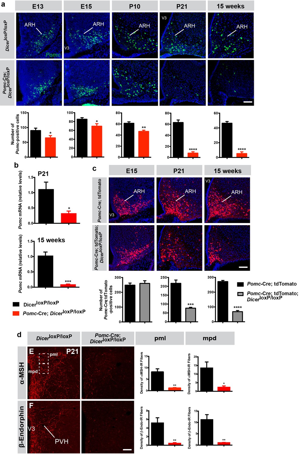
Reduced number of Pomc mRNA-expressing cells in mice lacking Dicer in POMC neurons.
(a) Representative images and quantification of Pomc mRNA-expressing cells in the arcuate nucleus (ARH) of E13 (n = 6 per group), E15 (n = 6 – 8 per group), P10 (n = 4 per group), P21 (n = 4 per group) and 15-week-old (n = 3 – 4 per group) DicerloxP/loxP and Pomc-Cre; DicerloxP/loxP male mice. (b) Relative expression of Pomc mRNA in the mediobasal hypothalamus of P21 and 15-week-old DicerloxP/loxP and Pomc-Cre; DicerloxP/loxP male mice (n = 3 – 4 per group). (c) Representative images and quantification of Pomc-Cre cells genetically labeled by tdTomato in the ARH of E15 (n = 5 – 7 per group), P21 (n = 4 per group) and 15-week-old (n = 3 – 4 per group) Pomc-Cre and Pomc-Cre; DicerloxP/loxP male mice. (d) Representative images and quantification of the density of α-MSH- and β-endorphin-immunopositive fibers (n = 3 – 4 per group) in the posterior magnocellular (pml) and medial parvicellular (mpd) parts of the paraventricular nucleus (PVH) of P21 DicerloxP/loxP and Pomc-Cre; DicerloxP/loxP male mice. Scale bars, 100 μm (a, b, c). Values are shown as mean ± SEM. *p≤0.05 versus DicerloxP/loxP (a, b, d); **p≤0.01 versus DicerloxP/loxP (a, d); ***p≤0.001 versus DicerloxP/loxP (b), versus Pomc-Cre; tdTomato (c); ****p≤0.0001 versus DicerloxP/loxP (a), versus Pomc-Cre; tdTomato (c). Statistical significance was determined using 2-tailed Student’s t test. V3, third ventricle.
Figure 2—figure supplement 1
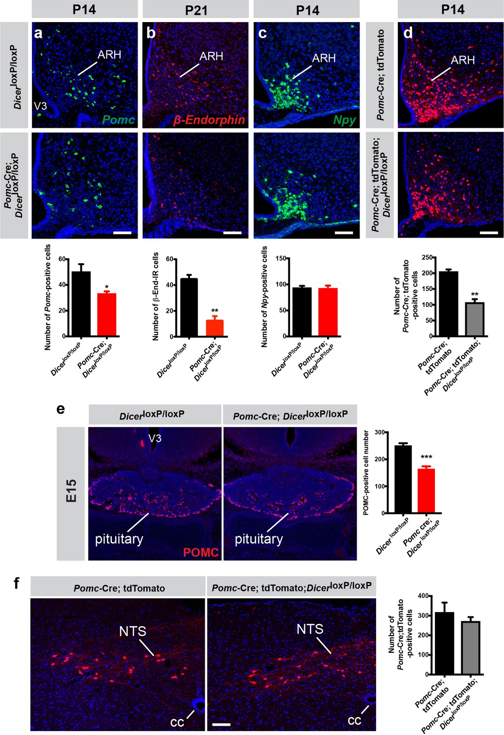
Reduced number of POMC neurons in the arcuate nucleus of Pomc-Cre; DicerloxP/loxP mice.
Representative images and quantification of (a) Pomc mRNA-, (b) β-endorphin-, and (c) Npy mRNA-expressing cells of P14 and P21 DicerloxP/loxP and Pomc-Cre; DicerloxP/loxP male mice (n = 4 per group). (d) Representative images and quantification of Pomc-Cre; tdTomato + cells in the ARH of P14 Pomc-Cre and Pomc-Cre; DicerloxP/loxP male mice (n = 3 per group). (e) Representative images and quantification of POMC-immunopositive cells in the pituitary of DicerloxP/loxP and Pomc-Cre; DicerloxP/loxP E15 embryos (n = 5 – 6). (f) Representative images and quantification of Pomc-Cre tdTomato + cells in the NTS of P21 Pomc-Cre and Pomc-Cre; DicerloxP/loxP male mice (n = 4 per group). ARH, arcuate nucleus of the hypothalamus; cc, central canal; NTS, solitary nucleus; V3, third ventricle. Scale bars, 100 μm (a–e). Values are shown ± SEM. *p≤0.05 versus DicerloxP/loxP (a, e); **p≤0.01 versus DicerloxP/loxP (b), versus Pomc-Cre; tdTomato (d), ***p≤0.001 versus DicerloxP/loxP (e). Statistical significance was determined using 2-tailed Student’s t test (a–f).
Figure 3
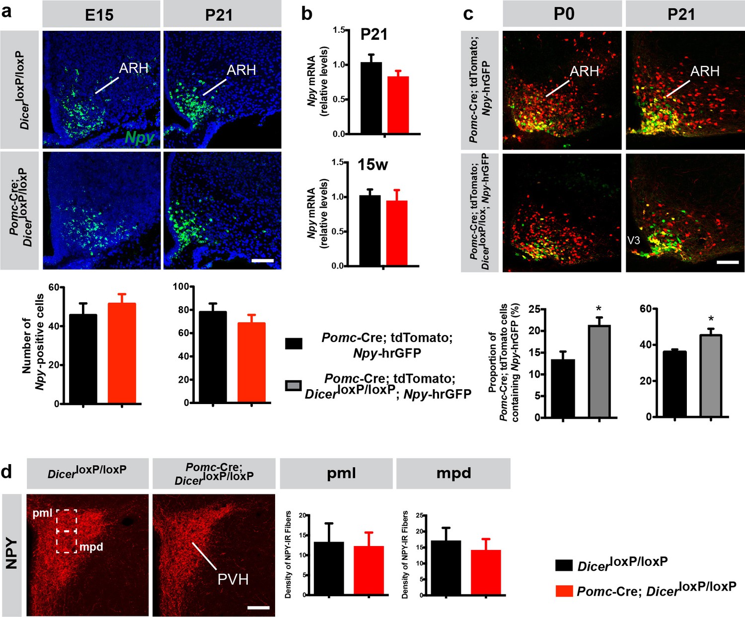
Loss of Dicer in POMC neurons favors the differentiation of Pomc-expressing progenitors into NPY neurons.
(a) Representative images and quantification of Npy mRNA-expressing cells in the arcuate nucleus (ARH) of E15 (n = 5–6 per group) and P21 (n = 4 per group) DicerloxP/loxP and Pomc-Cre; DicerloxP/loxP male mice. (b) Relative expression of Npy mRNA in the mediobasal hypothalamus of P21 and 15-week-old DicerloxP/loxP and Pomc-Cre; DicerloxP/loxP male mice (n = 3–4 per group). (c) Representative images and quantification of Pomc-Cre tdTomato + cells that co-express Npy-hrGFP in the ARH of P0 (n = 5–6 per group) and P21 (n = 3 per group) Pomc-Cre and Pomc-Cre; DicerloxP/loxP male mice. (d) Representative images and quantification of the density of NPY-immunopositive fibers (n = 3–4 per group) in the posterior magnocellular (pml) and medial parvicellular (mpd) parts of the paraventricular nucleus (PVH) of P21 DicerloxP/loxP and Pomc-Cre; DicerloxP/loxP male mice. Scale bars, 100 μm (a, c, d). Values are shown as mean ± SEM. *p≤0.05 versus Pomc-Cre; tdTomato; Npy-hrGFP (c). Statistical significance was determined using 2-tailed Student’s t test. V3, third ventricle.
Figure 4 with 1 supplement

miR-107 silencing modulates Pomc and Npy expression and impairs long-term glucose homeostasis.
Images and FACS isolation of (a) Pomc-eGFP+ and (b) Pomc-tdTomato+/Npy-hrGFP+ cells from E15 hypothalami (n = 43 – 53 per group). Arrow heads point to double-labeled cells. Relative expression of (c) Pomc, (d) Npy, and (g) miR-103 and miR-107 mRNA in sorted Pomc-eGFP, Npy-hrGFP, and Pomc-Cre; tdTomato; Npy-hrGFP cells at E15 (n = 2 – 4 per group). (e) Scatterplots and (f) histograms showing the miRNA fold change in E15 Pomc-tdTomato/Npy-GFP+ cells compared with E15 Pomc-eGFP+ cells (n = 1 per group, pool of 4 – 7 samples). Relative expression of hypothalamic (h) miR-107, (j) Pomc, and (l) Npy mRNA in E15 embryos injected with antagomirs against miR-107 (Ant-107) or vehicle at E12 (n = 5 – 18 per group). Correlation between hypothalamic (i) Pomc and (k) Npy mRNA expression and miR-107 expression in E15 embryos injected with Ant-107 at E12 (n = 10 – 12 per group). (m) Relative expression of miR-107 mRNA in hypothalamic embryonic explants incubated with Ant-107 or control Ant-Scr (n = 10 – 13 per group). (n) Images and quantification of Pomc-Cre tdTomato + cells that express Npy-hrGFP in hypothalamic embryonic explants incubated with Ant-107 or control Ant-Scr (n = 5 – 6 per group). (o) Quantification of Pomc-Cre tdTomato + cells that express Pomc-eGFP in hypothalamic embryonic explants incubated with Ant-107 or control Ant-Scr (n = 3 – 5 per group). (p) Experimental overview. (q) Pre- and post-weaning growth curves (body weights) of male mice injected with locked nucleic acids against miR-107 (LNA-107) or scrambled control LNA (LNA-Scr) at E12 (n = 4 – 10 per group). (r) Body composition of 15-week-old male mice injected with LNA-107 or control LNA-Scr at E12 (n = 7 – 10 per group). (s) Glucose tolerance test of 10-week-old male mice injected with LNA-107 or control LNA-Scr at E12 (n = 4 – 6 per group). Dark blue line represents animals with more than 28% reduction in the number of POMC neurons. (t) Correlation between the glucose area under the curve during GTT and number of POMC neurons in 10-week-old male mice injected with LNA-107 at E12 (n = 8 animals). Dark blue squares represent animals with more than 28% reduction in the number of POMC neurons. Scale bars, 100 μm (a, b) and 40 μm (n). Data are presented as mean ±SEM. *p≤0.05 versus vehicle (j, l), versus Ant-Scr (o); **p≤0.01 versus E15 Pomc-eGFP (c), versus Ant-Scr (n), versus LNA-Scr (s); ***p≤0.001 versus Ant-Scr (m); ****p≤0.0001 versus vehicle (h). Statistical significance was determined using 2-tailed Student’s t test (h, j, l–o), linear regression (i, k, t), 1-way ANOVA followed by Tukey’s post hoc test (c, d, g), and 2-way ANOVA followed by Bonferroni’s post hoc test (q, r, s). ARH, arcuate nucleus of the hypothalamus; V3, third ventricle.
Figure 4—figure supplement 1
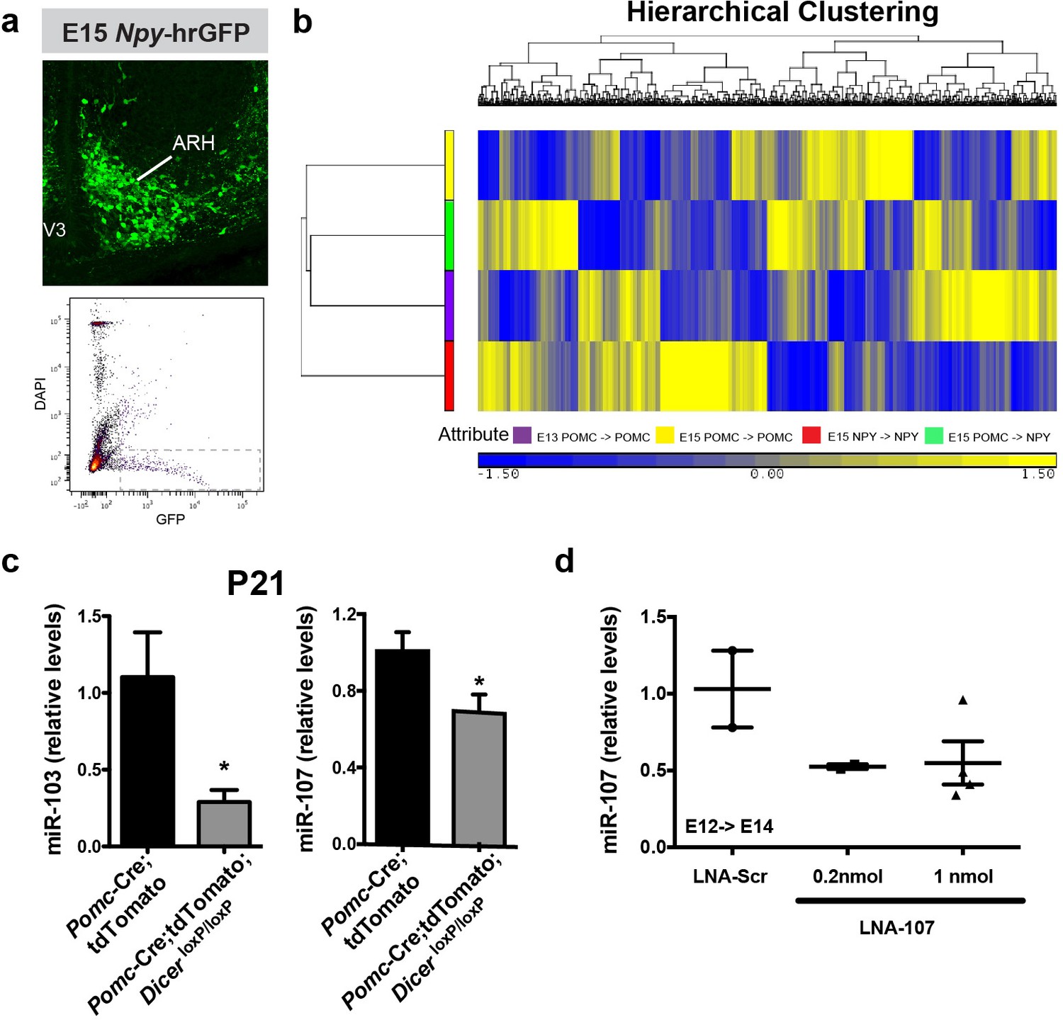
Identification and manipulation of miR-107 expression in POMC neurons.
(a) Representative images and FACS isolation of Npy-hrGFP+ cells from microdissected hypothalamic tissue at E15 (n = 6 per group). (b) Heatmap diagram illustrating the differential expression of miRNAs in sorted Pomc-eGFP cells at E13 and Pomc-eGFP, Npy-hrGFP and Pomc-Cre; tdTomato; Npy-hrGFP cells at E15. (c) Relative expression of miR-103 and miR-107 in sorted tdTomato + cells of P21 Pomc-Cre and Pomc-Cre; DicerloxP/loxP male mice (n = 4 per group). (d) Relative expression of miR-107 mRNA in the hypothalamus of E14 embryos injected with locked nucleic acid (LNA) against miR-107 (LNA-107) or scrambled control LNA against miR-690 at E12 (n = 2 – 4 per group). Scale bar, 100 μm (a). Data are presented as mean ± SEM. *p≤0.05 versus DicerloxP/loxP (c). ARH, arcuate nucleus of the hypothalamus; V3, third ventricle.
Additional files
-
Supplementary file 1
List of miRNAs up- and down-regulated in sorted Pomc-Cre; tdTomato; Npy-hrGFP cells versus E15 Pomc-eGFP cells derived from E15 embryos
- https://doi.org/10.7554/eLife.40429.009
-
Transparent reporting form
- https://doi.org/10.7554/eLife.40429.010
Download links
A two-part list of links to download the article, or parts of the article, in various formats.
Downloads (link to download the article as PDF)
Open citations (links to open the citations from this article in various online reference manager services)
Cite this article (links to download the citations from this article in formats compatible with various reference manager tools)
Central Dicer-miR-103/107 controls developmental switch of POMC progenitors into NPY neurons and impacts glucose homeostasis
eLife 7:e40429.
https://doi.org/10.7554/eLife.40429


