Primed Track, high-fidelity lineage tracing in mouse pre-implantation embryos using primed conversion of photoconvertible proteins
Abstract
Accurate lineage reconstruction of mammalian pre-implantation development is essential for inferring the earliest cell fate decisions. Lineage tracing using global fluorescence labeling techniques is complicated by increasing cell density and rapid embryo rotation, which hampers automatic alignment and accurate cell tracking of obtained four-dimensional imaging data sets. Here, we exploit the advantageous properties of primed convertible fluorescent proteins (pr-pcFPs) to simultaneously visualize the global green and the photoconverted red population in order to minimize tracking uncertainties over prolonged time windows. Confined primed conversion of H2B-pr-mEosFP-labeled nuclei combined with light-sheet imaging greatly facilitates segmentation, classification, and tracking of individual nuclei from the 4-cell stage up to the blastocyst. Using green and red labels as fiducial markers, we computationally correct for rotational and translational drift, reduce overall data size, and accomplish high-fidelity lineage tracing even for increased imaging time intervals – addressing major concerns in the field of volumetric embryo imaging.
https://doi.org/10.7554/eLife.44491.001eLife digest
A mouse embryo starts with one cell, which divides to create identical daughters that quickly start to multiply. Within three to four days, certain cells begin to specialize and take on specific roles. Scientists want to track these early events to understand how they give rise to an individual formed of huge numbers of cells organized in specialized tissues. To do so, researchers genetically manipulate embryos so that each cell produces fluorescent molecules that ‘glow’ under light. These embryos are grown inside a special microscope for several days. Images are taken regularly and then processed by specialized software that automatically tracks the fluorescent cells and their daughters over time. This helps reconstruct the history of each cell, and which structures they give rise to.
However, many embryos move and turn around between images, and so software packages often lose track of which cell was which. Taking images more frequently is not possible because each imaging event exposes the embryo to light, which can damage its fragile cells.
To address this problem, Welling, Mohr et al. made embryonic cells produce a special fluorescent marker, which is normally green but can be converted to red. Then, a technique known as primed conversion was used so that only one cell in a four-cell embryo would glow red. Welling, Mohr et al. designed a piece of software, baptized ‘primed Track’, that can use this red cell (and its daughters) to reorient the embryo during image analysis and reliably identify and match any mother cell to its daughters. The new approach means the experiments require fewer imaging events, but also fewer embryos because even the ones that move a lot can be studied. This should help scientists look into how early life processes give rise to specialized cells, and even explore the fate of cells in other tissues.
https://doi.org/10.7554/eLife.44491.002Introduction
Accurate lineage tracing and precise tracking of single cells in pre-implantation embryos are essential for a mechanistic understanding of the first cell fate decisions during mammalian development (Welling et al., 2016; Pantazis and Bollenbach, 2012). Selective plane illumination microscopy (SPIM) has the potential to play a major role in achieving comprehensive, non-invasive imaging of mammalian pre-implantation development. During these early steps of development, a major fraction of embryos (n = 9/19, 45% in this study) exhibit confounding rotational and spatial drift (Videos 1, 2 and 3), which often leads researchers to exclude these embryos from analysis, drastically decreasing efficiency, losing valuable data, and potentially biasing downstream results (Strnad et al., 2016; Motosugi et al., 2005). While high-imaging rates have helped to overcome these challenges for samples like zebrafish embryos, they demand increased data storage capacities. Moreover, higher frame rates increase photodamage from laser overexposure and are hence less applicable for highly sensitive mouse embryos (Strnad et al., 2016; Takenaka et al., 2007).
Video of a developing embryo before drift correction imaged every 15 mintes.
Timelapse video of an example embryo, which shows strong spatial and rotational drift before drift correction. pr-mEosFP fluorescence (green) and primed converted pr-mEosFP fluorescence (red). Scale bars, 15 µm; framerate: one frame every 15 min.
Video of another developing embryo before drift correction images every 7.5 min.
Timelapse video of an example embryo, which shows strong spatial and rotational drift before drift correction. pr-mEosFP fluorescence (green) and primed converted pr-mEosFP fluorescence (red). Scale bars, 10 µm; framerate: one frame every 7.5 min.
Video of another developing embryo before drift correction images every 5 min.
Timelapse video of an example embryo, which shows strong spatial and rotational drift before drift correction. pr-mEosFP fluorescence (green) and primed converted pr-mEosFP fluorescence (red). Scale bars, 30 µm; framerate: one frame every 5 min.
Labeling strategies using green-to-red photoconvertible fluorescent proteins (pcFPs) allow for visualization of both the entire population of cells in green and a selected population in red. This combination of global and sparse labeling yields great potential for facilitating lineage tracing and trophectoderm (TE) and inner-cell-mass (ICM) fate assignments after photoconversion (Kurotaki et al., 2007). However, to our knowledge these sparse labels have not been combined with SPIM - presumably because photoconversion has been limited by the need for axially unconfined, potentially photodamaging, intense violet light (Post et al., 2005). Our recent report of a novel photochemical mechanism called “primed conversion” overcomes this long-standing problem by using dual-wavelength illumination with blue 488nm and far-red 730nm laser light instead (see Mohr and Pantazis, 2018) for a review). Importantly, primed conversion allows for confined photoconversion of small volumes in three dimensions (3D) by selectively intersecting the two laser beams in a common focal spot, yielding axial confinement unachievable by 405 nm photoconversion (Dempsey et al., 2015; Mohr et al., 2016). The discovery of the mechanism responsible for primed conversion enabled the rational engineering of primed convertible (“pr-”) variants of most pcFPs (Mohr et al., 2017; Turkowyd et al., 2017) with improved brightness and photostability, essential properties for long-term imaging in a SPIM (Mohr et al., 2017).
Here, we show that primed conversion of single pr-pcFP-labeled cells in early stages of mouse development allows for computational correction of spatial and rotational drift, which minimizes uncertainties in tracking and lineage tracing. Accurate tracking is achievable even for larger imaging intervals further reducing laser exposure to the sensitive specimen.
Results and discussion
H2B-pr-mEosFP-labeled cells primed converted at the 4-cell stage can be visualized up to the blastocyst stage
Previously, we and others found that pr-pcFP variants based on the Eos-family of Anthozoa-derived pcFPs efficiently undergo primed conversion and exhibit high levels of photostability and brightness (Mohr et al., 2017; Turkowyd et al., 2017). In order to assess which protein of the Eos-family is most suitable for long-term cell tracking and lineage tracing experiments in mouse embryos, we directly compared pr-mEos2 and pr-mEosFP. We injected mouse zygotes with mRNAs encoding for the histone fusions H2B-pr-mEos2 or H2B-pr-mEosFP and imaged them at different stages to observe their developmental progression. Embryos injected with mRNA encoding for H2B-pr-mEosFP showed no visible signs of developmental impairment, similar to un-injected control embryos (Figure 1—figure supplement 1a and 1b). In contrast, H2B-pr-mEos2-injected embryos showed partly divided, seemingly connected nuclei and prematurely arrested in development (n=30/30) (Figure 1—figure supplement 1b). This apparent inability to separate the nuclei during cell division is likely due to a residual tendency of mEos2 to oligomerize, as proposed previously (Zhang et al., 2012). As a consequence, we identified primed convertible mEosFP (pr-mEosFP) as the optimal fluorescent protein variant for in vivo primed conversion in the mouse embryo followed by long-term imaging.
Next, we investigated whether a single round of green-to-red photoconversion at the four-cell stage would create a sufficiently large pool of red-converted protein that could be followed throughout development until the blastocyst stage. For this purpose, we performed confined primed conversion in a confocal system as previously described (Mohr et al., 2016) to photoconvert a single nucleus of an H2B-pr-mEosFP expressing embryo at the four-cell stage. Primed converted embryos were then transferred and monitored for 60 hours during early embryo development in a custom built SPIM suitable for long term imaging of mouse embryos (Figure 1a). To compensate for signal dilution of the H2B-pr-mEosFP signal over time primarily due to cell division, the laser power was gradually increased throughout the imaging sessions. Embryos subjected to photoconversion of a single cell developed normally and the red daughter cells of the initially primed converted cell were clearly distinguishable from non-converted green cells up to the blastocyst stage (Figure 1b; Figure 1—figure supplement 2a). In addition, primed conversion itself did not impede the development of photoconverted embryos compared to non-converted control embryos (Figure 1—figure supplement 2b).

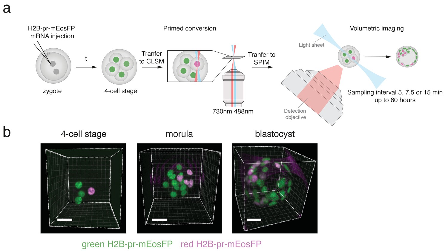
H2B-pr-mEosFP injected embryos develop to the blastocyst stage.
(a) Experimental setup: Zygotes are injected with H2B-pr-mEosFP mRNA. At the 4-cell stage confined primed conversion of a single nucleus is performed using intersecting 488 nm and 730 nm lasers. The embryos are transferred to an inverted SPIM for non-invasive imaging of their development up to the blastocyst stage. Images are taken every 5, 7.5 or 15 min. (b) Embryos injected with mRNA encoding H2B-pr-mEosFP and converted at the four-cell-stage develop normally and maintain visibility of the red label up to the early blastocyst stage. pr-mEosFP fluorescence (green) and primed converted pr-mEosFP fluorescence (magenta). N ≥ 200 embryos out of ≥10 independent experiments. Scale bar, 20 µm.
Dual labeling of pre-implantation embryos greatly facilitates automatic segmentation, tracking, and lineage tracing
As cells converted at the four-cell stage can be visualized up to the blastocyst stage, we wondered whether such sparsely labeled subsets of cells could aid computational reorientation and automated lineage tracing in embryos that exhibit dramatic spatial and rotational drift (Videos 1, 2 and 3). Of note, while we initially imaged our embryos with time intervals of 7.5 or 15 minutes, we found that increased sampling frequency did not recover successful lineage tracing for rotating embryos: the percentage of embryos showing spatial and rotational drift prohibitive of automated lineage tracing in our experiments with 5-minute imaging time intervals (=50%, n=8) was similar to those imaged with larger time intervals (=45% embryos imaged every 7.5 or 15 minutes, n=11). To accomplish accurate tracking, we developed a computational pipeline, referred to as “primed Track”, for automated segmentation, cell tracking, and lineage tracing. Primed Track uniquely takes advantage of the sparse red cell population to correct for spatial and rotational drift as well as to simplify lineage reconstruction (Figure 2a). In the 5-dimensional (5D, that is 3 spatial dimensions, time, color) imaging data, cells were first segmented based on size, shape, and fluorescence taking into account both color channels. The use of increasing laser power to compensate for red signal dilution mainly due cell division resulted however in increasing background fluorescence. To discern red signal from increasing background signal, we took advantage of the dual nuclear labeling that allowed us to identify weaker fluorescent red nuclei at advanced time points by their overlap with the green signal in which lower autofluorescence was detected (Figure 2a, left column). Background signal that was falsely segmented in the green channel due to increased illumination could be excluded by ignoring spots detected outside of a defined radius of the embryo. The ability to select parameters that match the brightness, size, and shape of the embryos combined with fluorescence information of two channels makes the segmentation both robust and flexible for use in different experiments. In addition, the dual color information allowed for cell distinction in instances otherwise rendered ambiguous through high cell density and proximity of nuclei. For instance, we were able to distinguish nuclei that would have been identified as a single nucleus even after manual validation (Figure 2—figure supplement 1a-c).

Primed track results in efficient lineage reconstruction of embryos with high spatial and rotational drift.
(a) Overview of the pipeline used for reliable automated segmentation, tracking, and lineage tracing of the imaged embryos; (1) Segmentation: low thresholds are used for the spot detection in both the green and red channel to enable detection of dimmer cells at later developmental time points. Incorrectly segmented spots are excluded by defined filters: (i) exclusion of spots outside of a defined radius of the embryo, (ii) replacement of incorrectly segmented double spots by one spot per one nucleus, and (ii) exclusion of red spots that do not colocalize with green nuclear spots. (2) Tracking: Spatial drift as well as rapid embryo rotation complicates tracking nuclei over prolonged time windows. The segmented nuclei are used for defining reference frames based on the center of mass of the green nuclei and the orientation of the red nuclei. The alignment of the references frames of each time point compensates the spatial and rotational drifts. (3) Lineage tracing: Automated lineage tree reconstruction can make false connections when cells are dividing. By separating the calculation of the lineage trees in the photoconverted red channel from the green channel, the less complex datasets for each channel result in more consistent lineage tracing. pr-mEosFP fluorescence (green) and primed converted pr-mEosFP fluorescence (magenta) overlaid with segmentation results (green and Magenta spheres); Scale bar, 20 µm (b) Lineage trees from the same embryo (corresponding to Videos 1 and 4) reconstructed from segmented nuclei before correction for rotational and translational drift (left), after correction for rotational and translational drift for the red channel (second left), after correction for rotational and translational drift for the green channel minus the spots that colocalize with the red spots (second right), and after final manual lineage reconstruction (right). The embryo was imaged every 15 min.
-
Figure 2—source data 1
Summary of challenges with state-of-the-art segmentation and tracking tools.
- https://doi.org/10.7554/eLife.44491.013
In a second step, the embryo was positioned at its fluorescence center of mass, cropped and rotated, such that the red center of mass was oriented to the same side of the embryo in every time frame to compensate for rotational and spatial drift (Figure 2a, middle column; Videos 4, 5 and 6). The resulting high-quality 5D cropped and registered datasets were reduced to only 34±11% of the original size (Figure 2—figure supplement 2). The automatic tracking of a realigned embryo resulted in greatly improved lineage tracing fidelity compared to a naïve state-of-the-art lineage-tracing algorithm that was not able to reconstruct a lineage tree from rotating and spatially drifting embryos imaged with a time interval of 15, 7.5 or 5 minutes (Bitplane Imaris cell lineage package) (Figure 2b; Figure 2—figure supplement 2). Of note, none of the existing state-of-the-art lineage tracing tools such as Ilastik, TrackMate and the TGMM software (Amat et al., 2014) were designed to compensate for heavy rotational and spatial drift and are therefore incapable of calculating lineage trees from these embryos (Figure 2—source data 1). Separating the green and red channels to generate two less complex datasets during lineage reconstruction further increased the fidelity of lineage tracing versus a dataset consisting of the green channel alone (Figure 2a, right column; Figure 2—figure supplement 3). We assessed the power of primed Track by comparing the lineage trees obtained i) without corrections, ii) after embryo realignment with all algorithmic corrections, and iii) after final manual review by calculating the total distance between these lineage trees (see Materials and methods for more details) (Zhang and Shasha, 1989). We were able to recover all rotating embryos that we acquired using this image analysis pipeline and the resulting lineage trees required a minimal amount of time for manual corrections (0.5-1.5 hours per lineage tree).
Primed Track allows for decreased sampling frequency
The observation that the registration of embryos based on the dual labeling with primed Track allows for reliable lineage tracing despite heavy embryo rotation suggests that decreasing the imaging frequency will have limited effect on lineage tracing ability. To test the robustness of primed Track, we removed time points from datasets from both rotating as well as non-rotating embryos to examine lineage tracing capacity at a decreased sampling rate. Naturally, larger imaging time intervals increase embryo displacement in consecutive time points and exacerbate the accurateness of lineage tracing (Figure 3). Using primed Track to correct for spatial and rotational drift results in reliable reconstruction of the lineage trees even at imaging time intervals of 30 minutes for a rotating embryo and 40 minutes for a non-rotating embryo (Figure 3; Figure 3—figure supplement 1). While the registration of non-drifting embryos does not increase lineage tracing accuracy for high 5-minute sampling rates, the gain of fidelity in lineage tree reconstruction of these embryos greatly benefits from our presented approach when the imaging frequency is reduced (Figure 3). However, in general, one should keep in mind that sampling rates above 40 minutes will decrease the possibility to precisely infer cell divisions and assign daughter cells to their correct mother. Still, the opportunity to reduce laser exposure while maintaining accurate tracking and lineage tracing potential offers a great advantage for long-term imaging experiments of sensitive specimen.
Summary and conclusion
In summary, primed Track enables fast, automated, high fidelity lineage tracing of mammalian pre-implantation development combined with reduced illumination time and data volume, key considerations for handling and analyzing data by the biological community (Pantazis and Supatto, 2014). A recently published study presents a compelling image analysis framework that enables the long-term tracking of cells during gastrulation and early organogenesis in the post-implantation embryo (McDole et al., 2018). Primed Track complements such efforts by enabling accurate fate mapping of mouse pre-implantation embryos.
The ability to correct for both spatial and rotational drift overcomes the previous requirement to exclude spinning embryos from the analysis using primed Track. Furthermore, primed conversion of photoconvertible proteins in combination with primed Track enables the experimenter to still achieve reasonable lineage tracing quality with datasets acquired at lower sampling rate.
The timescales and intensities at which the fluorescent signal of photoconvertible proteins can be observed depend on the expression system (i.e. stable vs. transient expression, promotor choice) as well as the stability of the fusion protein. While we present tracking and lineage tracing of embryos labeled with a relatively highly-expressed and stable H2B-pr-pcFP fusion protein, it is important to take into consideration that low abundant protein fusions may require higher illumination power for visualization, potentially impacting sample integrity. Such cases will in particular benefit from our primed Track pipeline, as it facilitates imaging with longer time intervals while preserving high fidelity in cell tracking and lineage tracing.
In the future, implementing primed conversion to take place inside a SPIM used for volumetric imaging will allow for repeated manual or automatic primed conversion of nuclei once the red fluorescence signal intensity drops below a user-defined threshold. Such pulse-chase experiments can then be extended even longer, ultimately being only limited by the rate of new green pr-pcFP synthesis. The combination of confined primed conversion of pr-pcFPs with primed Track will allow researchers to get more accurate insight into the dynamic processes responsible for cell fate decisions in the early mammalian embryo.
Materials and methods
Molecular cloning and mRNA preparation
View detailed protocolThe coding sequences for pr-mEosFP and pCS2+-H2B-pr-EosFP were obtained by PCR amplification from pQE32-pr-mEosFP (Addgene No. 99213) and pRSET-pr-mEos2 (gift from Dominique Bourgeois) and cloned into pCS2+-H2B-Dendra2 using AgeI and SnaBI, hence replacing the Dendra2 coding sequence to obtain pCS2+-H2B-pr-EosFP and pCS2+-H2B-pr-Eos2. mRNA was synthesized using the mMESSAGE mMACHINE kit (ThermoFisher Scientific), followed by poly-A-tailing (ThermoFisher Scientific), and purified using a Qiagen RNAeasy kit according to manufacturer guidelines.
mRNA microinjection of mouse preimplantation embryos and ex utero culture up to four-cell stage
Request a detailed protocolC57Bl/6 wild-type females (Janvier Labs, France) were superovulated by hormone priming, mated to C57Bl/6 males (RRID:IMSR_JAX:000664), and mated females were euthanized by CO2 asphyxiation. Embryos were recovered by flushing oviducts as described previously (Mohr et al., 2016; Plachta et al., 2011). Embryos were cultured at 37°C and 5% CO2 in KSOM + AA medium covered with mineral oil. mRNA constructs were microinjected into the pro-nucleus at 50 ng/µl or in both cells in two-cell stage embryos, following standard protocols. All these experiments were approved by the veterinary authority of the canton Basel Stadt, Switzerland.
Confined primed conversion of single nuclei in mouse embryos
View detailed protocolConfined primed conversion of single nuclei was performed on mouse embryos at the four-cell stage as previously described in great detail (Mohr et al., 2016).
Volumetric imaging of mouse pre-implantation embryos
View detailed protocolRight after confined primed conversion was performed, the four-cell stage embryos were transferred to a pre-equilibrated, custom built inverted SPIM setup suitable for long term imaging of mouse embryos and continuously cultured/imaged until they reached blastocyst stage. For each embryo, a z-stack consisting of 80 planes, 3 μm apart, was acquired every 5, 7.5 or 15 min.
Mouse embryo lineage tracing
View detailed protocolTo establish a reference, mouse embryos were lineage traced using the state-of-the-art Imaris lineage tracing package (Bitplane, CH). The automated high-fidelity mouse embryo drift correction and lineage-tracing algorithm described here is explained in detail below.
Detailed description of primed Track
5D movies of photoconverted mouse embryos were processed with the following pipeline using a custom MATLAB code implemented in Imaris (Bitplane, CH). All codes of primed Track can be downloaded from this code repository: https://git.bsse.ethz.ch/scu_public/primed_track (copy archived at https://github.com/elifesciences-publications/primed_track) (Ponti, 2018).
Cell segmentation
View detailed protocol1.Detect green and red cells using the Spot detector in Imaris. Use low threshold to segment all cells even at the cost of including spurious spots. Allow spot radius to be adapted to more accurately fit the volume of the segmented cell. Bright but very small spots can easily be filtered out during segmentation.
First validation
View detailed protocolUse the green spot positions to estimate the embryo diameter and discard green spots that are likely to be outside of the embryo. The radius of the embryo is roughly estimated as the median of all maximal inter-spot distances. A user-defined multiplicative factor can optionally compensate for estimation errors and prevent cells at the boundary of the embryo to be discarded should this constraint be too stringent.
Search for spots that occur within a small defined distance from a spot in the same channel, discard all wrongly segmented double spots on one nucleus and replace them by one new spot.
Discard all spurious red spots that do not colocalize with a green spot. Note that due to the equilibrium between protonated and de-protonated chromophore, green to red photoconversion of pcFPs is never exhaustive and will always retain a green population, rendering this quality control step possible.
A red spot discarded during the first validation can optionally be recovered if there is a valid red spot in previous time point within a user-defined search radius. This adjustment compensates for remaining miss-segmentations in the green channel.
Embryo alignment (drift and rotational correction)/Cropping
View detailed protocolImaris Reference Frame Objects are created in MATLAB for each time point: their origin is set at the position of the center of mass (COM) of the green spots and their orientation is given by the vector ΔCOM = COMred - COMgreen. This correction still has one degree of freedom. The rotation angle around the reference frame axis is obtained by comparing the positions of the green spots at timepoints t and t - 1 over 360 1-degree rotations and by choosing the angle that minimizes the cell drift between time points. The resampling is performed in Imaris.
Crop data to the smallest bounding volume.
Second validation and subsetting
View detailed protocolTo pick up red cells that were not recovered previously, re-run the first validation on the re-aligned embryo.
Create a new spot object that contains the subset of green cells that do not colocalize with red cells.
Lineage tree reconstruction
View detailed protocolImaris’ Lineage module is used to track the cells over time and reconstruct their lineage tree. The subsetting in the previous step allows us to reduce the complexity of the lineage tracing problem by breaking it down into two simpler, computationally less expansive, disjoint problems.
Comparative analysis of lineage trees
Request a detailed protocolTo assess the power of our newly created algorithm, we sought to compare the lineage trees obtained with i) no corrections, ii) after embryo realignment with all algorithmic corrections, and iii) after final manual review. We quantified the effects of the corrections and validations on the quality of the lineage trees by calculating the total distance between the lineage trees using the implementation of the tree Zhang-Shasha edit distance algorithm (Zhang and Shasha, 1989) by Tim Henderson and Steve Johnson (Henderson and Johnson, 2013). The zss algorithm assigns a (user-defined) cost for each node insertion, removal, and update necessary to transform an ordered tree into another, and gives therefore a quantitative measure of dissimilarity of the two trees. Small tracking differences between corrected and uncorrected trees, however, can result in quite large tree distances if the zss algorithm is applied to the complete trees. A correction that relinks one cell to its mother cell in just one time point causes the whole branch to be flagged as incorrect, and the longer the branch, the higher the distance between the trees. In other words, the earlier the tracking error occurs, the larger the distance; yet, only the first time point in the track is incorrect, and its penalty should be the same whether it happens at the beginning of the time series or the end.
To circumvent these issues, we applied the algorithm to a condensed version of the lineage trees. The condensed tree retains only the branch points of the original lineage tree (i.e. the cell divisions). Also, each branch point stores information about the original number of child nodes in its branches (i.e. the number of time points the daughter cells were tracked until their next cell division). The distance between condensed trees will flag positions where cell divisions were tracked incorrectly and tracks that have different lengths, without causing an explosion in the reported distance.
Since our acquisitions started at the four-cell stage, we aimed to build a tree for each of the original four cells (one containing the progeny of the primed converted cell). The final, manually curated lineage was used as ground truth to quantify the effects of the various algorithmic correction steps. The sets of trees across correction schemes were assigned to each other by minimizing the spatial and temporal distance of their origins. After condensation, their pairwise distances were calculated. All distances were summed to give the total lineage tree difference. In addition, spurious trees that resulted from bad segmentation and tracking were not used for the distance calculation, since they already indirectly affected the difference of the tree from which they were erroneously detached.

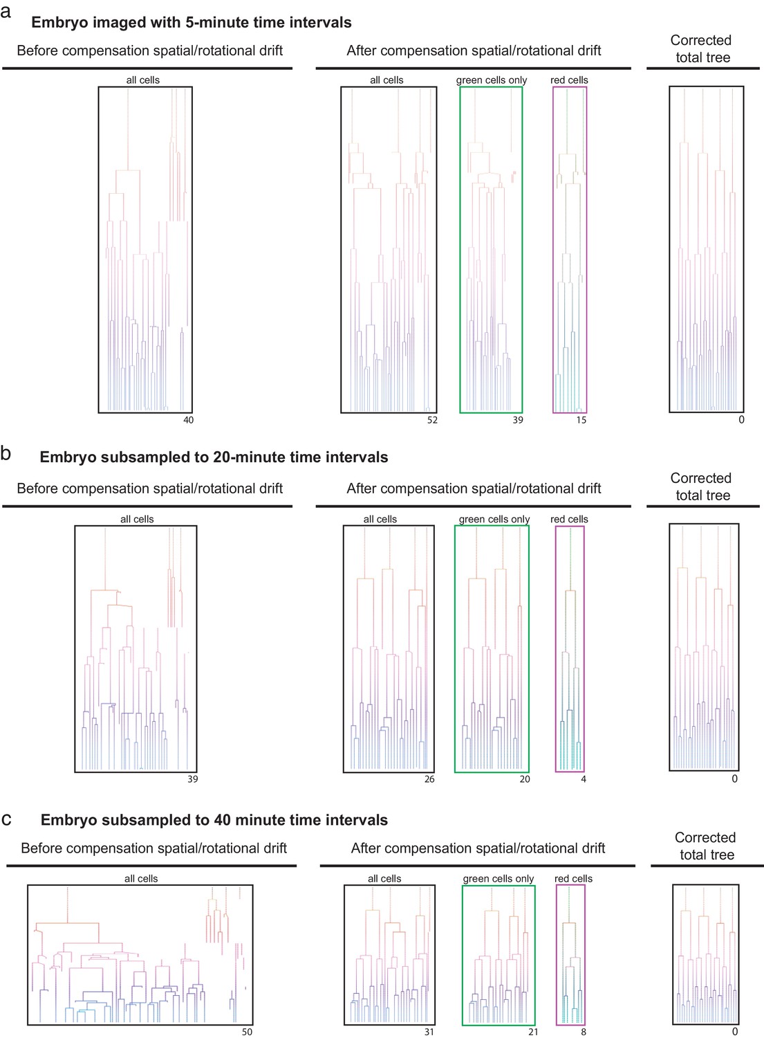
Efficient lineage reconstruction using primed Track is still achieved at large imaging time intervals An originally non-rotating embryo imaged with 5 min.
(a) time intervals were subsampled to 20 min (b) and 40 min (c) time intervals. Lineage tracing was performed on the non-processed dataset as well as after correction for spatial and rotational drift using the presented approach. The numbers displayed below each lineage tree indicate the distance to the final correct lineage tree.
Video of a developing embryo (same as in Video 1) after drift correction.
Timelapse video of the example embryo from Video 1 after drift correction. pr-mEosFP fluorescence (green) and primed converted pr-mEosFP fluorescence (red). Corresponding lineage trees are displayed in Figure 2d. Scale bars, 15 µm; framerate: one frame every 15 min.
Video of a developing embryo (same as in Video 2) after drift correction.
Timelapse video of the example embryo from Video 2 after drift correction. pr-mEosFP fluorescence (green) and primed converted pr-mEosFP fluorescence (red). Corresponding lineage trees are displayed in Figure 2—source data 1. Scale bars, 10 µm; framerate: one frame every 7.5 min.
Video of a developing embryo (same as in Video 3) after drift correction.
Timelapse video of the example embryo from Video 2 after drift correction. pr-mEosFP fluorescence (green) and primed converted pr-mEosFP fluorescence (red). Corresponding lineage trees are displayed in Figure 2—source data 1. Scale bars, 30 µm; framerate: one frame every 5 min.
Data availability
All codes of primed Track can be downloaded from this code repository: https://git.bsse.ethz.ch/scu_public/primed_track (copy archived at https://github.com/elifesciences-publications/primed_track).
References
-
In vivo single-cell labeling by confined primed conversionNature Methods 12:645–648.https://doi.org/10.1038/nmeth.3405
-
Rational engineering of photoconvertible fluorescent proteins for Dual-Color fluorescence nanoscopy enabled by a Triplet-State mechanism of primed conversionAngewandte Chemie International Edition 56:11628–11633.https://doi.org/10.1002/anie.201706121
-
Primed conversion: the new kid on the block for photoconversionChemistry - a European Journal 24:8268–8274.https://doi.org/10.1002/chem.201705651
-
Polarity of the mouse embryo is established at blastocyst and is not prepatternedGenes & Development 19:1081–1092.https://doi.org/10.1101/gad.1304805
-
Advances in whole-embryo imaging: a quantitative transition is underwayNature Reviews Molecular Cell Biology 15:327–339.https://doi.org/10.1038/nrm3786
-
Oct4 kinetics predict cell lineage patterning in the early mammalian embryoNature Cell Biology 13:117–123.https://doi.org/10.1038/ncb2154
-
A general mechanism of photoconversion of Green-to-Red fluorescent proteins based on blue and infrared light reduces phototoxicity in Live-Cell Single-Molecule imagingAngewandte Chemie International Edition 56:11634–11639.https://doi.org/10.1002/anie.201702870
-
Symmetry breaking in the early mammalian embryo: the case for quantitative single-cell imaging analysisMolecular Human Reproduction 22:172–181.https://doi.org/10.1093/molehr/gav048
-
Simple fast algorithms for the editing distance between trees and related problemsSIAM Journal on Computing 18:1245–1262.https://doi.org/10.1137/0218082
Decision letter
-
Jeremy NathansReviewing Editor; Johns Hopkins University School of Medicine, United States
-
Marianne E BronnerSenior Editor; California Institute of Technology, United States
In the interests of transparency, eLife includes the editorial decision letter and accompanying author responses. A lightly edited version of the letter sent to the authors after peer review is shown, indicating the most substantive concerns; minor comments are not usually included.
[Editors’ note: a previous version of this study was rejected after peer review, but the authors submitted for reconsideration. The first decision letter after peer review is shown below.]
Thank you for submitting your manuscript "High fidelity lineage tracing in mouse pre-implantation embryos using primed conversion of photoconvertible proteins" for consideration by eLife. Your article has been reviewed by three experts and the evaluation has been overseen by a Senior and Reviewing Editor. While the reviewers found the work interesting, the number of substantive questions raised was such that we feel we must reject it. We hope that the reviewer's comments below will be useful to you in revising the manuscript for submission elsewhere. We apologize for not being able to deliver better news, and we hope that you will continue to consider eLife for future submissions.
Reviewer #1:
In their manuscript, Pantazis and colleagues demonstrate a combined optical/computational method for reducing the effects of translational and rotational drift in pre-implantation mouse embryo lineage recordings. Primed conversion is used to introduce a sparse second color (red fluorescent nuclei), which is used as a fiducial to reduce drift and an additional quality check on the derived lineages. Although the authors convincingly demonstrate that their method does reduce the effects of drift and thus computational error in their experiments, I am unconvinced that their method is either necessary or generally important for this particular biological application.
While the problem the authors address is a real one, their method does not appear a significant improvement over previous work – in particular the groundbreaking method of Lars Hufnagel and Jan Ellenberg (Strnad et al., 2016). In that manuscript, Hufnagel and Ellenberg performed similar recordings at higher spatiotemporal resolution than reported here. In particular, the temporal sampling in Hufangel and Ellenberg was performed every 5 minutes, 1.5-3x faster than the 7.5 minute and 15 minute recordings performed by Pantazis. One has to wonder if the increased temporal sampling is in fact the dominant source of error in reconstructing lineages – if Pantazis et al. had simply recorded faster, would they have encountered the same degree of drift/error? Hufnagel and Ellenberg claimed a 100% tracking accuracy in their manuscript (for the embryos they ultimately select for lineage analysis) – if this is really the case, I have to wonder why Pantazis et al. did not simply adopt the previous tracking approach. Pantazis et al. compare their computational pipeline to Bitplane Imaris, but the real state-of-the-art comparison is to Hufnagel and Ellenberg. How does the new tracking pipeline presented by Pantazis compare to the coherent point drift method described in this previous work? It is also never explicitly spelled out how many datasets the new method 'rescues', i.e. of the 5/11 embryos that exhibited severe translation/rotational drift, how many were 'recoverable' in the new method? What is the fraction of embryos that are now fully trackable? Because the authors of the current manuscript have failed to put their method into context against previous work, it is difficult to properly assess the impact of their method.
The authors also assert that their method might allow less dosing of the sample (presumably due to the worsened temporal sampling they report) and that their pipeline results in smaller datasets due to the tighter cropping that results. Neither assertion is particularly compelling – (i) I am not convinced that in fact lowering the temporal resolution is advantageous as it seems this makes the tracking problem harder; (ii) the original data sizes they report of ~5GB are hardly massive by today's standards. In summary, I am concerned that the authors' paper constitutes a kind of 'straw man', i.e. they are attacking a problem that has been satisfactorily addressed by previous work. A thorough, statistical comparison of their method to Hufnagel and Ellenberg's would go a long way to convincing me of the value of their method.
Other comments:
The authors are to be commended for comparing H2B-pr-mEosFP to H2B-pr-mEos2. However, I would like to see more evidence for their assertion that their photoconverted embryos develop normally, especially since the primed conversion operation itself intrinsically introduces additional dose. In the previous work by Hufnagel and Ellenberg, 'the tracked embryos had a division timing and number of ICM cells comparable to those of in vitro-cultured embryos… and healthy pups were born after transfer of the imaged embryos into pseudopregnant females…'. Were similar controls done here? What is the additional dose introduced by the primed conversion on the confocal microscope, relative to the light sheet illumination dose used for imaging? The authors image from 4 cell to blastocyst, yet it seems that in previous work it is possible to image from zygote onwards. Is the 4 cell stage necessary due to the increased light sensitivity at earlier stages?
Reviewer #2:
Welling et al. present a combined reverse genetic/optical and computational approach to extract developmental lineages from pre-implantation mouse embryos. The genetic trick relies on photo-convertible proteins that are converted on a confocal set-up and later imaged using light sheet. The computational pipeline extending Imaris achieves proper segmentation, image alignment and uses the total and photo-converted nuclei to improve unsupervised lineaging.
This work has potential, however, for me, it falls short of being a minimal publishable unit. The photoconversion approach has already been published by the authors. What remains is a useful technique that would however fit better into Materials and methods section of a paper focusing on the biology that can be done with this approach. I see the benefits of being able to use the rotating embryos previously excluded from similar analysis (Strnad et al.). However, that is a very niche problem and the pipeline lacks general applicability. The segmentation enhancement is completely dependent on the precise experiment described here, no new algorithm has been presented. Similarly, the re-orientation of the rotating embryos is done using very basic core functions of Imaris. The authors do show that it benefits the analysis of their specific data, however I doubt it will be generally applicable. The comparison of the performance of the Imaris tracker applied to uncorrected and corrected data is a straw man comparison. The Imaris tracker was not developed for tracking lineages in embryos that are fast rotating and therefore it, of course, fails spectacularly.
In order to make the paper work as a methods paper, it would have to be significantly expanded. On the hardware side, the photoconversion would need to happen at one microscope (something the authors clearly intend to do). On the software side, the tracker would need to be benchmarked against existing state of the art tracking solutions such as Ilastik, TrackMate and the Keller pipeline. In addition, the authors would need to show that it is also applicable to other lineaging problems.
Last but not least, the submission contains no code. There is insufficient details provided to reproduce the work, even inside such user friendly software as Imaris is. There is a mention of some MATLAB code that is stringing together the Imaris functionality. At least that needs to be put on github to make this work useful for others. In the current form, it has no impact.
Reviewer #3:
In the short paper entitled "High fidelity lineage tracing in mouse pre-implantation embryos using primed conversion of photoconvertible proteins" the authors use photoconversion of an EosFP by 'primed conversion' to follow by 3D SPIM imaging the cell lineage. In this very limited example the authors propose a potentially promising way of tracking cell fate. However I believe that it currently has a number of issues that should be addressed.
1) Novelty. The novelty here is only mediocre. The photoconversion of EosFP by a 488→730nm illumination pulse has been reported (Mohr, Argast and Pantazis, 2016). Similarly, lineage tracking has been done before (Kurotaki et al., 2007 and others). The novelty is using SPIM here for longer-term tracking, but unfortunately while the potential was there the illumination for both channels was done with the same objective (see point #2).
2) Implementation. The real power of this method should be to focally limit which cells, or region thereof, is getting photoconverted, by launching the light through objectives situated at 90 degrees. Unfortunately, the authors choose to illuminate/activate the cell through only a single objective and thus lose a potential major benefit of the technique. It would have been really neat, and more powerful, to do the activation at a later stage when it would be otherwise difficult to activate only a single cell. In my opinion, doing the activation by cross-beams and in a condition that would be impossible to achieve by a single beam is essential here, and would improve the novelty. The authors ironically discuss axial confinement of the dual activation yet fail to do so and exploit it in the experiments. This must be done.
3) Robustness of the data. It is unclear how many times this experiment was performed. Only once? To show that the technique is robust, more experiments are needed, with statistics. The authors mention that the photoconverted embryos were healthy, but from how many experiments?
4) Other reporters. The authors should show the technique for other reporters, such as in the cell cytosol, or membrane, to generalize the concept.
5) Ambiguity of assignment. It is unclear how long a single lineage can be tracked. The S/N seemed to be high at the later stages. Can the authors better quantify showing the accuracy of assignment in each stage, with statistics.
https://doi.org/10.7554/eLife.44491.021Author response
[Editors’ note: the author responses to the first round of peer review follow.]
To address the reviewers’ concerns, we performed a set of additional experiments. Specifically, we compared our image analysis pipeline, now referred to as “primed Track”, with three state-of-the-art existing algorithms for cell tracking and lineage tracing. We now show that these algorithms perform poorly in recovering rotating embryos, whereas primed Track faithfully returns high fidelity lineage trees. Primed Track is unique in its ability to perform a comprehensive cell lineage analysis by including rotating embryos, which had to be excluded in previous studies. While we intended to also test the full code developed by Strnad et al., 2016, we were not able to retrieve the full code necessary to repeat their work. We strongly believe that the current comparison to three other state-of-the-art algorithms is sufficient to demonstrate the marked potential of primed Track. We now also show that primed Track is not limited to recovering drifting embryos but also has general value for long-term imaging of sensitive specimen, since primed Track retains the ability for high fidelity tracking and lineage tracing when imaging time intervals are significantly increased (i.e. from 5min to 40min). The opportunity to reduce laser exposure offers a unique advantage for long-term imaging experiments of sensitive specimen, in which single cell tracking and lineage tracing would otherwise not be possible. These additional results demonstrate that primed Track provides superior fidelity in tracking and lineage tracing of drifting specimen over existing algorithms and further allows for increased imaging time intervals, reducing detrimental effects of phototoxicity. We believe that our revised manuscript will be of large interest to the readers of eLife with a particular appeal to the volumetric imaging and bioimage informatics community.
Reviewer #1:
In their manuscript, Pantazis and colleagues demonstrate a combined optical/computational method for reducing the effects of translational and rotational drift in pre-implantation mouse embryo lineage recordings. Primed conversion is used to introduce a sparse second color (red fluorescent nuclei), which is used as a fiducial to reduce drift and an additional quality check on the derived lineages. Although the authors convincingly demonstrate that their method does reduce the effects of drift and thus computational error in their experiments, I am unconvinced that their method is either necessary or generally important for this particular biological application.
While the problem the authors address is a real one, their method does not appear a significant improvement over previous work – in particular the groundbreaking method of Lars Hufnagel and Jan Ellenberg (Strnad et al., 2016). In that manuscript, Hufnagel and Ellenberg performed similar recordings at higher spatiotemporal resolution than reported here. In particular, the temporal sampling in Hufangel and Ellenberg was performed every 5 minutes, 1.5-3x faster than the 7.5 minute and 15 minute recordings performed by Pantazis. One has to wonder if the increased temporal sampling is in fact the dominant source of error in reconstructing lineages – if Pantazis et al. had simply recorded faster, would they have encountered the same degree of drift/error?
We would like to thank reviewer #1 for pointing out this concern. Increased sampling should indeed result in increased lineage tracing fidelity. To test if increased sampling resolves the observed spatial and rotational drift, we performed two sets of light-sheet experiments with 5-minute time intervals. We found that the number of embryos that showed a dramatic spatial/rotational drift did not change when the imaging time intervals are shortened. In fact, the percentage of embryos showing significant rotational and spatial drift when imaged with 5-minute time intervals (4/8 embryos) was similar to the embryos imaged with 7.5-minute or 15-minute time intervals (5/11 embryos).
Hufnagel and Ellenberg claimed a 100% tracking accuracy in their manuscript (for the embryos they ultimately select for lineage analysis) – if this is really the case, I have to wonder why Pantazis et al. did not simply adopt the previous tracking approach. Pantazis et al. compare their computational pipeline to Bitplane Imaris, but the real state-of-the-art comparison is to Hufnagel and Ellenberg. How does the new tracking pipeline presented by Pantazis compare to the coherent point drift method described in this previous work?
Ellenberg and colleagues indeed published an elegant two-step segmentation and tracking pipeline that reliably reconstructed lineages from pre-implantation embryos. However, this method was developed for embryos that were imaged at 5-minute time intervals and that did not display rotational or spatial drift. It is worth noting that Ellenberg and colleagues reported that they excluded a significant fraction of embryos (n = 6 out of 19 embryos) that ‘[…] rotated too rapidly for automatic alignment’ (Strnad et al., 2016). In addition, Ellenberg and colleagues reported that even lineage trees reconstructions from non-rotating embryos required manual corrections.
In our manuscript, we show that we are able to reliably reconstruct lineages from embryos that display heavy spatial and rotational drift using an image analysis pipeline (referred to as “primed Track”) that takes advantage of the dual labeling in the embryos, an approach that would otherwise not have been possible. Importantly, we now show that primed Track even allows for the reliable reconstruction of lineages from embryos imaged with larger time intervals. This achievement provides unique opportunities for long-term imaging of sensitive and/or dim specimen that require higher laser intensities for visualization of fluorescent signals while limiting potential effects on developmental progression.
It is also never explicitly spelled out how many datasets the new method 'rescues', i.e. of the 5/11 embryos that exhibited severe translation/rotational drift, how many were 'recoverable' in the new method? What is the fraction of embryos that are now fully trackable?
We were able to recover 100% of the rotating embryos. This accomplishment is due to the fact that embryos were labeled with an optimized photoconvertible protein, pr-mEosFP. It provided sufficient contrast for robust labeling up to the blastocyst stage due to its superior brightness and photostability when compared to previously employed fluorescent protein versions. Consequently, primed Track correctly segmented all labeled nuclei and realigned the embryos, allowing for the reliable reconstruction of the lineage trees. In both realigned embryos and non-rotating embryos, occasional wrong daughter cell assignment after cell division required limited manual correction to obtain maximal fidelity.
Because the authors of the current manuscript have failed to put their method into context against previous work, it is difficult to properly assess the impact of their method.
To compare our work to other existing state-of-the-art lineage tracing tools, we tested the performance of the Ilastik, the TrackMate and the TGMM algorithm for segmentation and lineage tracing developed by the Keller lab on our data. We intended to also test the algorithm developed by Ellenberg and colleagues, yet the code provided in the published paper turned out to be incomplete; the authors were notified of this discrepancy.
We found that all sophisticated segmentation and tracking tools were not able to reconstruct correct lineage trees from rotating embryos. In pre-implantation embryos, daughter cells experience dramatic repositioning relative to their division plane. Due to these dynamics upon cell division, Ilastik, TrackMate and TGMM had difficulties in correctly assigning daughter cells to their respective mother cells after cell division. Combined with the substantial spatial and rotational drift of pre-implantation embryos, tracking of cells became uncertain for tested lineage tracing tools. In addition, both Ilastik and TrackMate showed limitations in the correct segmentation of individual cells when the cell density in the embryo increased.
In primed Track we overcame these limitations: the Imaris spot detection tool was enhanced with additional customizable parameters that disabled the segmentation of background signal (i.e. signal outside a certain radius of the embryos) and that required the overlap of segmented red spots with green spots in order to distinguish real nuclear signal from falsely segmented background signal. A concise summary of the main limitations of tested algorithms is included in Figure 2—figure supplement 3 in the revised manuscript.
The authors also assert that their method might allow less dosing of the sample (presumably due to the worsened temporal sampling they report) and that their pipeline results in smaller datasets due to the tighter cropping that results. Neither assertion is particularly compelling – (i) I am not convinced that in fact lowering the temporal resolution is advantageous as it seems this makes the tracking problem harder; (ii) the original data sizes they report of ~5GB are hardly massive by today's standards. In summary, I am concerned that the authors' paper constitutes a kind of 'straw man', i.e. they are attacking a problem that has been satisfactorily addressed by previous work. A thorough, statistical comparison of their method to Hufnagel and Ellenberg's would go a long way to convincing me of the value of their method.
We thank reviewer #1 for raising these important points that we would like to address as follows:
1) Mouse pre-implantation embryos are very sensitive to laser illumination. Despite the fact that specimen imaged with a SPIM are only excited in the focal plane, we found that embryos arrest before reaching the blastocyst stage upon higher laser intensities and/or higher sampling intervals. Specifically, we saw that the exact laser intensities that allowed the development of 50% of the embryos to the blastocysts stage at imaging intervals of 7.5 minutes, did not allow any of the embryos (n=10) to develop past the 8-cell to morula stage when sampled every 5 minutes. In order for us to be able to image our embryos with a higher sampling frequency, we had to decrease the power of laser illumination. The possibility to image with larger time intervals is therefore of particular importance for embryos that possess weaker fluorescent signal and need higher laser dosage for detection. Examples include but are not limited to endogenously tagged embryos expressing a fluorescent protein fusion at physiological amounts as opposed to high overexpression using potent promoters. To test the ability to perform lineage tracing in embryos imaged with larger time intervals, we subsampled both rotating and non-rotating embryos and applied primed Track to realign the embryos and reconstruct lineage trees. While we did see a small decrease in lineage tracing fidelity compared to embryos imaged with shorter time intervals, primed Track was able to still reconstruct lineages from both rotating and non-rotating embryos imaged every 30 or 40 minutes, respectively (see Figure 3 and Figure 3—figure supplement 1).
2)The sizes of the original data are dependent on the sampling rate of the embryos as well as on the duration of the time series. Therefore, the size of the original data ranged from 5GB to 25GB. While storage of large data does not pose a challenge these days, the execution speed of fundamental functions of available bioimage analysis software solutions can be severely affected by the size of the dataset. For instance, the need to load and analyze data sets for the optimization of image analysis parameters took several hours for 4D datasets on GPU-accelerated high-performance workstations. Consequently, cropping data imaged with larger time intervals to less than half of the original size greatly decreased the amount of time needed for analysis calculations.
Other comments:
The authors are to be commended for comparing H2B-pr-mEosFP to H2B-pr-mEos2. However, I would like to see more evidence for their assertion that their photoconverted embryos develop normally, especially since the primed conversion operation itself intrinsically introduces additional dose. In the previous work by Hufnagel and Ellenberg, 'the tracked embryos had a division timing and number of ICM cells comparable to those of in vitro-cultured embryos… and healthy pups were born after transfer of the imaged embryos into pseudopregnant females…'. Were similar controls done here? What is the additional dose introduced by the primed conversion on the confocal microscope, relative to the light sheet illumination dose used for imaging?
We thank reviewer #1 for pointing out this issue that we now address in detail in the revised manuscript. Before embryos were transferred to the light-sheet microscope for volumetric imaging, they were exposed to 20 seconds of continuous-wave 488nm and 730nm light required for effective confined primed conversion of H2B-pr-mEosFP. Normal development of H2B-pr-mEosFP injected embryos was evaluated by comparing the progression of primed converted and non-converted embryos to the blastocyst stage. Under these imaging conditions, the percentage of embryos that reached the blastocysts stage (see Figure 1—figure supplement 2B) was similar for both groups, indicating that neither confined primed conversion nor expression of H2B-pr-mEosFP affected their development.
The authors image from 4 cell to blastocyst, yet it seems that in previous work it is possible to image from zygote onwards. Is the 4 cell stage necessary due to the increased light sensitivity at earlier stages?
The reason to start imaging at the 4-cell stage was for practical reasons: due to absence of a transgenic line expressing H2B-pr-mEosFP protein, we injected embryos with H2B-prmEosFP mRNA at the zygote or 2-cell stage that rendered H2B-pr-mEosFP protein robustly visible for our lineage tracing approach mostly at the 4-cell stage (i.e. enough red contrast could be generated upon confined primed conversion for lineage tracing).
Reviewer #2:
[…] This work has potential, however, for me, it falls short of being a minimal publishable unit. The photoconversion approach has already been published by the authors. What remains is a useful technique that would however fit better into Materials and methods section of a paper focusing on the biology that can be done with this approach. I see the benefits of being able to use the rotating embryos previously excluded from similar analysis (Strnad et al.). However, that is a very niche problem and the pipeline lacks general applicability.
We thank reviewer #2 for the comments and suggestions. In the revised manuscript we expanded on the applicability and features of our image analysis pipeline primed Track. Specifically, we have shown that primed Track is indeed unique in its ability to reliably realign rotating embryos, which is required for faithful reconstruction of lineage trees, a task that cannot be accomplished with currently existing state-of-the-art image analysis algorithms. Using primed Track, a significant amount of valuable early embryos (~45% in our dataset) can be restored and used for further analysis. In addition, we now prove that primed Track is able to reconstruct lineage trees from embryos sampled at a much lower temporal rate. Although the lower sampling density results in an increased displacement of individual cells in consecutive time points, primed Track allows for reliable lineage tree reconstruction of embryos imaged with larger time intervals.
Importantly, the opportunity to image with larger time intervals while maintaining the capacity to perform lineage tracing will have significant beneficial consequences for the developmental imaging community: (i) decreased phototoxicity in specimen that require higher laser intensities for visualization of relatively weak fluorescent signal of e.g. endogenous tagged proteins of interest, and (ii) extended volumetric imaging of sensitive specimen such as embryos and organoids where lineage tracing is an important tool for the understanding of principles of cell fate decision and self-organization, respectively.
The segmentation enhancement is completely dependent on the precise experiment described here, no new algorithm has been presented. Similarly, the re-orientation of the rotating embryos is done using very basic core functions of Imaris. The authors do show that it benefits the analysis of their specific data, however I doubt it will be generally applicable. The comparison of the performance of the Imaris tracker applied to uncorrected and corrected data is a straw man comparison. The Imaris tracker was not developed for tracking lineages in embryos that are fast rotating and therefore it, of course, fails spectacularly.
Like all other currently existing tracking and lineage tracing algorithms, Imaris itself does not provide a function that allows for the reorientation of segmented images and therefore is not able to track our non-corrected datasets. To address this need, we introduced primed Track, a code for calculating the center of mass of both the green channel and the red channel, which allows exact reorientation of drifting embryos. Our data demonstrate that primed Track is unique in enabling precise realignment of rotating embryos and can accomplish high fidelity tracking and lineage tracing in Imaris, essentially outperforming current gold standard algorithms for lineage tracing.
Moreover, we now provide proof that primed Track can accomplish robust lineage tracing of samples imaged with significantly larger time intervals, providing important benefits for volumetric imaging of sensitive specimen.
In order to make the paper work as a methods paper, it would have to be significantly expanded. On the hardware side, the photoconversion would need to happen at one microscope (something the authors clearly intend to do).
The goal of the current study was the development of an integrated image analysis algorithm that allows for high fidelity segmentation, tracking, and lineage tracing of specimen that display substantial spatial and rotational drift. While we are planning the implementation of primed conversion into a light-sheet microscope, we feel that this is out of the scope of the currently presented work. For more detail, please see our answer to point 2 of reviewer #3.
On the software side, the tracker would need to be benchmarked against existing state of the art tracking solutions such as Ilastik, TrackMate and the Keller pipeline.
We tested the performance of Ilastik, TrackMate and the TGMM algorithm for segmentation and lineage tracing developed by the Keller lab on our data and show now that these state-of-the-art tracking and lineage tracing algorithms all fail to reconstruct lineage trees from embryos that display substantial drift (summarized in Figure 2—figure supplement 3). More detailed information is provided in the third comment for reviewer #1.
In addition, the authors would need to show that it is also applicable to other lineaging problems.
Besides showing that our image analysis algorithm can be used to recover embryos that display significant spatial and rotational drift, we now also show that the use of our pipeline to reconstruct lineage trees is beneficial when working with lower sampling frequency. Timelapse imaging with longer time intervals will automatically result in lower quality lineage reconstruction, yet primed Track is able to realign embryos sampled with up to 40-minute time intervals (see Figure 3). The resulting lineage trees are of much higher fidelity than when these embryos are tracked and lineage traced using their original non-corrected data. The ability to reduce laser exposure while maintaining accurate tracking and lineage tracing abilities offers a great advantage for long-term imaging experiments of sensitive specimen.
Last but not least, the submission contains no code. There is insufficient details provided to reproduce the work, even inside such user friendly software as Imaris is. There is a mention of some MATLAB code that is stringing together the Imaris functionality. At least that needs to be put on github to make this work useful for others. In the current form, it has no impact.
To enable immediate dissemination of primed Track, we now provide a link in the manuscript to the entire code used for our bioimage analysis pipeline.
Reviewer #3:
In the short paper entitled "High fidelity lineage tracing in mouse pre-implantation embryos using primed conversion of photoconvertible proteins" the authors use photoconversion of an EosFP by 'primed conversion' to follow by 3D SPIM imaging the cell lineage. In this very limited example the authors propose a potentially promising way of tracking cell fate. However I believe that it currently has a number of issues that should be addressed.
1) Novelty. The novelty here is only mediocre. The photoconversion of EosFP by a 488→730nm illumination pulse has been reported (Mohr, Argast and Pantazis, 2016). Similarly, lineage tracking has been done before (Kurotaki et al., 2007 and others). The novelty is using SPIM here for longer-term tracking, but unfortunately while the potential was there the illumination for both channels was done with the same objective (see point #2).
While cell tracking and lineage tracing approaches of early mouse embryos in a light-sheet microscope have indeed been shown before, a large proportion of embryos were excluded from analysis due to significant rotational drift (31% in the study from Strnad et al., 45% in our study) limiting developmental studies. Here, we present primed Track, an image analysis pipeline that is able to reliably register photoconverted nuclei and reorient embryos to compensate for dramatic drift. Primed Track takes advantage of previous achievements of the lab: we labeled the nuclei of mouse embryos with a rationally engineered primed convertible protein, pr-mEosFP, which possess improved photostability and brightness, and performed non-toxic, confined primed conversion of one nucleus in a 4-cell stage embryo. Using this strategy, we accomplished both global (green) and sparse (red) labeling of nuclei in the embryos that served as fiducial markers for registration. As a result, we were able to recover lineage trees from all rotating embryos in our analysis, an achievement that is not possible with currently existing tracking and lineage tracing tools.
Moreover, in the revised manuscript we now show that the use of our pipeline to reconstruct lineage trees is beneficial when working with lower sampling frequency. Although time-lapse imaging with longer time intervals typically result in lower quality lineage reconstruction, primed Track can robustly and reliably realign embryos sampled with up to 40-minute time intervals (see Figure 3). The resulting lineage tree fidelity is superior to embryos that were cell tracked and lineage traced with original coordinates. The ability to reduce laser exposure while maintaining accurate tracking and lineage tracing potential offers a great advantage for longterm imaging experiments of sensitive specimen.
2) Implementation. The real power of this method should be to focally limit which cells, or region thereof, is getting photoconverted, by launching the light through objectives situated at 90 degrees. Unfortunately, the authors choose to illuminate/activate the cell through only a single objective and thus lose a potential major benefit of the technique. It would have been really neat, and more powerful, to do the activation at a later stage when it would be otherwise difficult to activate only a single cell. In my opinion, doing the activation by cross-beams and in a condition that would be impossible to achieve by a single beam is essential here, and would improve the novelty. The authors ironically discuss axial confinement of the dual activation yet fail to do so and exploit it in the experiments. This must be done.
Primed Track is able to reliably reconstruct lineage trees of specimen even when these specimen experience substantial drift. In order for primed Track to successfully accomplish this goal, single nuclei need to be precisely photoconverted in the developing mouse embryo.
To accomplish axially confined photoconversion, we took advantage of our previously developed advanced imaging modality ‘primed conversion’ (Dempsey et al., 2015). We achieved confined photoconversion of H2B-pr-mEosFP by axially confining primed conversion through the selective intersection of the priming 488nm and the converting 730nm beam in a common focal spot. To accomplish the laser intersection, we simply added a commercially available primed conversion filter cube before the objective aperture, thereby separating both beams until the focal plane in a commercial confocal laser-scanning microscope (CLSM) as previously described (Mohr et al., 2015). Importantly, this straightforward implementation allows for precise primed conversion of volumes much smaller than one nucleus in more crowded 3D environments.
Hence, effective primed Track does not depend on implementing primed conversion into a SPIM, as sufficient confinement can be accomplished with our approach. While the opportunity to perform both primed conversion and long-term volumetric imaging in the same microscope would be neat for confined photoconversion at later developmental stages, we feel that this request is beyond the scope of the presented bioimage informatics study. Consequently, we included this consideration in the Discussion of the manuscript.
3) Robustness of the data. It is unclear how many times this experiment was performed. Only once? To show that the technique is robust, more experiments are needed, with statistics. The authors mention that the photoconverted embryos were healthy, but from how many experiments?
We acquired time-lapse data from 19 embryos that underwent primed conversion and developed to blastocysts. Out of those embryos 9 displayed significant spatial and rotational drift. We were able to recover 100% of the rotating embryos. We tested the health of the prmEosFP injected embryos by comparing the development of primed converted and nonconverted embryos to the blastocyst stage in vitro. We did not find a difference in the percentage of embryos that reached the blastocysts stage for these two groups (see Figure 1—figure supplement 2B), indicating that primed conversion did not affect their development.
4) Other reporters. The authors should show the technique for other reporters, such as in the cell cytosol, or membrane, to generalize the concept.
We decided to perform our analysis using fluorescent histone labeling, as it is the gold standard for lineage tracing in various model organisms. Histone labeling stays associated with the cell’s DNA and does not diffuse upon cell division, providing precise spatiotemporal orientation of nuclei throughout the cell cycle. As its signal does not overlap with other cells, fluorescent histone labeling is an exclusive marker for individual cells. Furthermore, histones have a very long half-life (reviewed by Toyama et al., 2013), and dilution of contrast is predominantly due to cell division during mouse embryonic development.
We have not used primed Track with a cytoplasmic or a membrane label, because these labeling strategies pose several disadvantages compared to histone labels. For both membrane labeled cells as well as cytoplasmic labeled cells, precise photoconversion of individual cells will be problematic, because it will be difficult to discern the exact cell boundaries of two adjacent cells that are in contact with each other. Furthermore, cell division will cause diffusion of the fluorescent signal which will also suffer from higher turnover rates.
In addition, early embryos have a relatively large cytoplasm which will result in dilution of the fluorescent signal and a decrease in its detectability. Last but not least, the segmentation of individual cells will be much more complicated using cytoplasmic labels.
5) Ambiguity of assignment. It is unclear how long a single lineage can be tracked. The S/N seemed to be high at the later stages. Can the authors better quantify showing the accuracy of assignment in each stage, with statistics.
Ambiguity in segmentation and tracking of individual cells can potentially be caused i) by the increase in cell numbers that are densely packed at later developmental stages, and ii) by the diminishing photoconverted red fluorescent signal that gets diluted upon repeated cell divisions. Here, we were able to reliably segment and precisely track all nuclei up to the blastocyst stage in all embryos that we acquired, because we labelled embryos with an optimized photoconvertible protein, pr-mEosFP. It provided sufficient contrast for robust labeling up to the blastocyst stage due to its superior brightness and photostability when compared to previously employed fluorescent protein versions.
We compared the lineage trees of rotating embryos generated without corrections and using primed Track with manually corrected ground truth trees. We calculated the distance between these trees using a method that gives penalties for incorrect cell divisions and track length. While it was not possible to compare ground truth trees to lineage trees reconstructed from non-corrected datasets due to the lack of tree similarity, we showed that we could reliably reconstruct lineage trees from corrected datasets. Moreover, we were able to simplify the lineage tree reconstruction by separating the calculation of the trees from converted red cells from those of non-converted green cells. The details can be found in the Materials and methods section of our manuscript (in “Comparative analysis of lineage trees”).
https://doi.org/10.7554/eLife.44491.022Article and author information
Author details
Funding
Nederlandse Organisatie voor Wetenschappelijk Onderzoek
- Maaike Welling
Peter und Traudl Engelhorn Stiftung
- Maaike Welling
Howard Hughes Medical Institute
- Manuel Alexander Mohr
- Periklis Pantazis
Swiss National Science Foundation (POOP3_157531)
- Prisca Liberali
European Research Council (ERC-StG-758617)
- Prisca Liberali
European Research Council (ERC-AdG-695288)
- Antoine HFM Peters
Swiss National Science Foundation (31003A_144048)
- Periklis Pantazis
European Union Seventh Framework Programme (CIG-334552-SIEAVD)
- Periklis Pantazis
Royal Society (Wolfson Research Merit Award)
- Periklis Pantazis
The funders had no role in study design, data collection and interpretation, or the decision to submit the work for publication.
Acknowledgements
We thank all members of the Pantazis lab and especially M Haffner for discussion and advice. We thank U Nienhaus and K Nienhaus for discussions and advice as well as E Schreiter, and L Looger for discussions; WP Dempsey for feedback on the manuscript and C Morkunas for administrative management.
Ethics
Animal experimentation: All these experiments were approved by the veterinary authority of the canton Basel Stadt, Switzerland (Permit Number: 2561).
Senior Editor
- Marianne E Bronner, California Institute of Technology, United States
Reviewing Editor
- Jeremy Nathans, Johns Hopkins University School of Medicine, United States
Version history
- Received: December 18, 2018
- Accepted: December 24, 2018
- Version of Record published: January 21, 2019 (version 1)
Copyright
© 2019, Welling et al.
This article is distributed under the terms of the Creative Commons Attribution License, which permits unrestricted use and redistribution provided that the original author and source are credited.
Metrics
-
- 2,466
- Page views
-
- 238
- Downloads
-
- 4
- Citations
Article citation count generated by polling the highest count across the following sources: Crossref, PubMed Central, Scopus.
Download links
Downloads (link to download the article as PDF)
Open citations (links to open the citations from this article in various online reference manager services)
Cite this article (links to download the citations from this article in formats compatible with various reference manager tools)
Further reading
-
- Chromosomes and Gene Expression
- Developmental Biology
Imaging experiments reveal the complex and dynamic nature of the transcriptional hubs associated with Notch signaling.
-
- Cell Biology
- Developmental Biology
Cylicins are testis-specific proteins, which are exclusively expressed during spermiogenesis. In mice and humans, two Cylicins, the gonosomal X-linked Cylicin 1 (Cylc1/CYLC1) and the autosomal Cylicin 2 (Cylc2/CYLC2) genes, have been identified. Cylicins are cytoskeletal proteins with an overall positive charge due to lysine-rich repeats. While Cylicins have been localized in the acrosomal region of round spermatids, they resemble a major component of the calyx within the perinuclear theca at the posterior part of mature sperm nuclei. However, the role of Cylicins during spermiogenesis has not yet been investigated. Here, we applied CRISPR/Cas9-mediated gene editing in zygotes to establish Cylc1- and Cylc2-deficient mouse lines as a model to study the function of these proteins. Cylc1 deficiency resulted in male subfertility, whereas Cylc2-/-, Cylc1-/yCylc2+/-, and Cylc1-/yCylc2-/- males were infertile. Phenotypical characterization revealed that loss of Cylicins prevents proper calyx assembly during spermiogenesis. This results in decreased epididymal sperm counts, impaired shedding of excess cytoplasm, and severe structural malformations, ultimately resulting in impaired sperm motility. Furthermore, exome sequencing identified an infertile man with a hemizygous variant in CYLC1 and a heterozygous variant in CYLC2, displaying morphological abnormalities of the sperm including the absence of the acrosome. Thus, our study highlights the relevance and importance of Cylicins for spermiogenic remodeling and male fertility in human and mouse, and provides the basis for further studies on unraveling the complex molecular interactions between perinuclear theca proteins required during spermiogenesis.