Meiotic cellular rejuvenation is coupled to nuclear remodeling in budding yeast
- University of California, Berkeley, United States
Figures
Figure 1 with 2 supplements

Senescence factors are sequestered away from chromosomes in meiosis II and are subsequently eliminated.
(A) Schematic depiction of a young and aged budding yeast cell. (B) (left panel) Montage of a young cell (one generation old) with diffuse Hsp104-eGFP progressing through meiosis (UB9724). (right panel) Montage of an aged cell (seven generations old) containing protein aggregates labeled with Hsp104-eGFP progressing through meiosis (UB9724). Chromosomes were visualized with histone marker Htb1-mCherry. (C) (left panel) Montage of a young cell (0 generations old) with rDNA repeats, visualized with TetR-GFP binding to tetO arrays in the rDNA repeats, progressing through meiosis (UB17338). (right panel) Montage of an aged cell (nine generations old) containing rDNA circles, visualized with TetR-GFP binding to tetO arrays in the rDNA repeats, progressing through meiosis (UB17338). Chromosomes were visualized with histone marker Hta1-mApple. For B-C, the time point depicting anaphase II onset was defined as 0 min as indicated by the arrows. (D) Quantification depicting the timing of protein aggregate sequestration relative to the timing of anaphase II onset (median replicative age = 7, mean replicative age = 6.3 ± 1.5, n = 50 cells). (E) Quantification depicting the timing of rDNA circle sequestration relative to the timing of anaphase II onset (median replicative age = 8, mean replicative age = 8.2 ± 2.4, n = 50 cells). Scale bars, 2 μm.
-
Figure 1—source data 1
Numerical values corresponding to the graph in Figure 1D.
- https://doi.org/10.7554/eLife.47156.007
-
Figure 1—source data 2
Numerical values corresponding to the graph in Figure 1E.
- https://doi.org/10.7554/eLife.47156.008
Figure 1—figure supplement 1
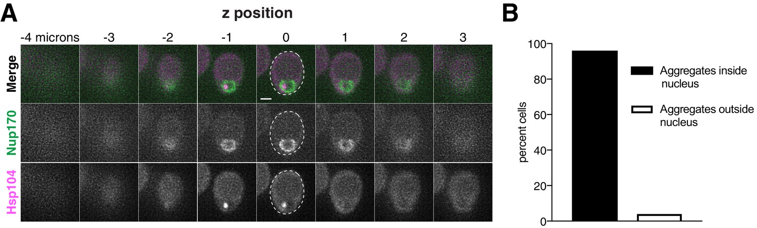
Age-induced protein aggregates are localized inside the nucleus prior to the meiotic divisions.
(A) Z-slices of an aged prophase I cell (seven generations old) depicting localization of NPCs, marked by Nup170-GFP, and protein aggregates, marked by Hsp104-mCherry (UB12975). (B) Quantification depicting frequency of pre-meiotic cells with protein aggregates inside the nucleus (median replicative age = 6, mean replicative age = 6.2 ± 2.1, n = 100 cells). Scale bar, 2 μm.
-
Figure 1—figure supplement 1—source data 1
Numerical values corresponding to the graph in Figure 1—figure supplement 1.
- https://doi.org/10.7554/eLife.47156.005
Figure 1—figure supplement 2

TetR-GFP is not sequestered in aged cells lacking the rDNA-tetO array.
Montage of an aged cell (nine generations old) containing TetR-GFP but lacking the tetO array (UB17509). Chromosomes were visualized with histone marker Hta1-mApple, and the time point depicting anaphase II onset was defined as 0 min as indicated by the arrow. Scale bar, 2 μm.
Figure 2
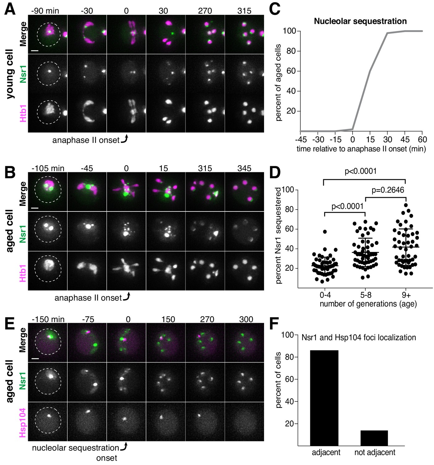
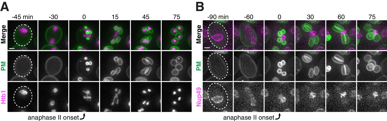
Nucleolar material is sequestered away from chromosomes during meiosis II in young and aged cells.
(A) Montage of a young cell (one generation old) with the nucleolar tag Nsr1-GFP progressing through meiosis (UB16712). (B) Montage of an aged cell (nine generations old) containing abnormal nucleolar material, labeled with Nsr1-GFP, progressing through meiosis (UB16712). For A-B, chromosomes were visualized with the histone marker Htb1-mCherry and the time point depicting anaphase II onset was defined as 0 min as indicated by the arrows. (C) Quantification depicting timing of Nsr1 sequestration relative to timing of anaphase II onset (median replicative age = 8, mean replicative age = 7.2 ± 2.4, n = 50 cells). (D) Quantification depicting the degree of Nsr1 sequestration in cells of different ages (n = 50 for cells with 0–4 doublings, n = 53 for cells with 5–8 doublings, and n = 49 for cells with nine or more doublings). The Mann-Whitney nonparametric test was used to test statistical significance, and data were generated from two biological replicates. (E) Montage of an aged cell (nine generations old) with the nucleolus marked by Nsr1-GFP and protein aggregates marked by Hsp104-mCherry progressing through meiosis (UB13299). For E, the time point depicting Nsr1-GFP sequestration was defined as 0 min as indicated by the arrow. (F) Quantification depicting the frequency of sequestered Hsp104-mCherry aggregates localizing adjacent to sequestered Nsr1-GFP (UB13299) immediately after nucleolar segregation (median replicative age = 7, mean replicative age = 6.7 ± 1.5, n = 100 cells). Adjacency was defined as sequestered Nsr1-GFP signal either neighboring or exhibiting partial overlap with sequestered Hsp104-mCherry signal in individual z-sections. Scale bars, 2 μm.
-
Figure 2—source data 1
Numerical values corresponding to the graph in Figure 2C.
- https://doi.org/10.7554/eLife.47156.012
-
Figure 2—source data 2
Numerical values corresponding to the graph in Figure 2D.
- https://doi.org/10.7554/eLife.47156.013
-
Figure 2—source data 3
Numerical values corresponding to the graph in Figure 2F.
- https://doi.org/10.7554/eLife.47156.014
Figure 3 with 6 supplements

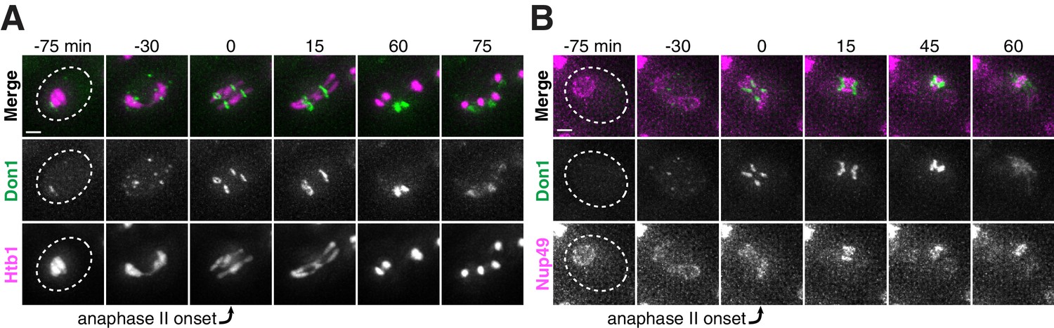
Nucleoporins from the core of the nuclear pore complex, but not the nuclear basket, are sequestered away from chromosomes during meiosis II and subsequently eliminated in young cells.
(A) A schematic depicting the different nucleoporins and subcomplexes that comprise the nuclear pore complex (NPC). Nup100 and Nup145N are not included in the schematic, since they represent linkers between different subcomplexes. Nomenclature and organization are from Beck and Hurt (2017); the schematic itself is adapted from Rajoo et al. (2018). Nucleoporins marked with an asterisk are sequestered away from chromosomes; nucleoporins marked with a pound sign return to dividing nuclei. For each nucleoporin, the observed phenotype was observed in all tetrads examined (n ≥ 25 tetrads). (B–G) Montages of cells with tagged nucleoporins from each subcomplex progressing through meiosis. Chromosomes were visualized with the histone marker Htb1-mCherry, and the first time point depicting anaphase II was defined as 0 min as indicated by the arrows. (B) Nup170-GFP, an inner ring complex nucleoporin (UB11513) (C) Nup120-GFP, a Y-complex nucleoporin (UB13499) (D) Pom34-GFP, a transmembrane nucleoporin (UB13503) (E) Nup82-GFP, a cytoplasmic nucleoporin (UB14652) (F) Nup49-GFP, a channel nucleoporin (UB13509) (G) Nup2-GFP, a nuclear basket nucleoporin (UB15305) (H) Montage depicting localization of Nup2-GFP, a nuclear basket nucleoporin, and Nup49-mCherry, a channel nucleoporin (UB15672). Scale bars, 2 μm.
Figure 3—figure supplement 1
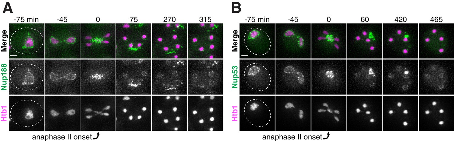
The inner ring complex nucleoporins Nup188 and Nup53 are sequestered away from chromosomes and subsequently eliminated.
(A-B) Montages of young cells with (A) Nup188-GFP (UB13505) and (B) Nup53-eGFP (UB3810) progressing through meiosis. Chromosomes were visualized with the histone marker Htb1-mCherry, and the time point depicting anaphase II onset was defined as 0 min as indicated by the arrows. Scale bars, 2 μm.
Figure 3—figure supplement 2
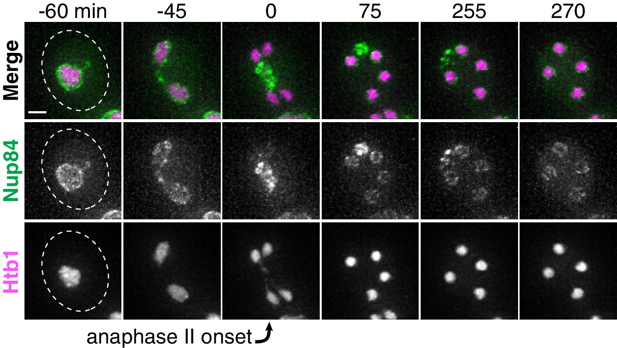
The Y-complex nucleoporin Nup84 is sequestered away from chromosomes and subsequently eliminated.
Montages of a young cell with Nup84-GFP (UB13497) progressing through meiosis. Chromosomes were visualized with the histone marker Htb1-mCherry, and the time point depicting anaphase II onset was defined as 0 min as indicated by the arrow. Scale bar, 2 μm.
Figure 3—figure supplement 3
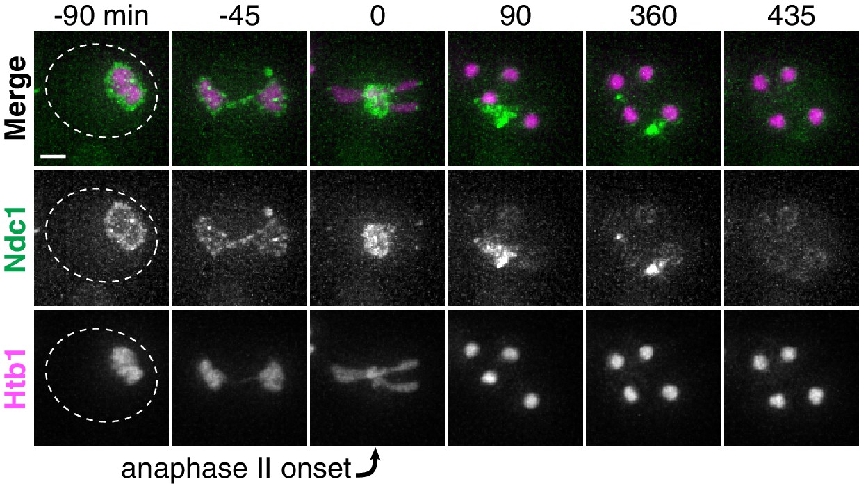
The transmembrane nucleoporin Ndc1 is sequestered away from chromosomes and subsequently eliminated.
Montages of a young cell with Ndc1-GFP (UB15301) progressing through meiosis. Chromosomes were visualized with the histone marker Htb1-mCherry, and the time point depicting anaphase II onset was defined as 0 min as indicated by the arrow. Scale bar, 2 μm.
Figure 3—figure supplement 4
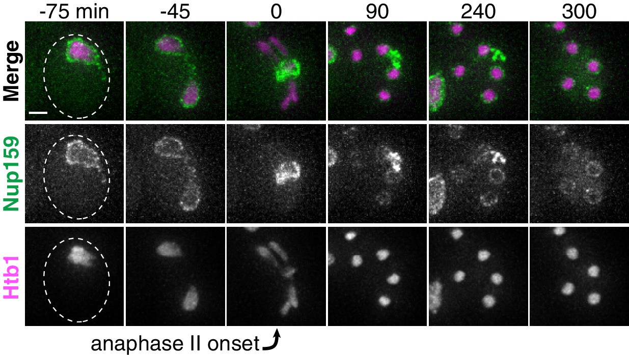
The cytoplasmic nucleoporin Nup159 is sequestered away from chromosomes and subsequently eliminated.
Montages of a young cell with Nup159-GFP (UB14650) progressing through meiosis. Chromosomes were visualized with the histone marker Htb1-mCherry, and the time point depicting anaphase II onset was defined as 0 min as indicated by the arrow. Scale bar, 2 μm.
Figure 3—figure supplement 5
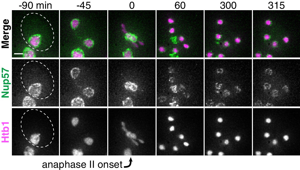
The channel nucleoporin Nup57 is sequestered away from chromosomes and subsequently eliminated.
Montages of a young cell with Nup57-GFP (UB14654) progressing through meiosis. Chromosomes were visualized with the histone marker Htb1-mCherry, and the time point depicting anaphase II onset was defined as 0 min as indicated by the arrow. Scale bar, 2 μm.
Figure 3—figure supplement 6
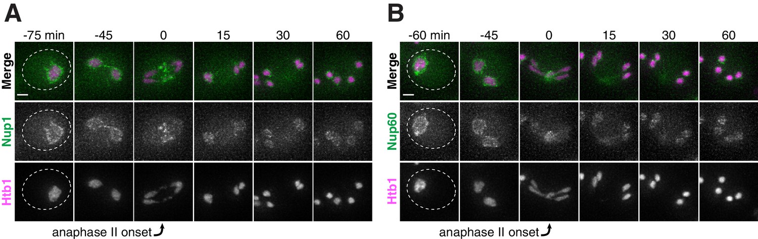
The nuclear basket nucleoporins Nup1 and Nup60 return to dividing nuclei during and after anaphase II.
(A-B) Montages of young cells with (A) Nup1-GFP (UB15303) and (B) Nup60-GFP (UB14646) progressing through meiosis. Chromosomes were visualized with the histone marker Htb1-mCherry, and the time point depicting anaphase II onset was defined as 0 min as indicated by the arrows. Scale bars, 2 μm.
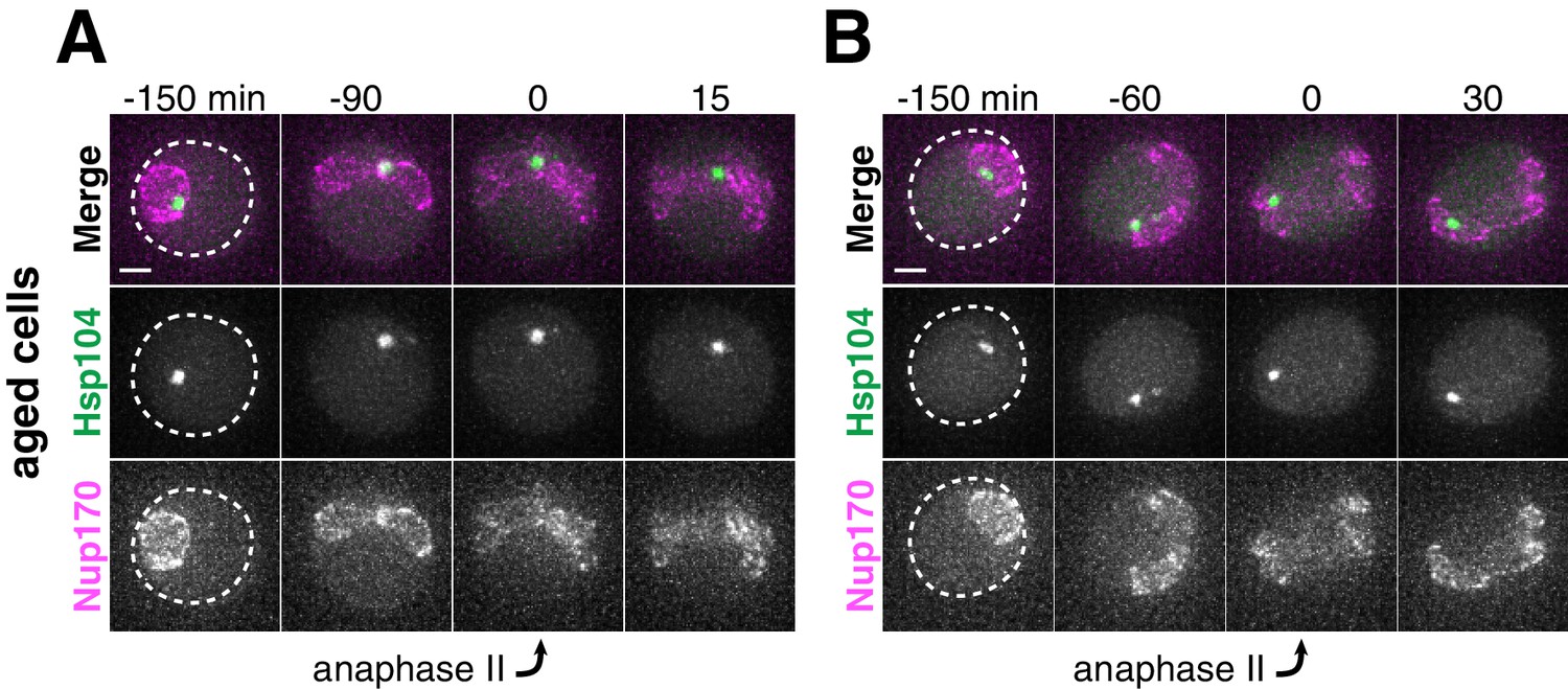
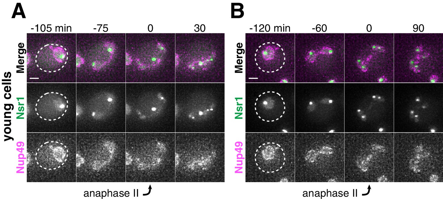
Figure 4 with 2 supplements
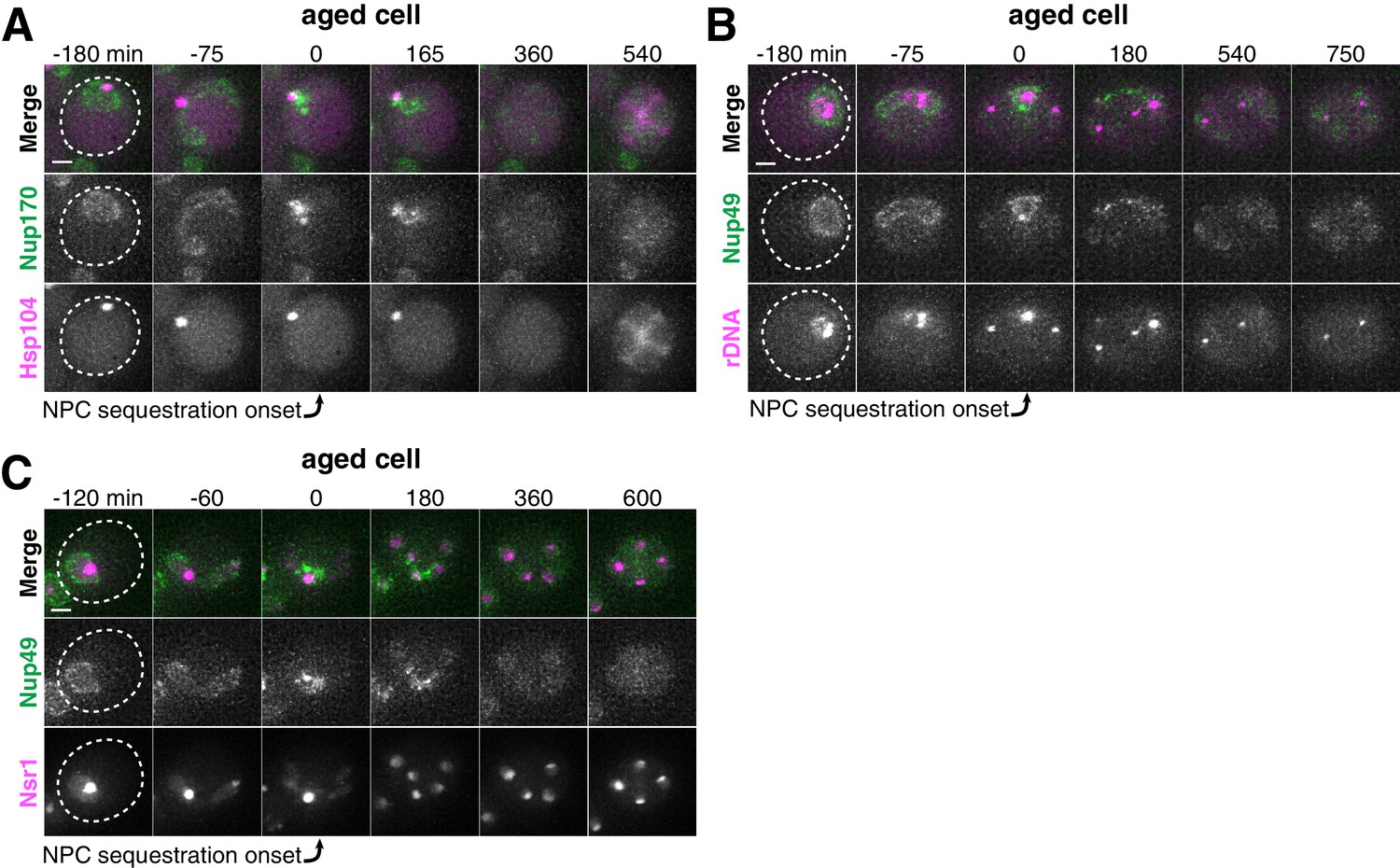
Age-dependent nuclear damage is sequestered with disposed NPCs during anaphase II.
(A) Montage of an aged cell (seven generations old) with protein aggregates, labeled with Hsp104-mCherry, and NPCs, labeled with Nup170-GFP, progressing through meiosis (UB12975). (B) Montage of an aged cell (nine generations old) with rDNA circles, marked by TetR-GFP binding to tetO arrays in the rDNA repeats, and NPCs, labeled with Nup49-mCherry, progressing through meiosis (UB17532). (C) Montage of an aged cell (seven generations old) with abnormal nucleolar material, marked by Nsr1-GFP, and NPCs, marked by Nup49-mCherry, progressing through meiosis (UB16708). The first time point depicting NPC sequestration was defined as 0 min as indicated by the arrows. Scale bar, 2 μm.
Figure 4—figure supplement 1
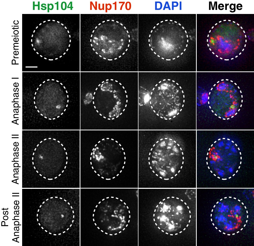
Protein aggregates are sequestered with disposed NPCs in fixed cells.
Maximum intensity projections of fixed premeiotic and meiotic cells depicting localization of NPCs, marked by Nup170-GFP, and protein aggregates, marked by Hsp104-mCherry (UB12975). Scale bar, 2 μm.
Figure 4—figure supplement 2

Nucleolar material is sequestered with disposed NPCs in fixed cells.
Maximum intensity projections of fixed premeiotic and meiotic cells depicting localization of NPCs, marked by Nup49-mCherry, and nucleolar material, marked by Nsr1-GFP (UB16708). Scale bar, 2 μm.
Figure 5 with 2 supplements
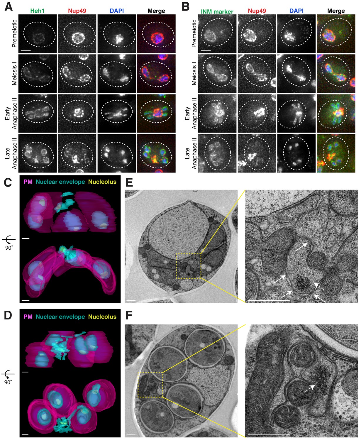
Nucleoporins are sequestered to a nuclear envelope-bound compartment during meiosis II.
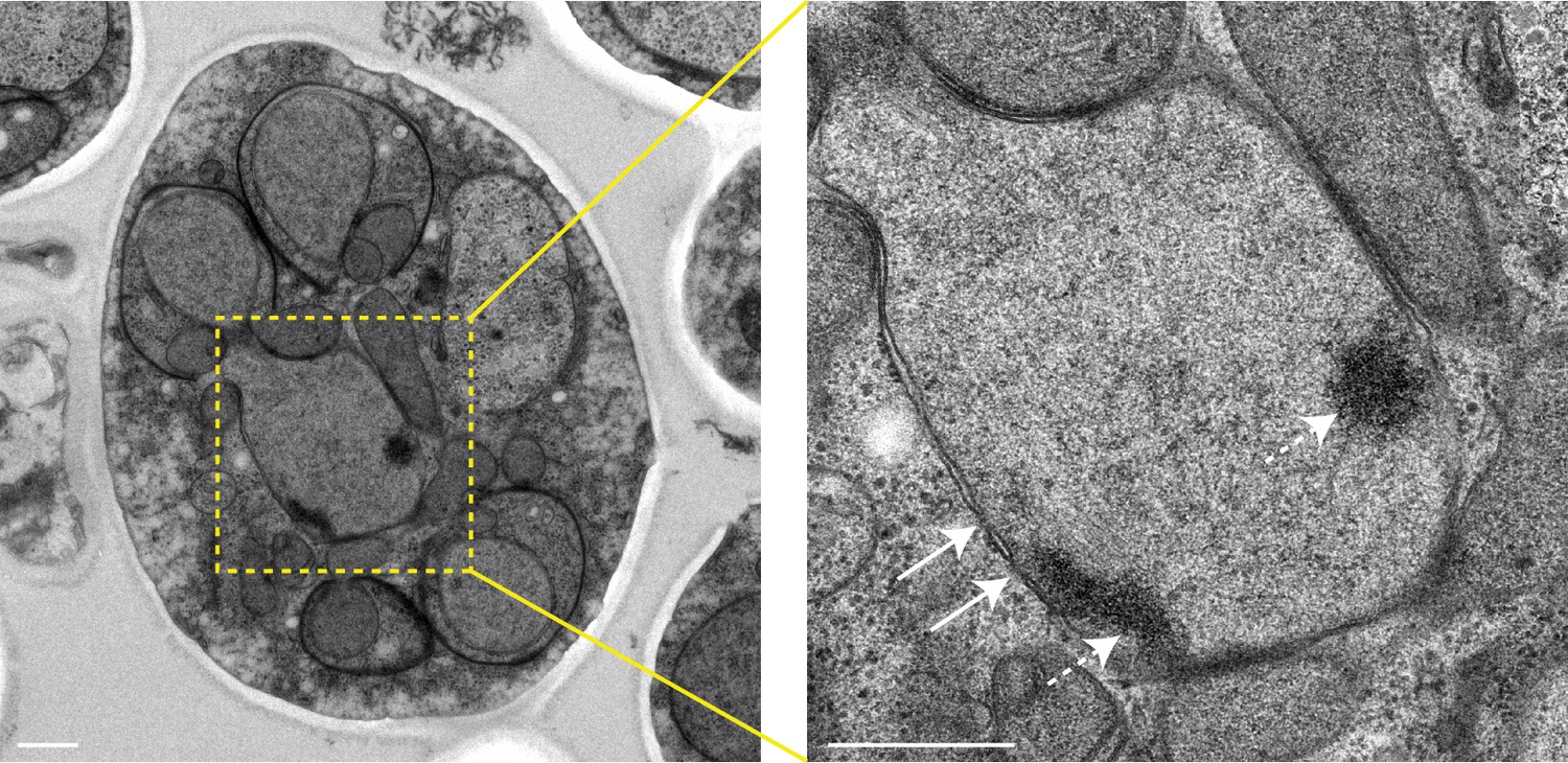
(A-B) Maximum intensity projections of fixed young cells depicting the localization of inner nuclear membrane proteins – (A) Heh1-3xeGFP (UB14391) and (B) the synthetic construct eGFP-h2NLS-L-TM (UB12932) from Meinema et al. (2011) – relative to the nucleoporin Nup49-mCherry and DAPI. Scale bars, 2 μm. (C–D) Reconstructions of (C) a young late anaphase II cell and (D) a young post-meiosis II cell from 70 nm serial TEM micrographs (UB11513). Gamete plasma membranes are depicted in magenta, the nuclear envelope is depicted in cyan, and nucleoli are depicted in yellow. Scale bars, 0.5 μm. (E–F) Electron micrographs of (E) a young late anaphase II cell and (F) a young post-meiosis II cell with insets depicting the nuclear envelope-bound region outside the gamete plasma membranes (UB11513). Solid arrows indicate NPCs; dashed arrows indicate nucleolar mass. Note that the electron micrographs in panel F come from the cell reconstructed in panel D. Scale bars, 0.5 μm.
Figure 5—figure supplement 1
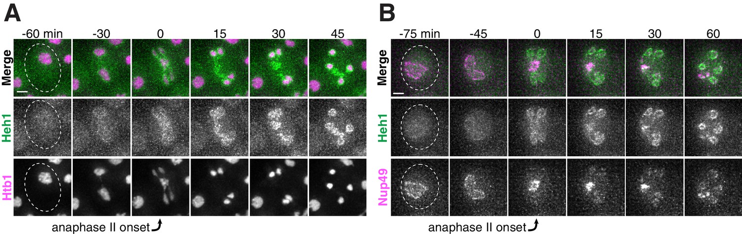
The inner nuclear membrane protein Heh1 localizes to the dividing nuclei and nucleoporin mass during anaphase II.
(A-B) Montages of young cells with a fluorescently-tagged inner nuclear membrane protein Heh1-3xeGFP and either (A) Htb1-mCherry, a histone marker (UB14393), or (B) Nup49-mCherry, a nucleoporin (UB14391). The time point depicting anaphase II onset was defined as 0 min as indicated by the arrows. Scale bars, 2 μm.
Figure 5—figure supplement 2
The nuclear envelope region outside of gamete plasma membranes contains NPCs and nucleolar material during early anaphase II.
Electron micrographs of a young early anaphase II cell and an inset of the nuclear envelope-bound region outside the gamete plasma membranes (UB11513). Solid arrows indicate NPCs; dashed arrows indicate nucleolar mass. Scale bars, 0.5 μm.
Figure 6 with 3 supplements

Core nucleoporins and age-dependent damage are excluded from developing gametes during meiosis II.
(A) Maximum intensity projections over 6 μm of fixed young cells depicting localization of the gamete plasma membrane marker yeGFP-Spo2051-91 relative to the nucleoporin Nup49-mCherry and DAPI (UB12342). (B) Maximum intensity projections over 8 μm of fixed young cells depicting localization of the leading edge complex tag Don1-GFP relative to the nucleoporin Nup49-mCherry and DAPI (UB12436). (C–D) Montages of cells with a protein aggregate tag, Hsp104-mCherry, and a marker of the gamete plasma membrane, yeGFP-Spo2051-91 (UB11821). (C) An aged cell (six generations old) that excluded its protein aggregate from developing gametes. (D) An aged cell (six generations old) that failed to exclude its protein aggregate from developing gametes. For C-D, the first time point depicting gamete plasma membrane nucleation was defined as 0 min as indicated by the arrows. Scale bars, 2 μm.
Figure 6—figure supplement 1
Dynamic localization of sequestered nucleoporins relative to gamete plasma membranes.
(A-B) Montages of young cells progressing through the meiotic divisions with a gamete plasma membrane marker yeGFP-Spo2051-91 and either (A) a histone marker Htb1-mCherry (UB12434), or (B) a nucleoporin marker Nup49-mCherry (UB12342). The time point depicting anaphase II onset was defined as 0 min as indicated by the arrows. Scale bars, 2 μm.
Figure 6—figure supplement 2
Dynamic localization of sequestered nucleoporins relative to the leading edge of gamete plasma membranes.
(A-B) Montages of young cells progressing through the meiotic divisions with the leading edge marker Don1-GFP and either (A) a histone marker Htb1-mCherry (UB12438), or (B) a nucleoporin marker Nup49-mCherry (UB12436). The time point depicting anaphase II onset was defined as 0 min as indicated by the arrows. Scale bars, 2 μm.
Figure 6—figure supplement 3
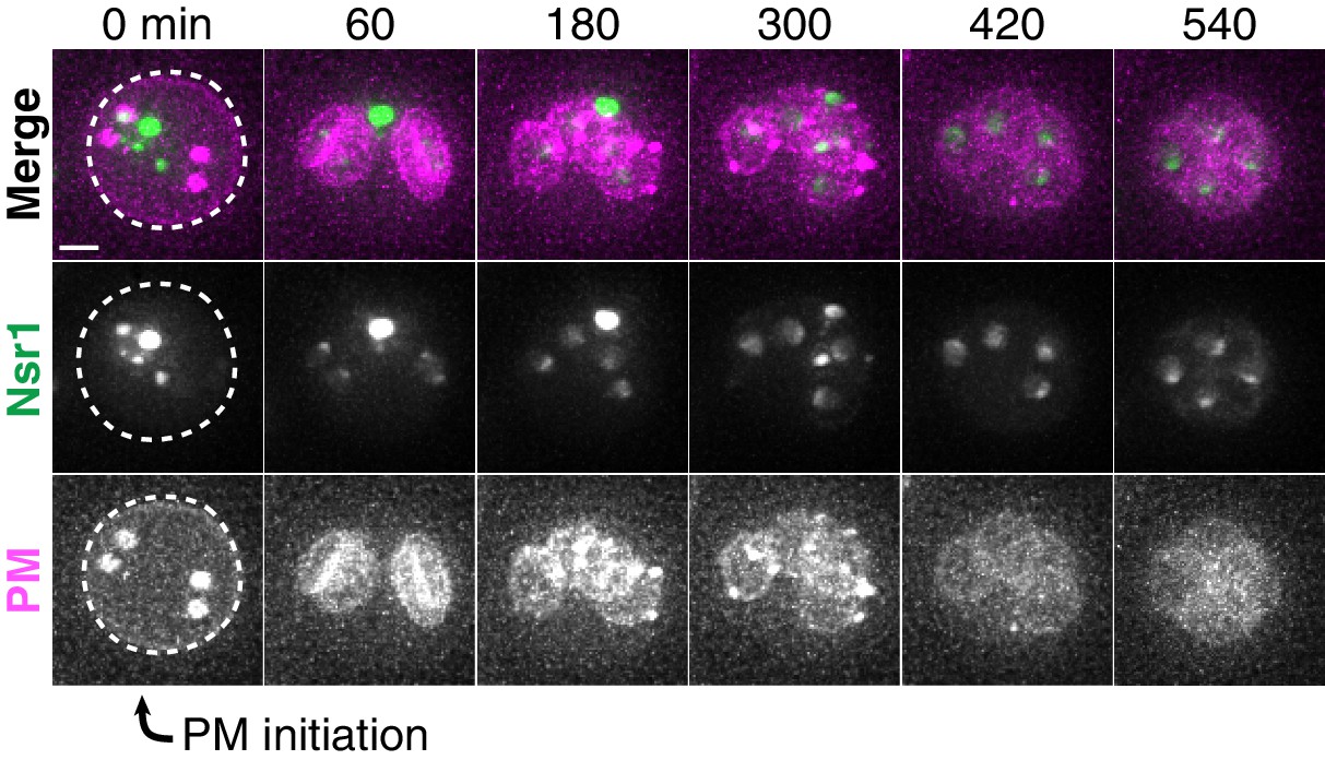
Nucleolar material in aged cells is excluded from the developing gametes.
Montage of an aged cell (seven generations old) excluding nucleolar material marked by Nsr1-GFP during meiosis II (UB16710). Gamete plasma membranes were marked by mKate-Spo2051-91. The time point depicting PM initiation was defined as 0 min as indicated by the arrow. Scale bar, 2 μm.
Figure 7
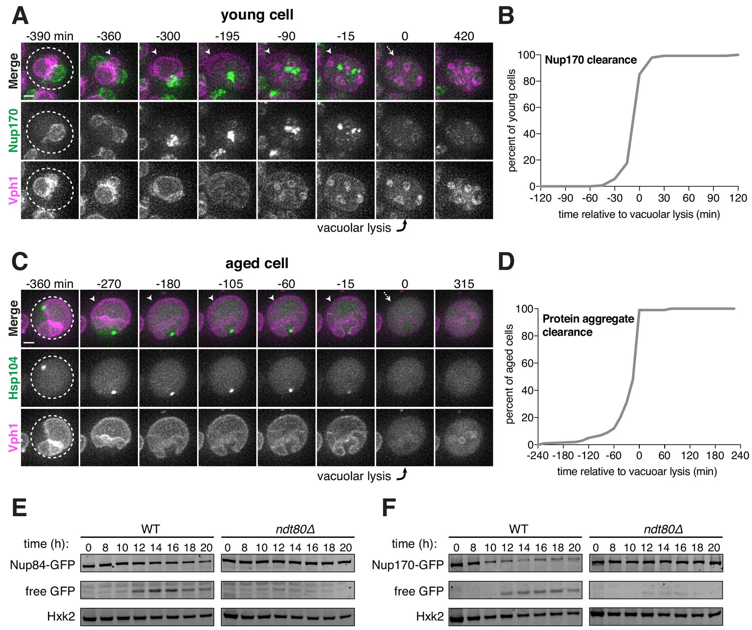
Core nucleoporins and protein aggregates are turned over coincident with vacuolar lysis.
(A) Montage of a young cell with an inner ring complex nucleoporin tag, Nup170-GFP, and a marker for the vacuole, Vph1-mCherry (UB15890). Images are maximum intensity projections over 6 μm; the first time point depicting vacuolar lysis was defined as 0 min as indicated by the arrow. (B) Quantification of the experiment in panel A. Timing of the excluded Nup170-GFP clearance relative to vacuolar lysis (n = 141 cells). (C) Montage of an aged cell (eight generations old) with a protein aggregate tag, Hsp104-mCherry, and a marker for the vacuolar membrane, Vph1-eGFP (UB12163). Images are maximum intensity projections over 8 μm; the first time point depicting vacuolar lysis was defined as 0 min as indicated by the arrow. (D) Quantification of the experiment in panel C. Timing of excluded Hsp104-mCherry clearance relative to vacuolar lysis (median replicative age = 6, mean replicative age = 5.9 ± 1.5, n = 100 cells). For panels A and C, solid arrows indicate the intact vacuolar membrane of the mother cell and dashed arrows indicate vacuolar permeabilization. For panels B and D, vacuolar lysis was scored as the time of vacuolar membrane disappearance. Scale bars, 2 μm. Immunoblot assay measuring degradation of (E) Nup84-GFP in wild type (UB13497) and ndt80Δ cells (UB19929) (F) Nup170-GFP in wild type (UB11513) and ndt80Δ cells (UB19927). Hxk2 levels were measured as a loading control.
-
Figure 7—source data 1
Numerical values corresponding to the graph in Figure 7B.
- https://doi.org/10.7554/eLife.47156.042
-
Figure 7—source data 2
Numerical values corresponding to the graph in Figure 7D.
- https://doi.org/10.7554/eLife.47156.043
Figure 8 with 2 supplements
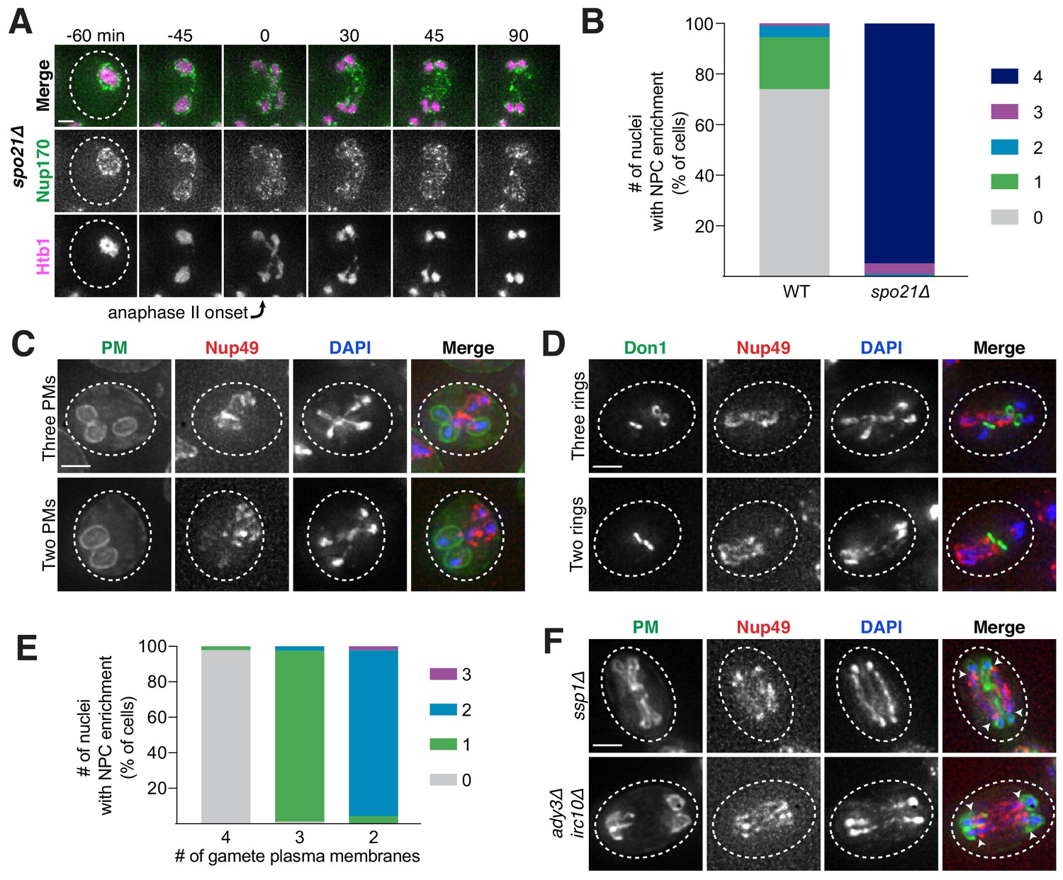
Gamete plasma membrane development is necessary for nucleoporin sequestration.
(A) Montage of inner ring complex nucleoporin Nup170-GFP localization relative to Htb1-mCherry in a young spo21Δ cell (UB13377). The first time point depicting anaphase II was defined as 0 min as indicated by the arrow. (B) Quantification of the experiment in panel A for spo21Δ and Figure 3B for WT. Number of nuclei enriched for nucleoporins following anaphase II in WT or spo21Δ cells (n = 108 cells for WT, n = 118 cells spo21Δ). (C–D) Maximum intensity projections of fixed young cells depicting (C) gamete plasma membrane yeGFP-Spo2051-91 (UB12342) or (D) leading edge Don1-GFP (UB12436) localization relative to nucleoporin Nup49-mCherry localization in low-carbon conditions that promoted the formation of fewer than four gamete plasma membranes. (E) Quantification of the experiment in panel C. Number of nuclei enriched for nucleoporins following anaphase II in cells with variable numbers of gamete plasma membranes (4 PMs: n = 48; 3 PMs: n = 80; 2 PMs: n = 46). (F) Maximum intensity projections of fixed young cells showing gamete plasma membrane (yeGFP-Spo2051-91) and nucleoporin (Nup49-mCherry) localization in mutants defective in leading edge complex formation, ssp1Δ (UB13473) or ady3Δ irc10Δ (UB13583). Arrowheads on merged images denote location of DAPI constrictions. Scale bars, 2 μm.
-
Figure 8—source data 1
Numerical values corresponding to the graph in Figure 8B.
- https://doi.org/10.7554/eLife.47156.049
-
Figure 8—source data 2
Numerical values corresponding to the graph in Figure 8E.
- https://doi.org/10.7554/eLife.47156.050
Figure 8—figure supplement 1
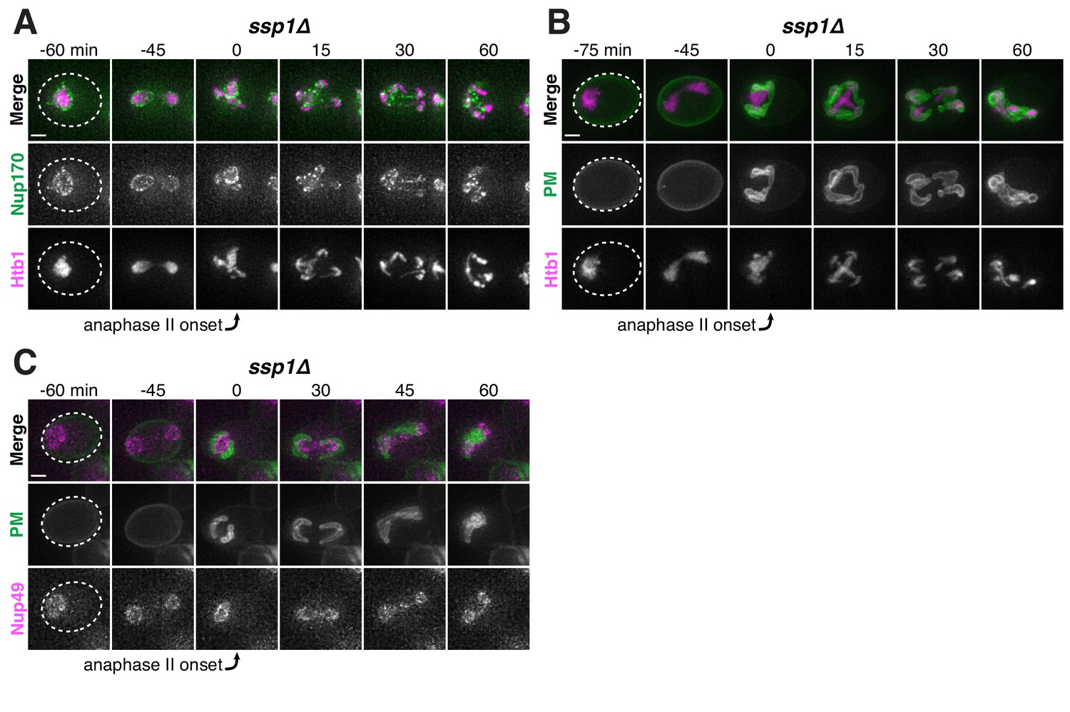
The leading edge complex member Ssp1 is required for proper gamete plasma membrane formation and nucleoporin sequestration.
(A-C) Montages of young ssp1Δ cells progressing through the meiotic divisions with the following tags: (A) nucleoporin Nup170-GFP and histone Htb1-mCherry (UB13373); (B) gamete plasma membrane yeGFP-Spo2051-91 and histone Htb1-mCherry (UB13475); (C) and gamete plasma membrane yeGFP-Spo2051-91 and nucleoporin Nup49-mCherry (UB13473). The time point depicting anaphase II onset was defined as 0 min as indicated by the arrows. Scale bars, 2 μm.
Figure 8—figure supplement 2
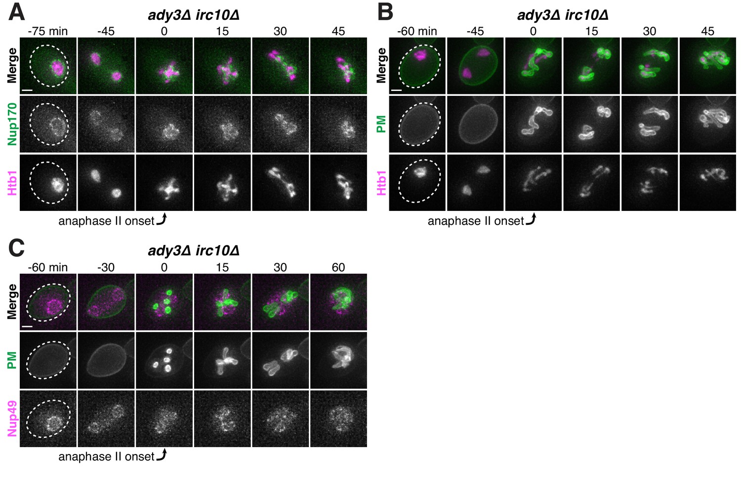
The leading edge complex members Ady3 and Irc10 are required for proper gamete plasma membrane formation and nucleoporin sequestration.
(A-C) Montages of young ady3Δ irc10Δ cells progressing through the meiotic divisions with the following tags: (A) nucleoporin Nup170-GFP and histone Htb1-mCherry (UB12465); (B) gamete plasma membrane yeGFP-Spo2051-91 and histone Htb1-mCherry (UB13585); (C) and gamete plasma membrane yeGFP-Spo2051-91 and nucleoporin Nup49-mCherry (UB13583). The time point depicting anaphase II onset was defined as 0 min as indicated by the arrows. Scale bars, 2 μm.
Figure 9 with 2 supplements
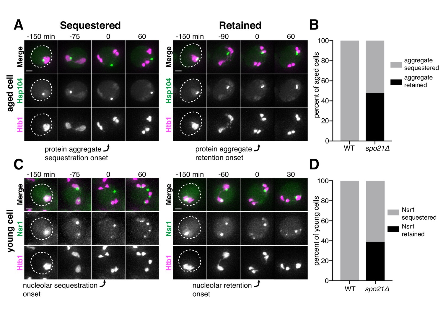
Protein aggregate and nucleolar sequestration is coupled to NPC sequestration via gamete plasma membrane development.
(A) Montages of aged spo21Δ cells in which protein aggregates marked by Hsp104-eGFP were either (left panel, five generations old) sequestered away from or (right panel, six generations old) retained by chromosomes during anaphase II (UB14418). (B) Quantification of protein aggregate retention in aged WT (UB9724) and spo21Δ cells (UB14418). Median replicative age = 7, mean replicative age = 6.5 ± 1.5, n = 100 for WT cells; median replicative age = 6, mean replicative age = 6.2 ± 1.2, n = 100 for spo21Δ cells. (C) Montages of young spo21Δ cells in which nucleolar material was either (left panel) sequestered away from or (right panel) retained by chromosomes during anaphase II (UB 14419). (D) Quantification of Nsr1 retention in young WT (UB15118) and spo21Δ cells (UB14419). n = 100 for WT cells, n = 100 for spo21Δ cells. For A and C, chromosomes were visualized with the histone marker Htb1-mCherry. For A, the first time point depicting protein aggregate sequestration or retention was defined as 0 min as indicated by the arrows. For C, the first time point depicting nucleolar sequestration or retention was defined as 0 min as indicated by the arrows. Scale bars, 2 μm.
-
Figure 9—source data 1
Numerical values corresponding to the graph in Figure 9B.
- https://doi.org/10.7554/eLife.47156.055
-
Figure 9—source data 2
Numerical values corresponding to the graph in Figure 9D.
- https://doi.org/10.7554/eLife.47156.056
Figure 9—figure supplement 1
Protein aggregates co-localize with NPCs during anaphase II in spo21Δ cells.
(A-B) Montages of aged spo21Δ cells with protein aggregates labeled with Hsp104-mCherry and NPCs labeled with Nup170-GFP (UB13568). (A) Montage of a cell that sequesters protein aggregates during meiosis II (seven generations old). (B) Montage of a cell that retains protein aggregates during meiosis II (six generations old). The time point depicting anaphase II was defined as 0 min as indicated by the arrows. Scale bars, 2 μm.
Figure 9—figure supplement 2
Nucleolar material co-localizes with NPCs during anaphase II in spo21Δ cells.
(A-B) Montages of young spo21Δ cells with nucleolar material labeled with Nsr1-GFP and NPCs labeled with Nup49-mCherry (UB14425). (A) Montage of a cell that sequesters an Nsr1-GFP punctum during meiosis II. (B) Montage of a cell that retains all Nsr1-GFP during meiosis II. The time point depicting anaphase II was defined as 0 min as indicated by the arrows. Scale bars, 2 μm.
Figure 10
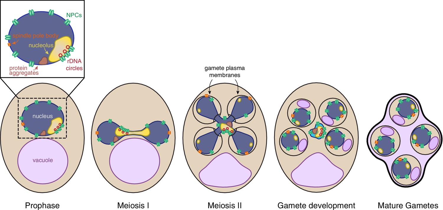
Nuclear rejuvenation during meiosis.
Aged yeast cells accumulate nuclear damage including extrachromosomal rDNA circles (red), nuclear-associated protein aggregates (brown), abnormal and enlarged nucleoli (yellow), and misorganized NPCs (green). During meiosis II, a nuclear envelope-bound compartment (light blue) containing much of this age-associated damage is formed and remains outside of the developing gametes. The material in the excluded compartment is turned over coincident with vacuolar lysis, completing rejuvenation of the gamete nuclei. Sequestration of the age-dependent damage away from gamete nuclei requires proper gamete plasma membrane development during anaphase II.
Author response image 1

Author response image 2
Videos
Video 1
Protein aggregates are sequestered away from chromosomes during meiosis II and subsequently eliminated.
An aged cell (seven generations old) with protein aggregates undergoing gametogenesis as depicted in Figure 1B. Protein aggregates were followed with Hsp104-eGFP and meiotic staging was followed using a histone marker Htb1-mCherry (UB9724). Movie frame rate, four frames per second. Scale bar, 2 μm.
Video 2
rDNA circles are sequestered away from chromosomes during meiosis II and subsequently eliminated.
An aged cell (nine generations old) containing tetO arrays in rDNA repeats undergoing gametogenesis as depicted in Figure 1C. rDNA was visualized with TetR-GFP and meiotic staging was followed using a histone marker Hta1-mApple (UB17338). Movie frame rate, four frames per second. Scale bar, 2 μm.
Video 3
Abnormal nucleolar material is sequestered away from chromosomes during meiosis II and subsequently eliminated.
An aged cell (nine generations old) with abnormal nucleolar material undergoing gametogenesis as depicted in Figure 2B. Nucleoli were followed with Nsr1-GFP and meiotic staging was followed using a histone marker Htb1-mCherry (UB16712). Movie frame rate, four frames per second. Scale bar, 2 μm.
Video 4
Core nucleoporins are sequestered away from chromosomes during meiosis II and subsequently eliminated.
A young cell with a tagged inner ring complex nucleoporin, Nup170-GFP, during gametogenesis as depicted in Figure 3B (UB11513). Meiotic staging was followed using a histone marker, Htb1-mCherry. Movie frame rate, four frames per second. Scale bar, 2 μm.
Video 5
Nuclear basket nucleoporins are not sequestered with core nucleoporins during meiosis II.
A young cell with a tagged nuclear basket nucleoporin, Nup2-GFP, and a tagged channel nucleoporin, Nup49-mCherry, during gametogenesis as depicted in Figure 3H (UB15672). Movie frame rate, four frames per second. Scale bar, 2 μm.
Video 6
A nuclear envelope-bound compartment remains outside of developing gametes during meiosis II.
Serial TEM micrographs of a young cell during late meiosis II used to construct the model shown in Figure 5C (UB11513). Section thickness, 70 nm. Scale bar, 0.5 μm.
Video 7
A nuclear envelope-bound compartment remains outside of developing gametes during meiosis II.
Reconstruction of a young cell during late meiosis II from 70 nm serial TEM micrographs as depicted in Figure 5C (UB11513). Plasma membranes are depicted in magenta, the nuclear envelope is depicted in cyan, and nucleoli are depicted in yellow. Scale bar, 0.5 μm.
Video 8
A nuclear envelope-bound compartment remains outside of gametes during gamete development.
Serial TEM micrographs of a young cell during gamete development used to construct the model shown in Figure 5D (UB11513). Section thickness, 70 nm. Scale bar, 0.5 μm.
Video 9
A nuclear envelope-bound compartment remains outside of gametes during gamete development.
Reconstruction of a young cell during gamete development from 70 nm serial TEM micrographs as depicted in Figure 5D (UB11513). Plasma membranes are depicted in magenta, the nuclear envelope is depicted in cyan, and nucleoli are depicted in yellow. Scale bar, 0.5 μm.
Video 10
Protein aggregates excluded from developing gametes are eliminated.
An aged cell (six generations old) with protein aggregates undergoing gametogenesis as depicted in Figure 6C. Protein aggregates were followed with Hsp104-mCherry and gamete plasma membrane formation was followed using yeGFP-Spo2051-91 (UB11821). Movie frame rate, four frames per second. Scale bar, 2 μm.
Video 11
Protein aggregates inherited by developing gametes are not eliminated.
An aged cell (six generations old) with protein aggregates undergoing gametogenesis as depicted in Figure 6D. Protein aggregates were followed with Hsp104-mCherry and gamete plasma membrane formation was followed using yeGFP-Spo2051-91 (UB11821). Movie frame rate, four frames per second. Scale bar, 2 μm.
Video 12
Core nucleoporins are eliminated coincident with vacuolar lysis.
A young cell with an inner ring complex nucleoporin tag, Nup170-GFP, and a marker for the vacuole, Vph1-mCherry, undergoing gametogenesis as depicted in Figure 7A (UB15890). Movie frame rate, four frames per second. Scale bar, 2 μm.
Video 13
Protein aggregates are eliminated coincident with vacuolar lysis.
An aged cell (eight generations old) with protein aggregates undergoing gametogenesis as depicted in Figure 7C. Protein aggregates were followed with Hsp104-mCherry and vacuolar lysis was followed using vacuolar membrane marker Vph1-eGFP (UB12163). Movie frame rate, four frames per second. Scale bar, 2 μm.
Video 14
Gamete plasma membrane development is necessary for nucleoporin sequestration.
A young spo21Δ cell with inner ring complex nucleoporin tag Nup170-GFP and histone marker Htb1-mCherry undergoing gametogenesis as depicted in Figure 8A (UB13377). Movie frame rate, four frames per second. Scale bar, 2 μm.
Additional files
-
Supplementary file 1
Strain table.
- https://doi.org/10.7554/eLife.47156.058
-
Supplementary file 2
Primers used for strain construction.
- https://doi.org/10.7554/eLife.47156.059
-
Supplementary file 3
Plasmids used for strain construction.
- https://doi.org/10.7554/eLife.47156.060
-
Supplementary file 4
Imaging conditions.
Transmission, exposure time, and excitation/emission wavelengths are specified for each channel. Distance between z-sections and number of z-sections acquired are indicated.
- https://doi.org/10.7554/eLife.47156.061
-
Supplementary file 5
Meiotic septin and leading edge complex genes are not required for nuclear pore complex or protein aggregate sequestration.
Movies of strains with the indicated deletion, and either (1) a fluorescently tagged inner ring complex nucleoporin (Nup170-GFP) and a meiotic staging marker (Htb1-mCherry) or (2) a fluorescently tagged chaperone that marks age-induced protein aggregates (Hsp104-mCherry) and a gamete plasma membrane marker (yeGFP-Spo2051-91) were generated. For mutants with successful spore packaging, at least 25 tetrads were observed. For mutants with poor or unsuccessful spore packaging, at least 50 cells that proceeded through MII were observed and compared to wild type (UB11513 for Nup170-GFP; UB11821 for Hsp104-mCherry).
- https://doi.org/10.7554/eLife.47156.062
-
Transparent reporting form
- https://doi.org/10.7554/eLife.47156.063
Download links
A two-part list of links to download the article, or parts of the article, in various formats.
Downloads (link to download the article as PDF)
Open citations (links to open the citations from this article in various online reference manager services)
Cite this article (links to download the citations from this article in formats compatible with various reference manager tools)
Meiotic cellular rejuvenation is coupled to nuclear remodeling in budding yeast
eLife 8:e47156.
https://doi.org/10.7554/eLife.47156


