Giant extinct caiman breaks constraint on the axial skeleton of extant crocodylians
- University of Zurich, Palaeontological Institute and Museum, Switzerland
- Department of Comparative Biomedical Sciences, The Royal Veterinary College, United Kingdom
- Dipartimento di Scienze della Terra, Università di Torino, Italy
- Institut Català de Paleontologia Miquel Crusafont, Universitat Autònoma de Barcelona, Spain
- Museo Paleontológico de Urumaco, Venezuela
Figures
Figure 1 with 1 supplement

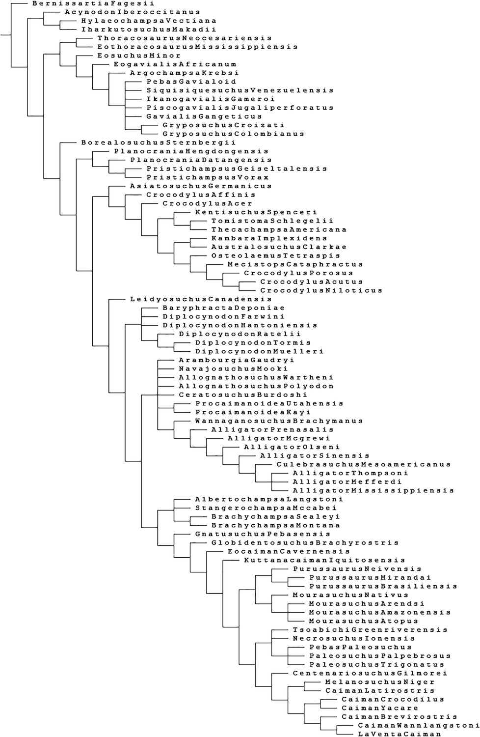
Phylogenetic analysis with updated scoring of Purussaurus mirandai based on AMU-CURS-541 (see Materials and methods for explanations).
Here only an excerpt of the Caimaninae clade of the strict consensus tree is shown to serve as phylogenetic framework of P. mirandai (in bold). Bremer support values are given above the branches. For the remainder of the topology see the results section and Figure 1—figure supplement 1 (see also Salas-Gismondi et al., 2015: supplementary fig. S6).
Figure 1—figure supplement 1
Full topology of the strict consensus tree of 20 most parsimonious trees (tree length = 687 steps; CI = 0.383, RI = 0.806) recovered in main analysis.
The scorings for P. mirandai were updated based on AMU-CURS-541. An excerpt of the Caimaninae clade is shown in Figure 1 in the main text.
Figure 2 with 1 supplement
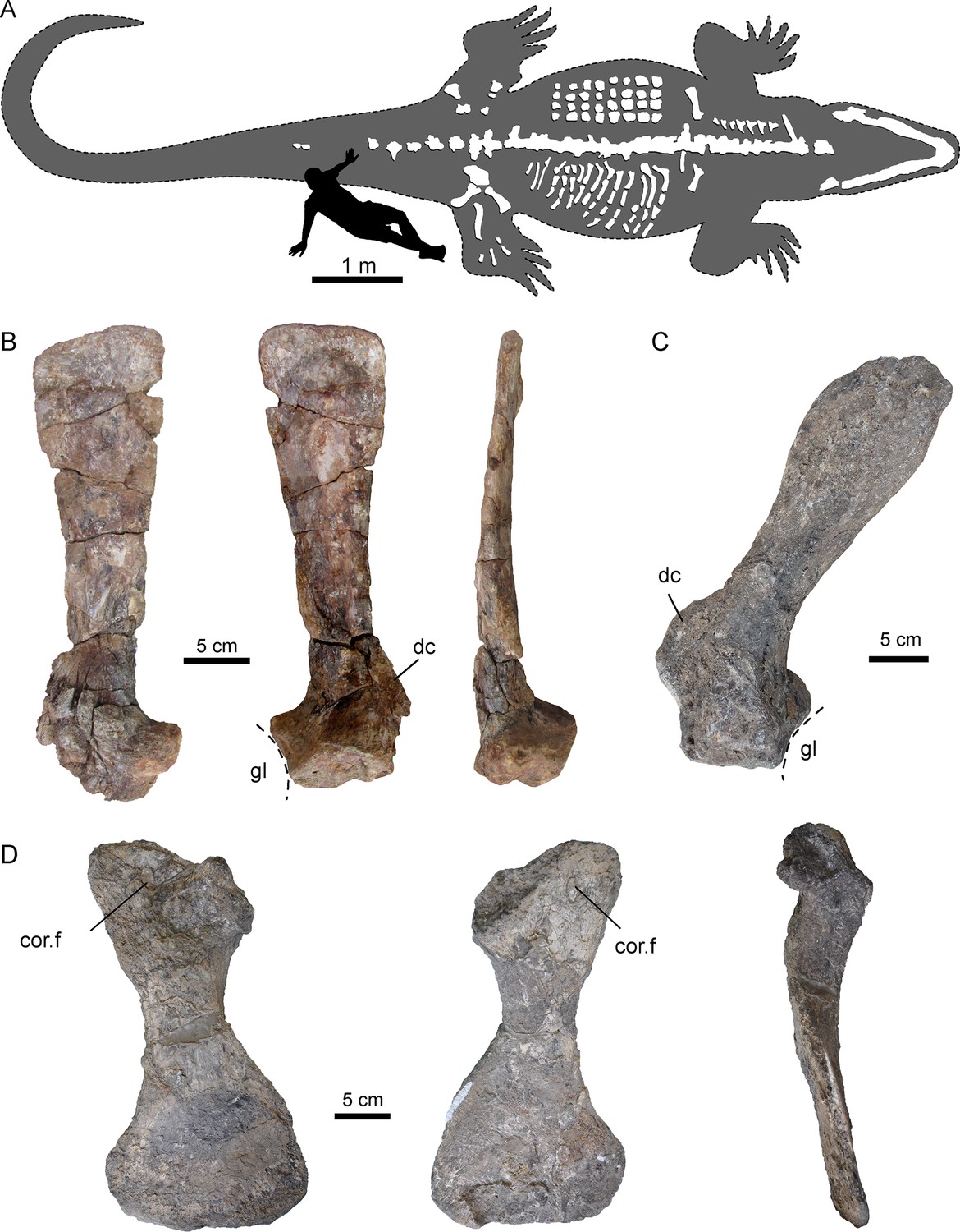
Selected pectoral bones of AMU-CURS-541 and UNEFM-CIAAP-1367 of Purussaurus from the Urumaco Formation of Venezuela.
(A) Interpretative reconstruction of the complete body outline of P. mirandai (AMU-CURS-541) showing the preserved and assembled postcranial bones and the lower jaw in tentative live position. Osteoderms (in upper part of trunk) and ribs (in lower part of trunk) are not in life position. The second author (OS) serves as scale (see Figure 2—figure supplement 1). (B) Left scapula of AMU-CURS-541 in lateral, medial, and posterior view. (C) Right scapula of Purussaurus cf. P. mirandai (UNEFM-CIAAP-1367) in medial view. (D) Right coracoid (UNEFM-CIAAP-1367) in dorsomedial, ventrolateral, and anterior view. Abbreviations: cor.f, coracoid foramen; dc, deltoid crest of scapula; gl, glenoid fossa.
Figure 2—figure supplement 1
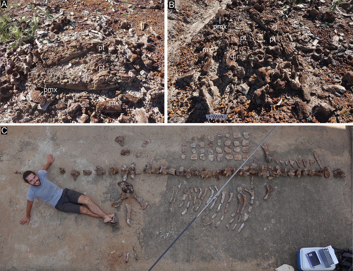
Specimen AMU-CURS-541 of Purussaurus mirandai in the field at ‘North of El Picache’ locality, Urumaco, Falcón state, Venezuela.
The lower jaw is still largely articulated, whereas fragments of the skull and postcranial elements are found scattered around it. (A) lower jaw in left laterodorsal view. (B) lower jaw in posterodorsal view. (C) Postcranial bones laid out in approximate life positions. Note that osteoderms (upper part) and ribs (lower part) have been placed separately next to the vertebral column. Abbreviations: j, jugal; mx, maxilla; pmx, premaxilla; pt, pterygoid; q, quadrate.
Figure 3 with 2 supplements
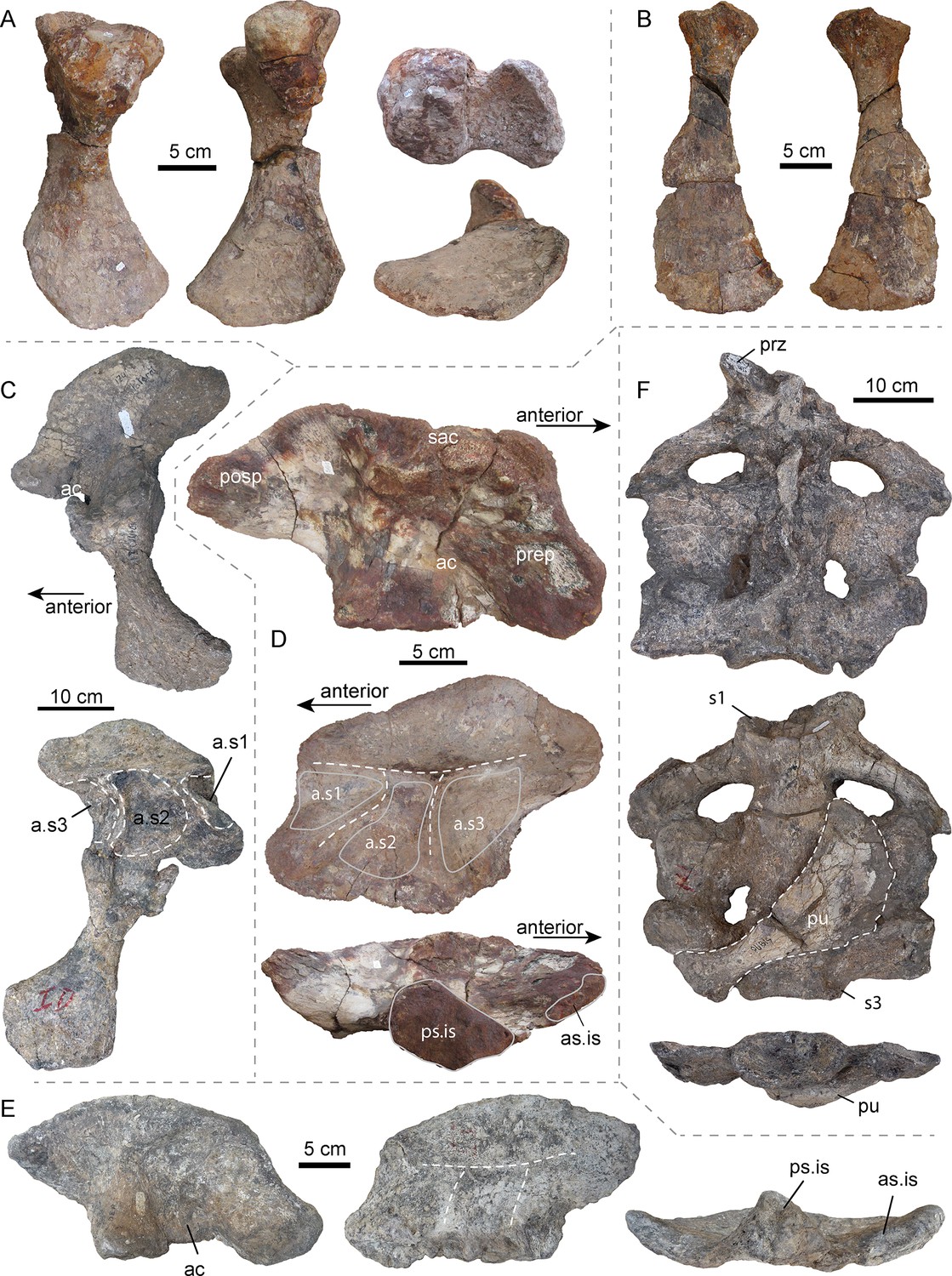
Selected pelvic and sacral bones of AMU-CURS-541 and other specimens of Purussaurus from the Urumaco Formation of Venezuela.
( A) Left ischium (AMU-CURS-541) in posterior, anterior, dorsal (above), and ventral (below) view. (B) Right pubis (AMU-CURS-541) in ventral and dorsal view. (C) Articulated left ilium and ischium (non-holotype, additional bones accessioned under UNEFM-CIAAP-1369) in lateral and medial view. Note the three large depressions separated by ridges, that is attachment sites for three sacral ribs (a.s1-a.s3). (D) Right ilium (AMU-CURS-541) in lateral, medial, and ventral view. (E) Right ilium (UNEFM-CIAAP-1367) in lateral, medial, and ventral view. Stippled white lines indicate the weathered ridges separating the articulation facets for the sacral ribs. (F) Sacral region (non-holotype, additional bones accessioned under UNEFM-CIAAP-1369) consisting of three sacrals in articulation in dorsal, ventral, and posterior view. Note the right pubis (in dorsal view) attached ventrally to the sacrals. Abbreviations: ac, acetabulum; a.s1-a.s3, attachment sites for three sacral ribs; as.is, anterior articular surface for ilium; posp, postacetabular process; prep, preacetabular process; prz, prezygapophysis; ps.is, posterior articular surface for ischium; pu, pubis; s1-s3: sacral vertebrae/ribs 1–3; sac, supraacetabular crest.
Figure 3—figure supplement 1
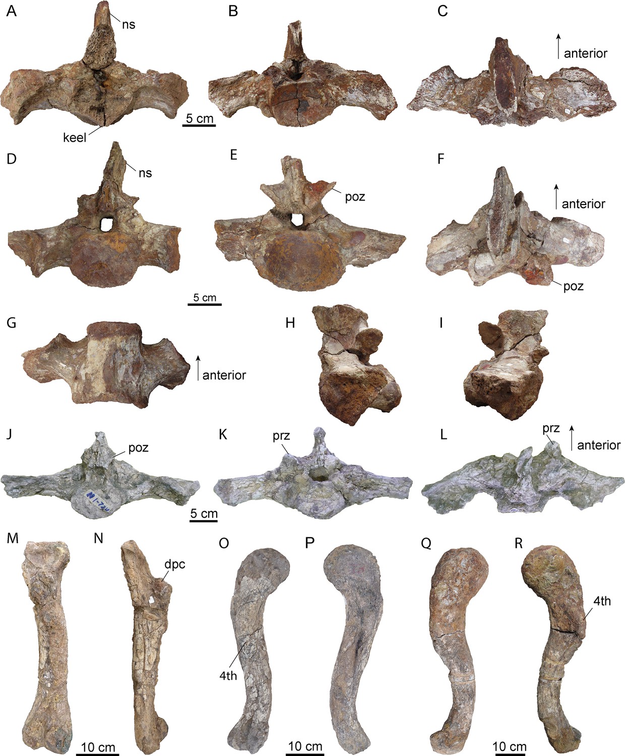
Selected sacral vertebrae and long bones of Purussaurus from the late Miocene Urumaco Formation.
Two sacral vertebrae (A–C): sacral 2 = primordial sacral 1; D–I): sacral 3 = primordial sacral 2) of Purussaurus mirandai (AMU-CURS-541). (A) anterior view. (B) posterior view. (C) dorsal view. (D) anterior view. (E) posterior view. (F) dorsal view. (G) ventral view. (H) left lateral view. (I) right lateral view. Sacral vertebra (identified as the dorsosacral) of Purussaurus (MCNC-URU-111–72V) in J) posterior K) anterior and L) dorsal view. Right humerus (UNEFM-CIAAP-1369, non-holotype material) in M) ventral and N) anterior view. Left femur of the holotype of Purussaurus mirandai (UNEFM-CIAAP-1369) in O) ventral and P) dorsal view. Right femur of Purussaurus sp. (AMU-CURS-20, associated with two dorsal vertebrae) in Q) dorsal and R) ventral view. Abbreviations: 4th, fourth trochanter; dpc, deltopectoral crest; ns, neural spine; poz, postzygapophysis; prz, prezygapophysis.
Figure 3—figure supplement 2
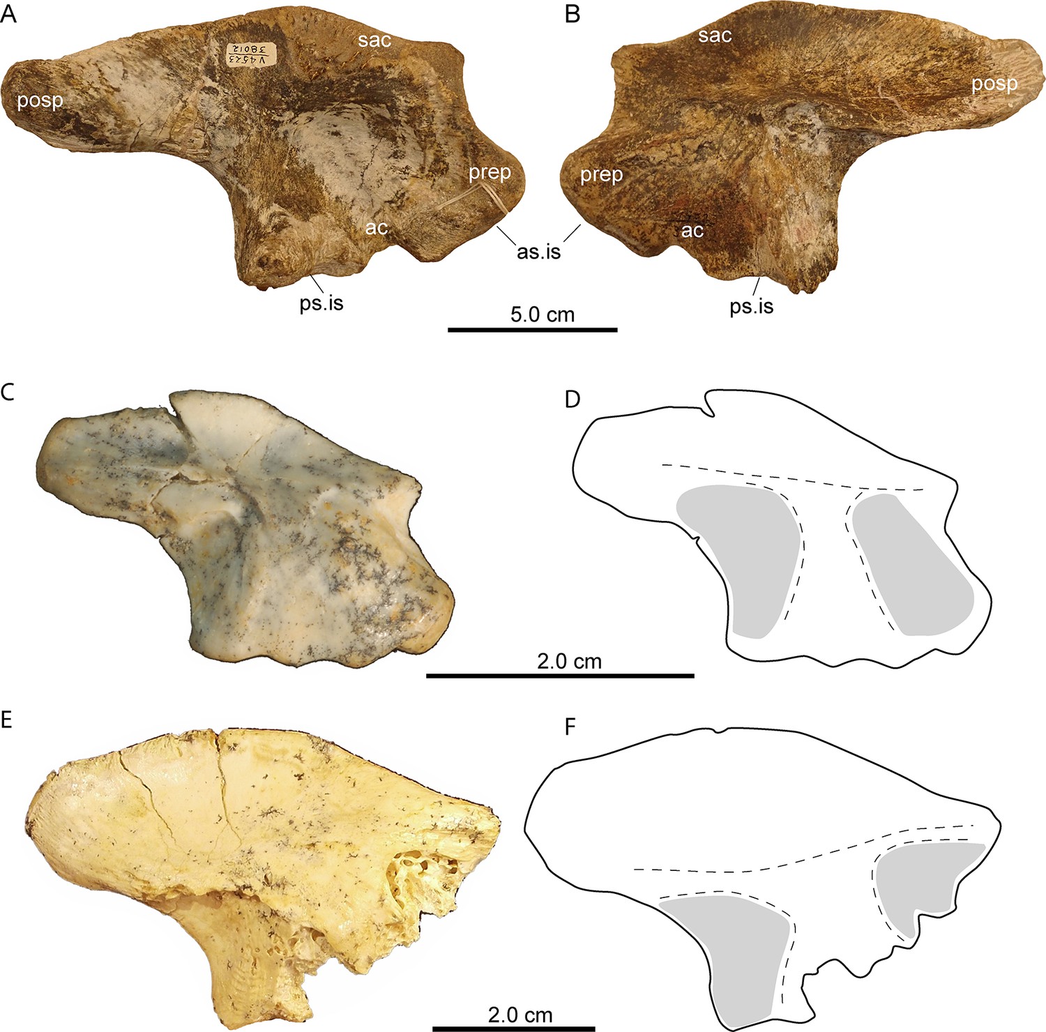
Right ilium (UCMP 38012) of Mourasuchus atopus, La Venta, Colombia [picture courtesy: P. Holroyd, Berkeley] and left ilia of mekosuchine ‘pelvic forms three and four’ (from Stein et al., 2017, figures 6B and 7B; images published under Creative Commons CC-BY 4.0).
(A) Lateral view. (B) medial view. (C) medial view of QM F41198 representing ‘pelvic form three’. (D) interpretative drawing of image in (C, E) medial view of QM F57913 representing ‘pelvic form four’. (F) interpretative drawing of image in (E). Note the space between the articular facets (in grey) in both interpretative drawings. Abbreviations: as.is, anterior articular surface for ischium; ac, acetabulum; posp, postacetabular process; prep, preacetabular process; ps.is, posterior articular surface for ischium; sac, supraacetabular crest.
Figure 4
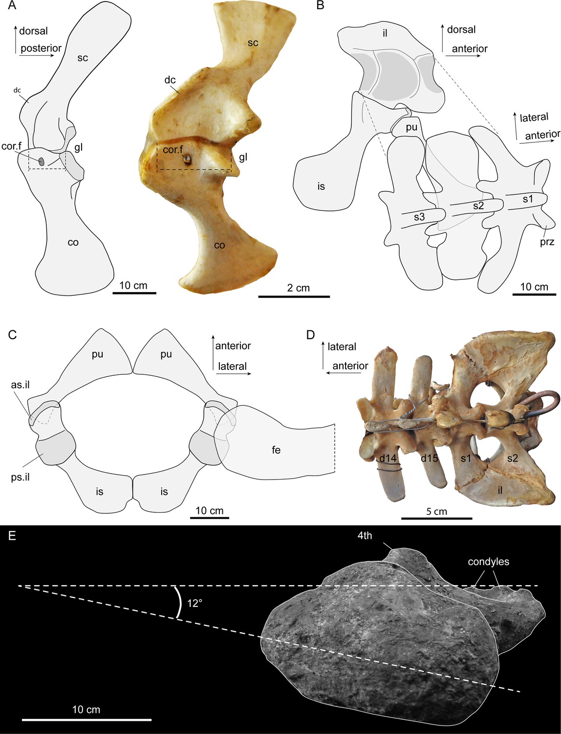
Interpretative sketches of girdle articulation in Purussaurus, based on the girdle bones of several specimens (AMU-CURS-541; UNEFM-CIAAP 1367; UNEFM-CIAAP-1369) in comparison to selected extant bones.
(A) Right pectoral girdle in medial view in comparison with a left (mirrored) pectoral girdle of an extant Crocodylus niloticus. Stippled lines indicate the width of the bony articulation between scapula and coracoid. (B) Left pelvic girdle in medial view and sacral vertebral portion in angled dorsal view. (C) Pubes and ischia (bones were mirrored for completion) and superimposed femoral head in dorsal view. (D) Dorsosacral transition of the vertebral column in an extant Caiman yacare. (E) Proximal view of femur revealing low torsion (12°) of the femoral head to the plane of the distal condyles. Abbreviations: as.il, anterior articular surface for ilium; 4th, fourth trochanter; co, coracoid; cor.f, coracoid foramen; d14-d15, the 14th and 15th dorsal vertebra; dc, deltoid crest; fe, femur; gl, glenoid fossa; il, ilium; is, ischium; pu, pubis; prz, prezygapophysis; ps.il, posterior articular surface for ilium; s1-s3, sacral vertebrae and ribs 1–3; sc, scapula.
Figure 5 with 1 supplement

Rendered 3D-models of a juvenile dwarf caiman Palaeosuchus palpebrosus (RVC-JRH-PP4; not to scale).
(A) Rendering of the complete specimen. (B) Model with only the axial and partial appendicular skeleton (in dorsal view) highlighted. Vertebrae are shown in light blue, except those of the sacral region, in which the dorsosacral in lavender and the true or primordial sacrals in dark blue. (C) Pectoral girdle elements in left lateral, anterior, and dorsal view. (D) Sacral region in left lateral, dorsal (and dorsal with femur included), and anterior view. Note asymmetry of the dorsosacral. The medial view of the right ilium reveals the ‘τ' ridges separating the two articular surfaces of the sacral ribs. The medial side of the left ilium (not shown) looks similar, because the sacralised rib of the dorsosacral articulated only slightly with the anterior margin of the ilium, leaving no deep articular scar. In the right femur, a torsional angle of 53.5° of the head to the plane of the distal condyles was measured. (E) Focus on the posterior dorsal, sacral and anterior caudal series of the skeleton in dorsal, ventral and right lateral view. The inferred asymmetrical shift of the domain boundaries of Hoxc8 and Hoxa11 leading to congenital malformation lumbosacral transitional vertebra in this specimen of P. palpebrosus is indicated. Abbreviations: ac, acetabulum; c1, first caudal vertebra; co, coracoid; d14, 14th dorsal vertebra; fe, femur; hu, humerus; ic, interclavicle; il, ilium; is, ischium; prz, prezygapophysis; pu, pubis; s1-s3: sacral vertebrae/ribs 1–3; sc, scapula; sr, sacral rib; tp, transverse process.
Figure 5—figure supplement 1
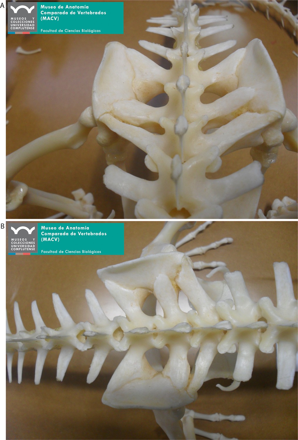
Close-up of the sacral region of specimen MACV-6139 of the extant dwarf caiman Palaeosuchus palpebrosus [picture courtesy: M. Padilla Cano, Madrid].
Additional files
Download links
A two-part list of links to download the article, or parts of the article, in various formats.
Downloads (link to download the article as PDF)
Open citations (links to open the citations from this article in various online reference manager services)
Cite this article (links to download the citations from this article in formats compatible with various reference manager tools)
Giant extinct caiman breaks constraint on the axial skeleton of extant crocodylians
eLife 8:e49972.
https://doi.org/10.7554/eLife.49972


