Yolk-sac-derived macrophages progressively expand in the mouse kidney with age
- Division of Nephrology, Department of Medicine, Duke University School of Medicine, United States
- Regeneration Next, Duke University, United States
- Department of Orthopedic Surgery, Duke University School of Medicine, United States
- Department of Orthopedic Surgery, Faculty of Medicine, University of Toyama, Japan
- Department of Cell Biology, Duke University School of Medicine, United States
- Department of Immunology, Duke University School of Medicine, United States
- Department of Molecular Genetics and Microbiology, Duke University School of Medicine, United States
Figures
Figure 1 with 2 supplements
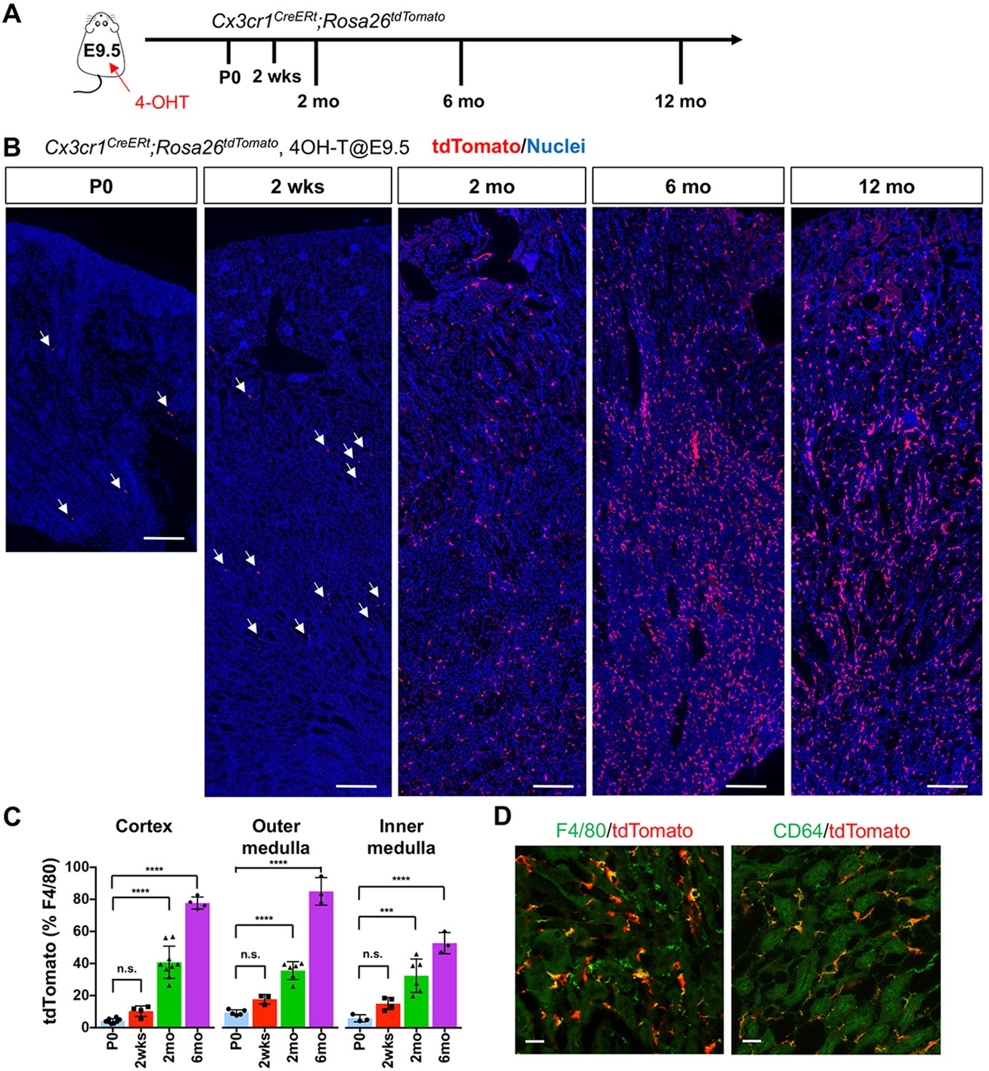
CX3CR1-positive yolk-sac macrophage descendants progressively expand in number in kidneys with age.
(A) Fate-mapping strategies of CX3CR1+ yolk-sac macrophages. 4-hydroxytamoxifen (4-OHT) was injected once into pregnant dams at 9.5 dpc and offspring analyzed at the indicated times (n = 4–6 for P0 to 6-month-old; n = 2 for 12-month-old). Yolk-sac macrophages and their progeny are irreversibly tagged with tdTomato. (B) Distribution of CX3CR1-lineage cells in postnatal kidneys. Arrows: CX3CR1-lineage cells. (C) Percentage of tdTomato+ to F4/80+ cells. Data are represented as means ± S.D. ***, p<0.001; ****, p<0.0001; n.s., not significant. (D) Confocal images of F4/80 and CD64 staining in aged kidneys (six mo) with CX3CR1-lineage tracing (n = 3). Scale bars: 200 μm in B; 20 μm in D.
-
Figure 1—source data 1
Percentage of tdTomato+ to F4/80+ cells.
- https://cdn.elifesciences.org/articles/51756/elife-51756-fig1-data1-v2.xlsx
Figure 1—figure supplement 1
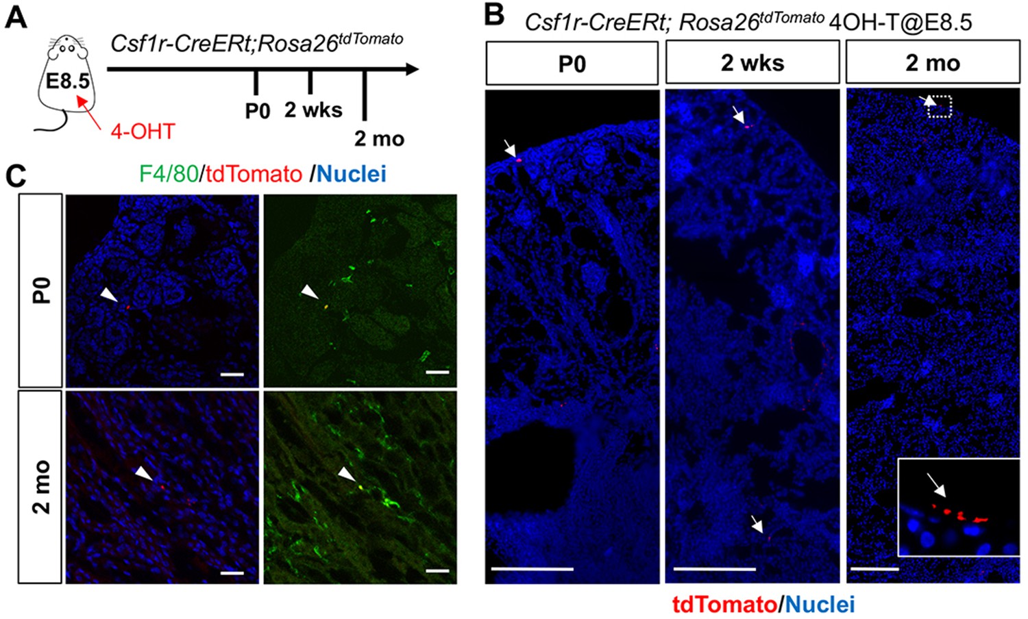
CSF1R-positive yolk-sac macrophage descendants do not expand in number in kidneys.
(A) Fate-mapping strategies of CSF1R+ yolk-sac macrophages. 4-hydroxytamoxifen (4-OHT) was injected once into pregnant dams at 8.5 dpc and offspring analyzed at the indicated times (n = 4–6). Yolk-sac macrophages and their progeny are irreversibly tagged with tdTomato. (B) Distribution of CSF1R-lineage cells in postnatal kidneys (n = 4). Arrows: CSF1R-lineage cells. Inset: higher magnification of dotted box. (C) Confocal images of F4/80 staining with CSF1R-lineage tracing (n = 3). Arrowheads: F4/80+CSFIR-lineage cells. Scale bars: 200 μm in B; and in 20 μm in C.
Figure 1—figure supplement 2
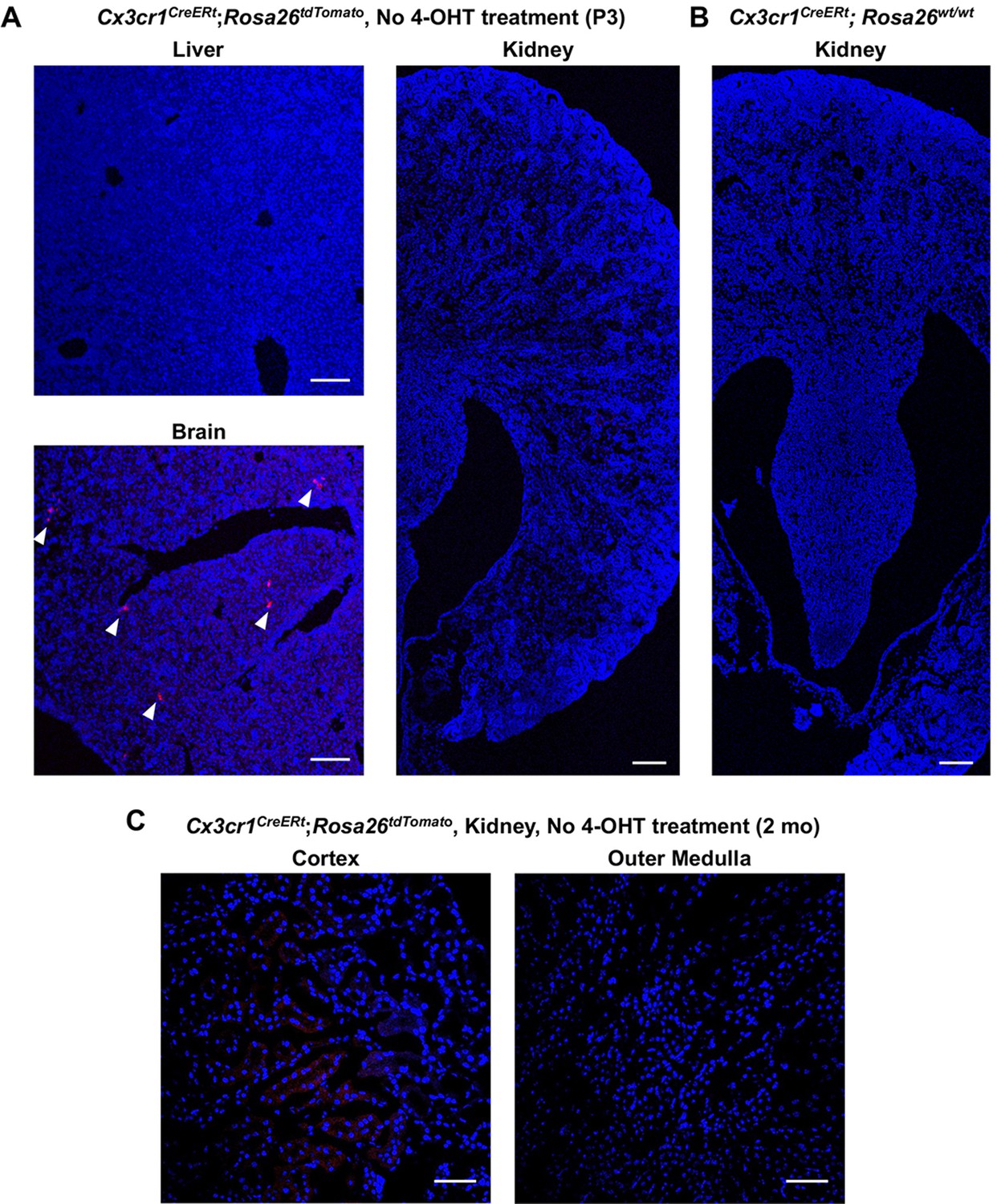
There is no basal Cre activity in kidneys without 4-hydroxytamoxifen (4-OHT) treatment.
Cx3cr1CreERt; Rosa26tdTomato/wt mice were used to determine tdTomato reporter activity in the absence of 4-OHT treatment. (A) While we observed a few tdTomato-expressing cells in the brain, there were no tdTomato-positive cells in the liver and kidney at P3 without 4-OHT treatment. Scale bars: 100 μm. (B) Cx3cr1CreERt; Rosa26wt/wt mice from the same litter were used as a negative control. (C) There were no tdTomato-positive cells in the kidney of 2-month-old Cx3cr1CreERt; Rosa26tdTomato/wt mice without 4-OHT treatment. Arrowheads: tdTomato-expressing cells. Scale bars: 100 μm in A and B; and 50 μm in C.
Figure 2

Age-dependent decrease of HSC-derived F4/80+ cells in the kidneys.
The Flt3-Cre; Rosa26tdTomato mouse line was used to examine the contribution of HSC-derived tissue-resident macrophages. (A) Distribution of F4/80+ Flt3-lineage cells in postnatal kidneys. The kidneys were analyzed at the indicated times (n = 4–5). HSC-derived cells and their progeny are irreversibly tagged with tdTomato. (B) Percentage of tdTomato+ F4/80+ to total F4/80+ cells. Note that the number of tdTomato+ F4/80+ cells decreases with age. Data are represented as means ± S.D. *, p<0.05; **, p<0.01.
-
Figure 2—source data 1
Percentage of tdTomato+ F4/80+ cells to total F4/80+ cells.
- https://cdn.elifesciences.org/articles/51756/elife-51756-fig2-data1-v2.xlsx
Figure 3
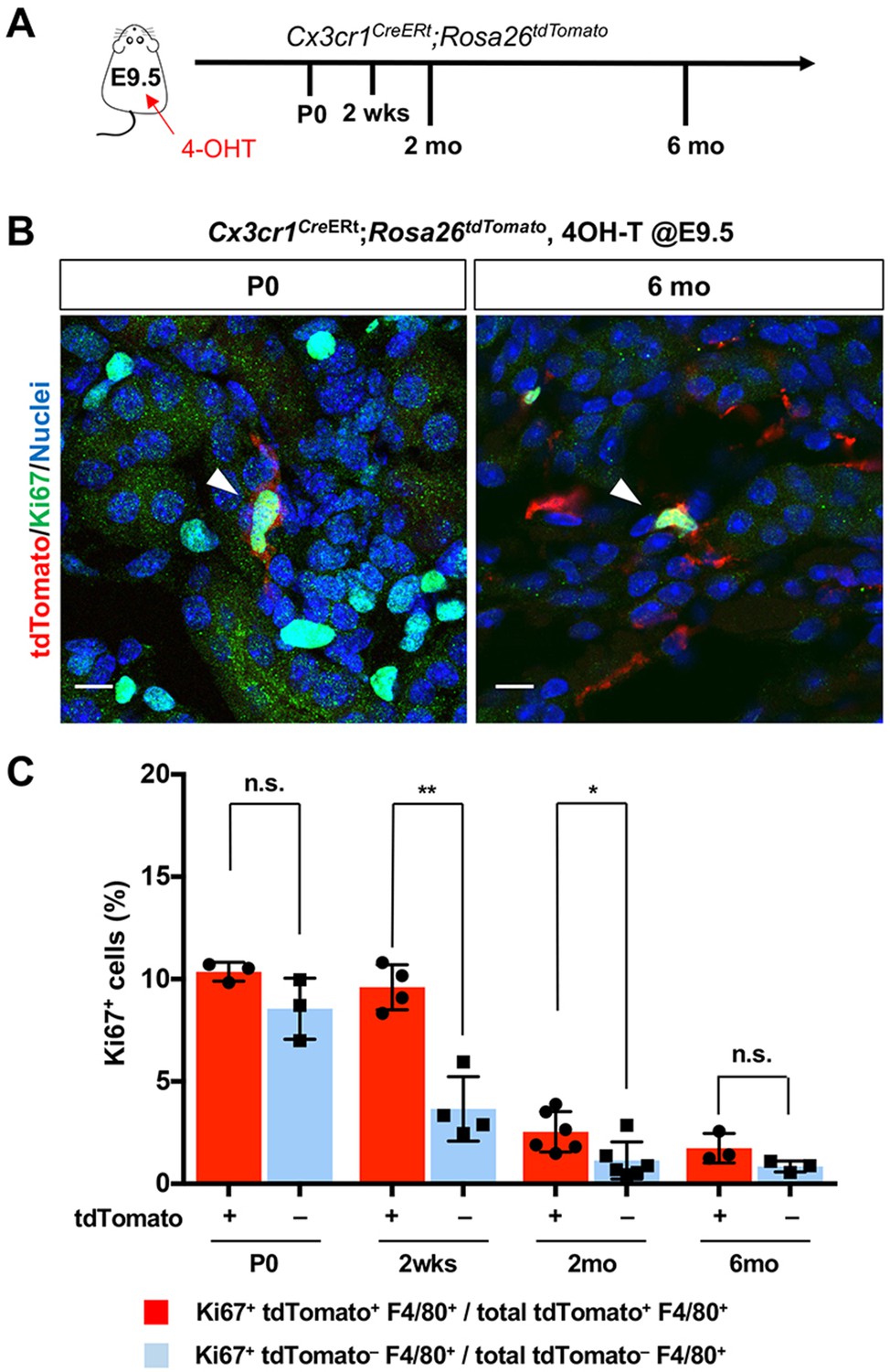
CX3CR1-positive yolk-sac macrophage descendants proliferate locally in the kidneys.
(A) Schematic of fate-mapping strategy. (B and C) CX3CR1-lineage macrophages proliferate in neonatal and aged kidneys. Cx3cr1CreERt; Rosa26tdTomato mice were treated with 4-hydroxytamoxifen (4-OHT) at E9.5 (n = 3–5). Arrowheads: Ki67+ CX3CR1-lineage cells. Percentage of Ki67+ proliferating cells are shown in C. Note that a higher percentage of CX3CR1-lineage F4/80+ cells (tdTomato+) are Ki67-positive compared to tdTomato– F4/80+ cells. Data are represented as means ± S.D. *, p<0.05; **, p<0.01; n.s., not significant. Scale bars: 10 μm.
-
Figure 3—source data 1
Percentage of Ki67+ proliferating F4/80+ cells.
- https://cdn.elifesciences.org/articles/51756/elife-51756-fig3-data1-v2.xlsx
Figure 4 with 1 supplement
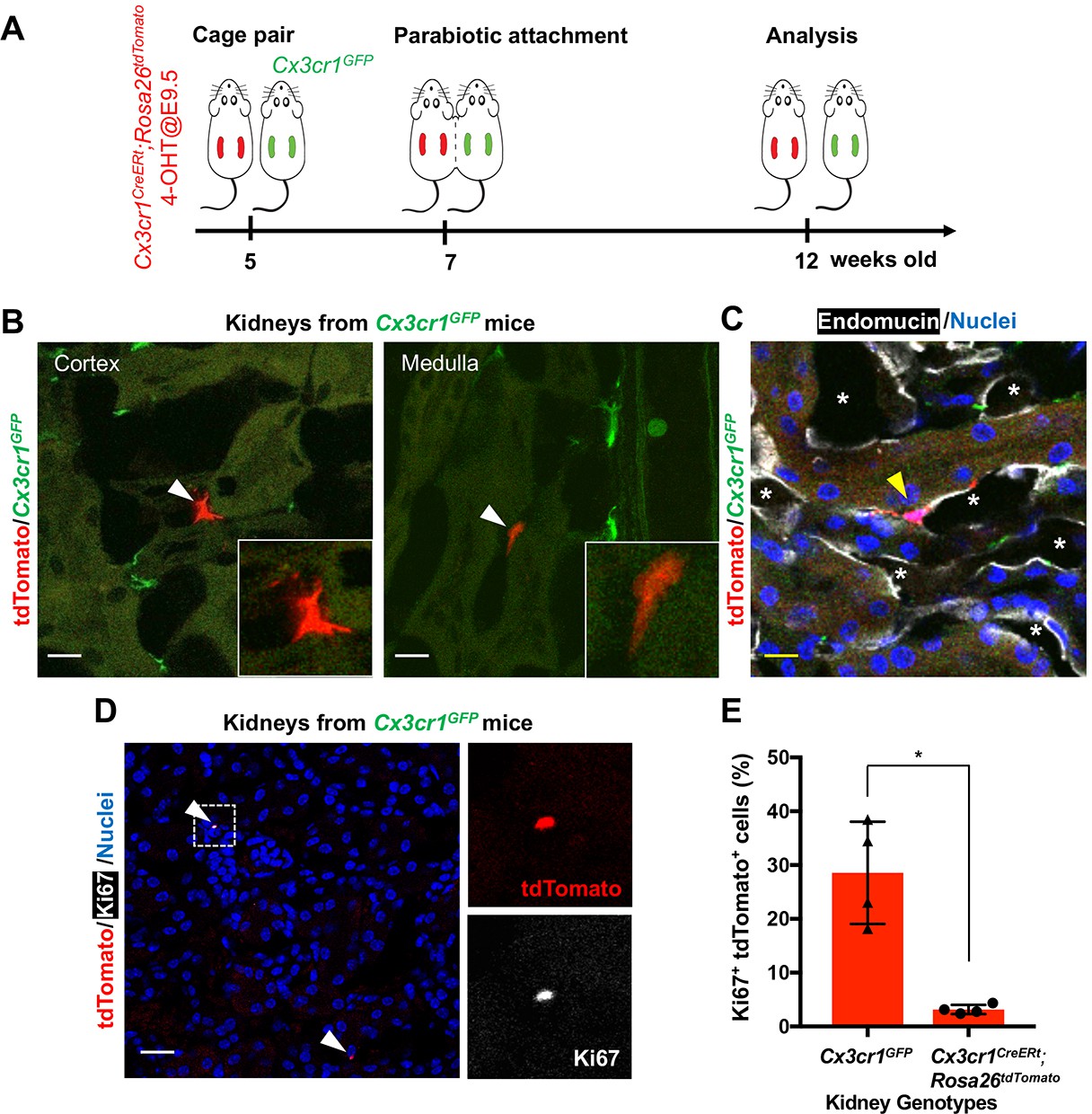
CX3CR1-positive yolk-sac macrophage descendants are recruited into adult kidneys from the circulation.
(A) Schematic of parabiotic experiments. (B and C) Localization of tdTomato+ CX3CR1-lineage cells in Cx3cr1GFP kidneys. The tdTomato-positive cells were lineage-labeled in Cx3cr1CreERt; Rosa26tdTomato mice in utero at E9.5. They migrated into the parabiont Cx3cr1GFP kidneys from circulation. Note that tdTomato+ cells were detected in extravascular interstitium (n = 4 per group). Endomucin; an endothelial cell marker. *, lumen of capillaries. Arrowheads, CX3CR1-lineage cells from circulation. (D and E) Circulation-derived CX3CR1-lineage cells proliferate in adult kidneys. Arrowheads, CX3CR1-lineage cells from circulation. GFP fluorescence was lost during the antigen retrieval process to detect Ki67. Percentages of Ki67+tdTomato+ cells relative to tdTomato+ cells in the kidneys of indicated genotype are shown in E (n = 4 per group). Note that tdTomato+ cells in Cx3cr1GFP kidneys are derived from circulation. Data are represented as means ± S.D. *, p<0.05. Scale bars: 10 μm in B and C; and in 20 μm in D. Legends for the Supplementary Figures.
-
Figure 4—source data 1
Percentages of Ki67+tdTomato+ cells relative to tdTomato+ cells in the kidneys of indicated genotypes.
- https://cdn.elifesciences.org/articles/51756/elife-51756-fig4-data1-v2.xlsx
Figure 4—figure supplement 1

There is no basal Cre activity in kidneys without 4-hydroxytamoxifen (4-OHT) treatment in the spleen of 2-month-old-mice.
Cx3cr1CreERt; Rosa26tdTomato/wt mice were used to determine tdTomato reporter activity in the absence of 4-OHT treatment. Note that 4-OH tamoxifen treatment at E9.5 effectively identifies CX3CR1-lineage cells (red) in the spleen of all animals received the drug (N = 7 out of 7 mice tested). Scale bars: 50 μm.
Author response image 1
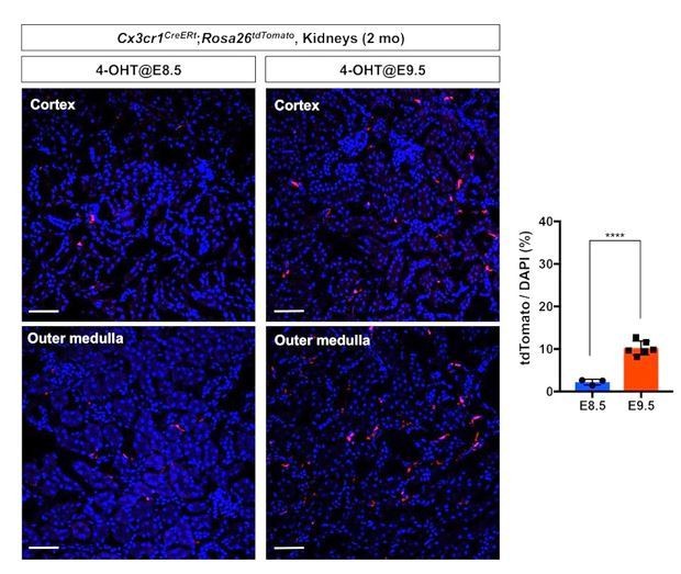
Comparison of fate-mapping strategies using different 4-OH tamoxifen treatment protocols.
4-hydroxytamoxifen (4-OHT) was injected once into pregnant dams at 8.5 dpc or 9.5 dpc, and the offspring were analyzed at 2 months of age. The first wave of pre-macrophages can be lineage traced with 4-OH tamoxifen injection at 8.5 dpc.
Author response image 2
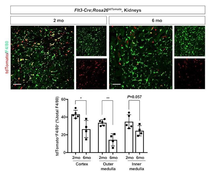
Age-dependent decline of HSC-derived F4/80-positive cells in kidneys.
The Flt3-Cre; Rosa26tdTomato mouse line was used to delineate the contribution of HSC-derived tissue resident macrophages. Please note that the number of tdTomato+ F4/80+ cells (yellow), which indicates HSC origin, decreases with age. *P<0.05, **P<0.01. Image-J was used for quantification. Flt3-Cre mice were kindly provided by Dr. K. Lavine (Washington University, St. Louis MO). Flt3-Cre mice were bred into the C57BL6/J background for 6 generations by the Shinohala lab.
Author response image 3
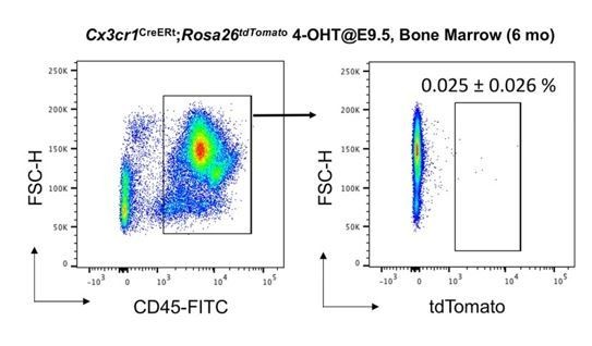
No increase of CD45+ tdTomato+ cells in bone marrow of 6 month-old Cx3cr1CreERt; Rosa26tdTomato mice.
We analyzed thenumber of tdTomato+ CD45+ cells in bone marrow (N=4). Compared to 2-month-old bone marrow (0.02% of CD45+ cells, Yahara et al., 2020), the number of tdTomato+CD45+ cells did not increase with age (0.025%).
Author response image 4
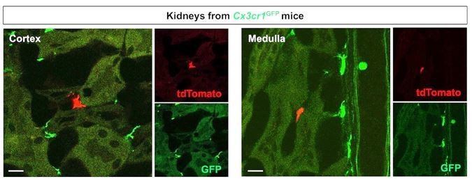
Merged and single color panels of Figure 4B.
The tdTomato-positive cells in the Cx3cr1GFP kidneys are derived from the parabiont animals (Cx3cr1CreERt; Rosa26tdTomato) through circulation. GFP-positive cells are resident Cx3cr1-expressing cells.
Author response image 5
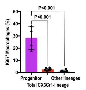
Circulation-derived CX3CR1-lineage cells have a profound proliferative capacity.
The percentage of Ki67+ cells is shown. Data are represented as means ± S.D. The mice were exposed to 4-OH tamoxifen in utero at E9.5. The tdTomato+ CX3CR1-lineage cells are descendants of yolk-sac macrophages. Data are derived and summarized from Figures 3 and 4 (see representative images in these figures). Progenitor, circulation-derived CX3CR1-lineage cells identified in Cx3cr1GFP kidneys; Total CX3CR1-lineage cells, macrophages derived from CX3CR1+ yolk-sac macrophages in the kidneys of Cx3cr1CreERt; Rosa26tdTomato mice; Other lineages, F4/80+tdTomato– macrophages in the kidneys of Cx3cr1CreERt; Rosa26tdTomato mice. Image-J was used for quantification.
Author response image 6

Morphological variation of CX3CR1-lineage cells.
The CX3CR1-lineage tdTomato-positive cells in the Cx3cr1GFP kidneys are derived from the parabiont animals (Cx3cr1CreERt; Rosa26tdTomato) through circulation. The Cx3cr1CreERt; Rosa26tdTomato mice were exposed to 4-OH tamoxifen in utero at E9.5. (A) Ki67+ CX3CR1-lineage cells are positive for DAPI (nuclear staining). (B) The CX3CR1-lineage tdTomato-positive cells exhibit oval and elongated shapes. Scale bars: 2 µm in A and 5 µm in B.
Tables
Key resources table
| Reagent type (species) or resource | Designation | Source or reference | Identifiers | Additional information |
|---|---|---|---|---|
| Genetic Reagent (M. musculus) | Cx3cr1CreERt | The Jackson laboratory | RRID:IMSR_JAX:020940 | |
| Genetic Reagent (M. musculus) | Csf1r-CreERt (aka, Csf1r-Mer-iCre-Mer) | The Jackson laboratory | RRID:IMSR_JAX:019098 | |
| Genetic Reagent (M. musculus) | Flt3-Cre | RRID:IMSR_EM:11790 | Flt3-Cre mice were bred into the C57BL6/J background for six generations by the Shinohara lab. | |
| Genetic Reagent (M. musculus) | Rosa26tdTomato | The Jackson laboratory | RRID:IMSR_JAX:007914 | |
| Genetic Reagent (M. musculus) | Cx3cr1GFP | The Jackson laboratory | RRID:IMSR_JAX:005582 | |
| Antibody | Anti-F4/80 (Rat monoclonal) | Bio-Rad (MCA497) | RRID:AB_2098196 | Clone C1:A3-1 IF: 1:100 |
| Antibody | Anti-CD64 (Rat monoclonal) | Bio-Rad (MCA5997) | RRID:AB_2687456 | Clone AT152-9 IF: 1:200 |
| Antibody | Anti-Endomucin (Rat monoclonal) | Abcam (ab106100) | RRID:AB_10859306 | Clone V.7C7.1 IF: 1:100 |
| Antibody | Anti-Ki67 (Rat monoclonal) | eBioscience (14-5698-82) | RRID:AB_10854564 | Clone SolA15 IF: 1:200 |
| Antibody | Anti-Ki67 (Rabbit monoclonal) | Thermo (MA5-14520) | RRID:AB_10979488 | Clone SP6 IF: 1:200 |
| Antibody | Anti-dsRed (Rabbit polyclonal) | Rockland (600-401-379) | RRID:AB_2209751 | IF: 1:200 |
| Software, algorithm | ImageJ | NIH, Bethesda, MD (Version 1.52P) | RRID:SCR_003070 | https://imagej.nih.gov/ij/ |
| Software, algorithm | GraphPad Prism | RRID:SCR_002798 | https://www.graphpad.com/scientific-software/prism/ |
Additional files
Download links
A two-part list of links to download the article, or parts of the article, in various formats.
Downloads (link to download the article as PDF)
Open citations (links to open the citations from this article in various online reference manager services)
Cite this article (links to download the citations from this article in formats compatible with various reference manager tools)
Yolk-sac-derived macrophages progressively expand in the mouse kidney with age
eLife 9:e51756.
https://doi.org/10.7554/eLife.51756


