Resident macrophages acquire innate immune memory in staphylococcal skin infection
- Institute for Immunodeficiency, Center for Chronic Immunodeficiency, Medical Center, Faculty of Medicine, University of Freiburg, Germany
- Faculty of Biology, University of Freiburg, Germany
- Center for Pediatrics and Adolescent Medicine, Medical Center, Faculty of Medicine, University of Freiburg, Germany
- Spemann Graduate School of Biology and Medicine (SGBM), Faculty of Biology, University of Freiburg and IMM-PACT Clinician Scientist Program, Faculty of Medicine, University of Freiburg, Germany
- Maurice Müller Laboratories (Department for Biomedical Research), Universitätsklinik für Viszerale Chirurgie und Medizin Inselspital, University of Bern, Switzerland
Figures
Figure 1 with 1 supplement
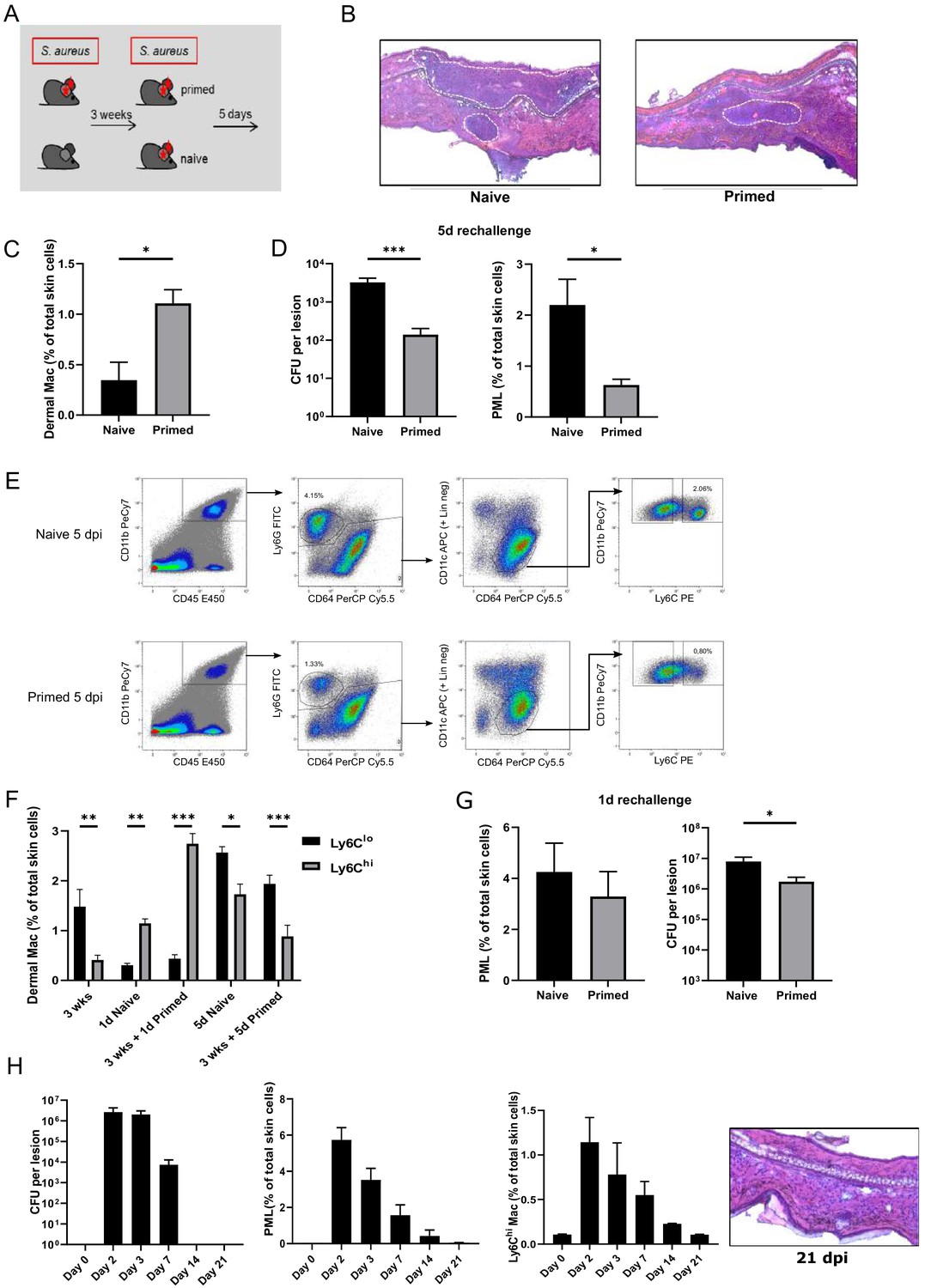
Intradermal S. aureus infection increases resistance to secondary infection.
(A, D) Wt mice were i.d. infected with S. aureus (107 CFU in 10 µl PBS (primed) or not (naive)), (re-)infected 3 weeks later with the same bacterial dosage and analyzed for bacterial load and skin infiltrating PML after an additional 5 days (n: eight biological replicates representing two independent experiments). (B) HE staining of cryosections from naïve and previously infected mice 5 days after (re-)infection. Dashed lines mark immune cell infiltrations. (C) Quantification of dermal Mφ in wt mice 3 weeks after primary infection and without secondary infection, data is gated on all CD64hi macrophages. (n: three biological replicates representing three independent experiments). (E) Representative FACS plots of the dermal myeloid cell composition in naïve and primed mice treated as in (A) 5 days after secondary infection (numbers represent % of total skin cells). (F) Frequency of Ly6Clo and Ly6Chi dermal Mφ 3 weeks after primary infection as well as after (re-)infection periods of 1 and 5 days in primed mice and mice infected for the first time (naïve). (G) Wt mice infected with S. aureus (107 CFU in 10 µl PBS) and (re-)infected 3 weeks later (primed) or for the first time (naïve) with the same amount of bacteria and analyzed for bacterial load and skin infiltrating PML after (re-)infection for 1 day (n: 7–8 biological replicates representing two independent experiments). (H) CFU counts, skin neutrophil and Ly6Chi dermal Mφ numbers of WT mice up to 21 days after i.d. S. aureus infection and without secondary infection. HE staining of cryosections from wt mice 3 weeks after i.d. S. aureus infection. Data were analyzed using either two tailed Mann-Whitney test or two-way ANOVA with Bonferroni’s multiple comparison test. Error bars are mean ± SEM. *p<0.05, **p<0.01, ***p<0.001.
Figure 1—figure supplement 1

Gating Strategy used to distinguish dermal macrophages.
(A) Representative FACS plots showing gating strategy used to identify tissue resident dermal macrophages in uninfected and 1 day post infection (naïve) wt mice. Resident dermal macrophages are defined as CD45hi CD11bhi CD64hi CD3lo CD11clo. (B) Representative FACS plots showing gating strategy used to distinguish Ly6C expression of dermal Mφ within the CD64hi population. (C) Bacterial load of wt mice injected with PBS or S. aureus and (re-)infected for an additional 5 days (n:8 biological replicates representing two independent experiments). Data were analyzed using either two tailed Mann-Whitney test. Error bars represent mean ± SEM, **p<0.01.
Figure 2 with 1 supplement
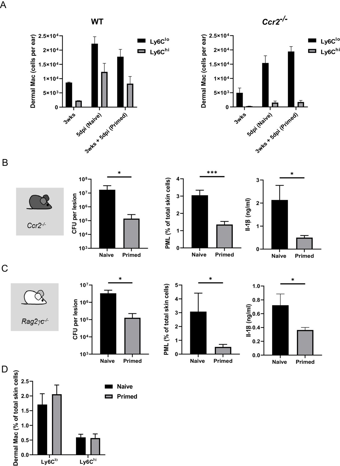
Innate memory does not depend on bone marrow-derived monocytes and on mature T, B, and NK cells.
(A) Quantification of Ly6Chi and Ly6Clo dermal Mφ in mice infected 3 weeks previously, naïve mice infected for the first time and primed mice, 5 days after i.d. (re-)infection with S. aureus in wt (left panel) and Ccr2-/- mice (right panel). (B) Ccr2-/- mice were i.d. infected with S. aureus (107 CFU in 10 µl PBS) and i.d. (re-)infected 3 weeks later (primed) or for the first time (naïve) with the same amount of bacteria and analyzed for bacterial load, skin infiltrating PMLs and skin IL-1β levels after additional 5 days (n: 11 biological replicates representing three independent experiments). (C) Rag2–/–γc–/– mice were i.d. infected with S. aureus (107 CFU in 10 µl PBS) and i.d. (re-)infected 3 weeks later (primed) or for the first time (naïve) with the same amount of bacteria and analyzed for bacterial load, skin infiltrating PML and skin IL-1β levels after additional 5 days (n: 5–7 biological replicates from two independent experiments). (D) Quantification of Ly6Chi and Ly6Clo dermal Mφ in naïve and primed Rag2–/–γc–/– mice 5 days after (re-)infection. Data were analyzed using two tailed Mann-Whitney test. Error bars are mean ± SEM. *p<0.05, ***p<0.001.
Figure 2—figure supplement 1

Bone marrow cells do not contribute to innate memory.
(A) Comparison of dermal Mφ numbers gated as in (Figure 1—figure supplement 1B) in wt and Ccr2-/- mice in steady state. (B) CD45.1 mice mice were i.d. injected with S. aureus (107 CFU in 10 µl PBS) or PBS. After 2 weeks, bone marrow from these CD45.1 mice mice was transferred into lethally irradiated naïve wt recipient mice and after additional 2 weeks recipient mice were infected with S. aureus (107 CFU in 10 µl PBS) for 5 days (n: five biological replicates).
Figure 3
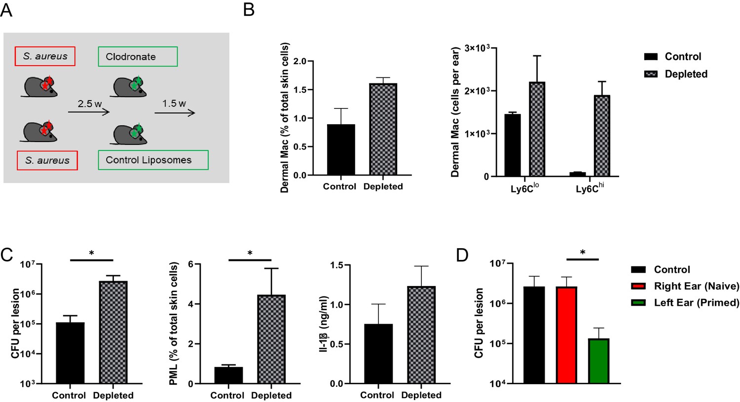
Dermal Mφ mediate innate memory.
(A) Wt mice were i.d.infected with S. aureus (107 CFU in 10 µl PBS) and 2.5 weeks later i.d. injected with either clodronate liposomes or control liposomes. (B) Quantification of CD64hi, Ly6Chi and Ly6Clo dermal Mφ from control and depleted mice treated as in (A). (C) Wt mice were treated as in (A) and re-challenged with S. aureus (107 CFU in 10 µl PBS) 1.5 weeks after clodronate treatment and analyzed for bacterial load, skin infiltrating PML and skin IL-1β levels after 5 days (n: eight biological replicates from two independent experiments). (D) Only the left ears of wt mice were i.d. infected with S. aureus for 3 weeks and the memory status of the corresponding right ear analyzed after additional infection of both ears for 5 days (n: 10–11 biological replicates from three independent experiments). Data were analyzed using two tailed Mann-Whitney test. Error bars are mean ± SEM. *p<0.05. (in D the groups right ear and left ear were compared).
Figure 4
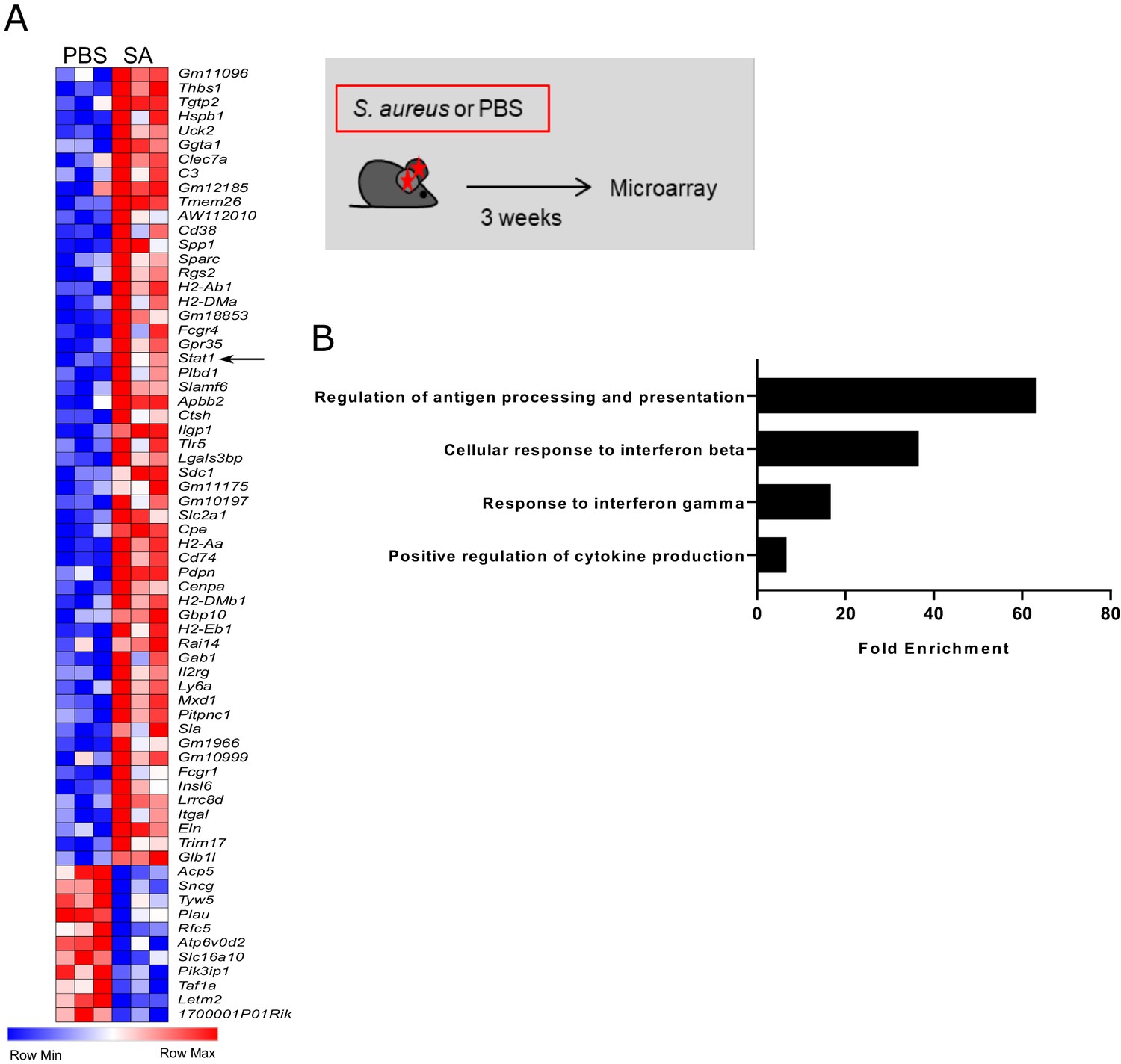
Innate memory signature in S. aureus challenged dermal Mφ.
(A) Heat map depicting differentially expressed genes between PBS and S. aureus treated Ccr2-/- mice 3 weeks after infection. Total CD64hi macrophages were sorted from ear skin as described in Figure 1—figure supplement 1A. (B) Gene ontology analysis of overrepresented terms in PBS vs S. aureus infected mice. (Data represents three biological replicates per condition in one experiment). Genes with a fold change above 2.0 and a student’s t test P value lower than 0.05 were analyzed using the Panther bioinformatics platform.
Figure 5 with 3 supplements
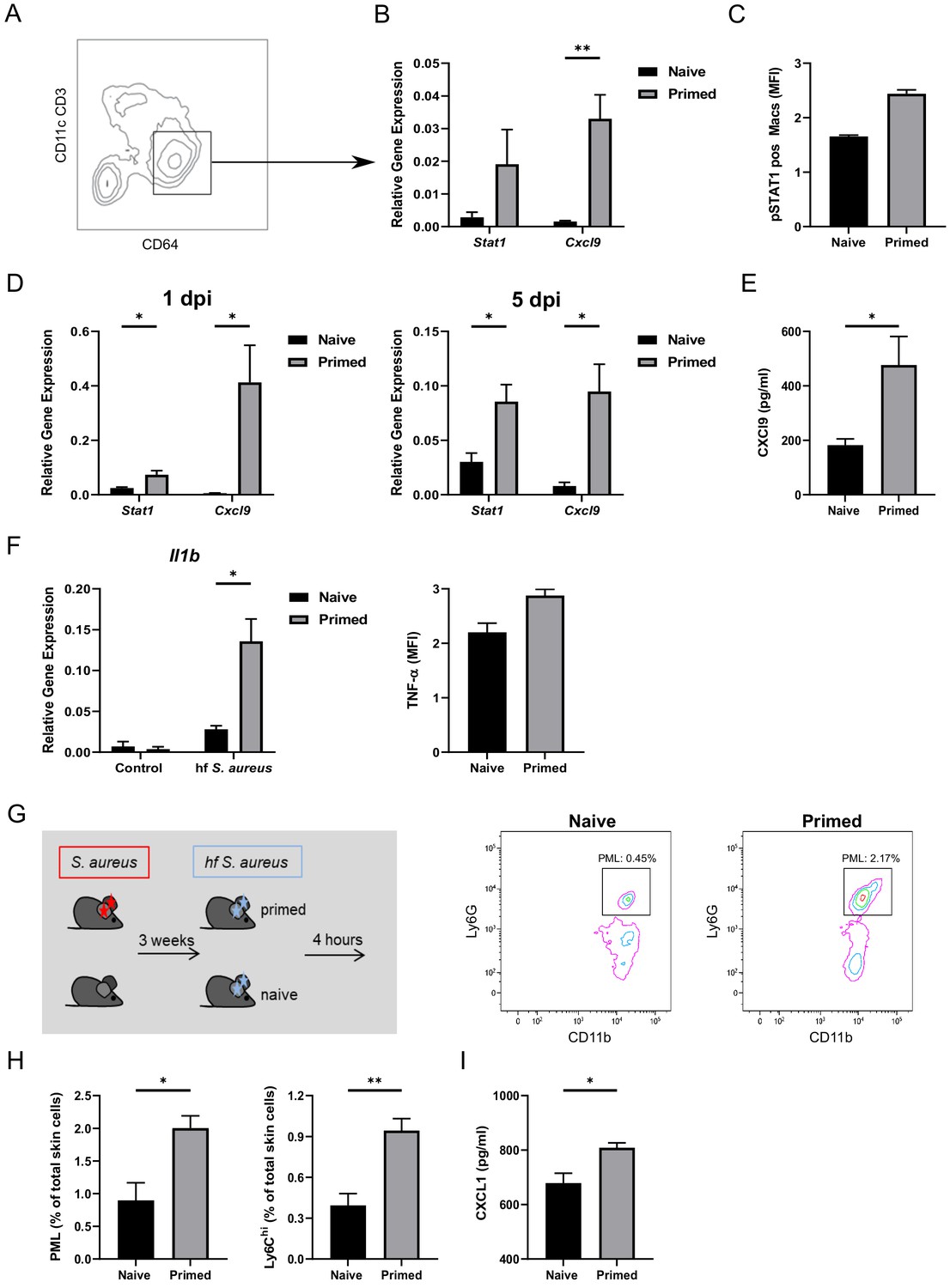
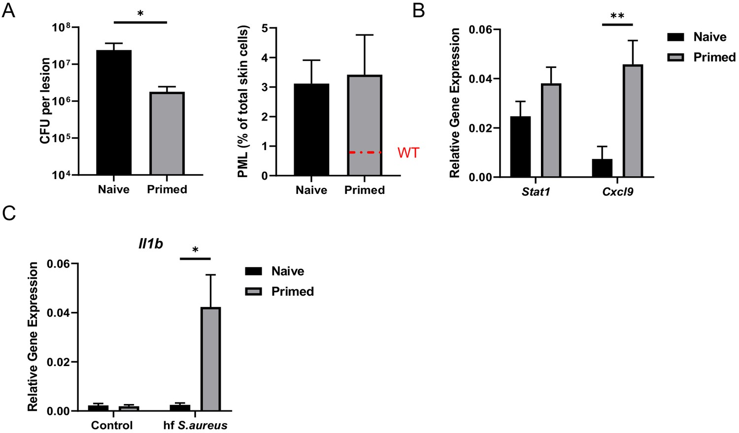
Dermal Mφ show a specific memory signature and increased responsiveness after (re-)infection ex vivo.
(A) Exemplary FACS plot of CD64hi dermal Mφ from wt mice 3 weeks after primary infection. (B) RNA expression of all CD64hi dermal Mφ (as in A) sorted from S. aureus infected wt mice (primed) or naïve mice, analyzed 3 weeks after infection (n: five biological replicates from two independent experiments). (C) Digested ear skin from naïve or infected (primed) mice was analyzed for phosphorylated STAT1 positive dermal Mφ by flow cytometry (n: two biological replicates from two independent experiments). (D) RNA expression of sorted dermal Mφ from primed or naïve wt mice following (re-)infection periods of 1 and 5 days (n: three biological replicates from one independent experiment). (E) CXCL9 ELISA of skin lysates of primed or naïve wt mice after 5 days of (re-)infection (n: eight biological replicates representing two independent experiments). (F) Dermal Mφ sorted either from S. aureus infected mice (primed) or naïve wt mice, were re-challenged ex vivo with fixed S. aureus. Expression of pro-IL-1β was determined by qPCR. Intracellular (i.c) TNF concentrations were determined by antibody staining and analysis by flow cytometry. (G–H) Wt mice were infected i.d. with S. aureus (107 CFU in 10 µl PBS) and (re-)infected i.d. 3 weeks later (primed) or for the first time (naïve) with fixed bacteria (108 CFU in 10 µl PBS) and analyzed for skin infiltrating PML and Ly6Chi dermal Mφ (n: eight biological replicates from two independent experiments). (I) CXCL1 ELISA of skin lysates from mice treated as in G. Data were analyzed using either two tailed unpaired T test (B, D and F) or with two tailed Mann-Whitney test (E, H and I). Error bars are mean ± SEM. *p<0.05, **p<0.01,.
Figure 5—figure supplement 1
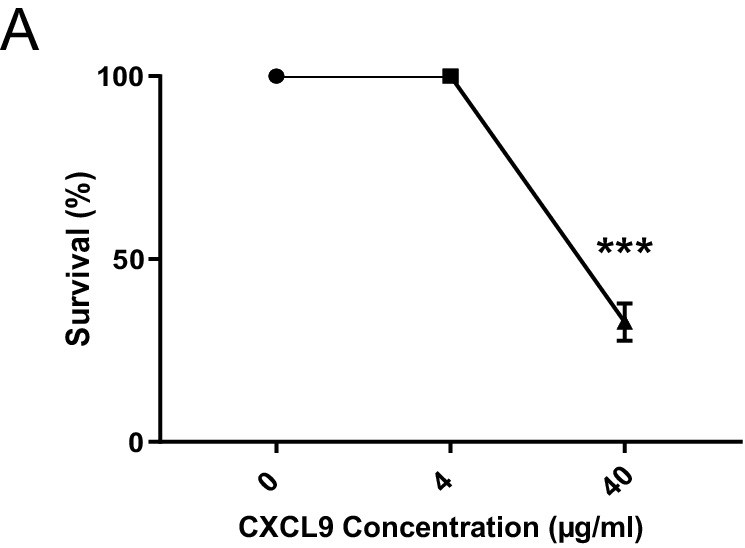
CXCL9 inhibits the growth of S. aureus Newman.
In vitro CXCL9 killing assay of S. aureus Newman (data represents three independent experiments). Data were analyzed using one way ANOVA with Tukey´s multiple comparison test. Error bars represent mean ± SEM, ***p<0.001.
Figure 5—figure supplement 2

Innate memory signature is lost in clodronate treated mice.
(A) Wt mice were i.d. infected with S. aureus (107 CFU in 10 µl PBS) and 2.5 weeks later i.d. injected with either clodronate liposomes or control liposomes. After an additional 1.5 weeks total dermal CD64hi Mφ were sorted and RNA expression analyzed by qPCR (data represents two independent experiments). (B) Lesion pathology of wt mice i.d. infected with S. aureus (107 CFU in 10 µl PBS) and i.d. (re-)infected 3 weeks later (primed) or for the first time (naïve) with fixed bacteria (108 CFU in 10 µl PBS).
Figure 5—figure supplement 3
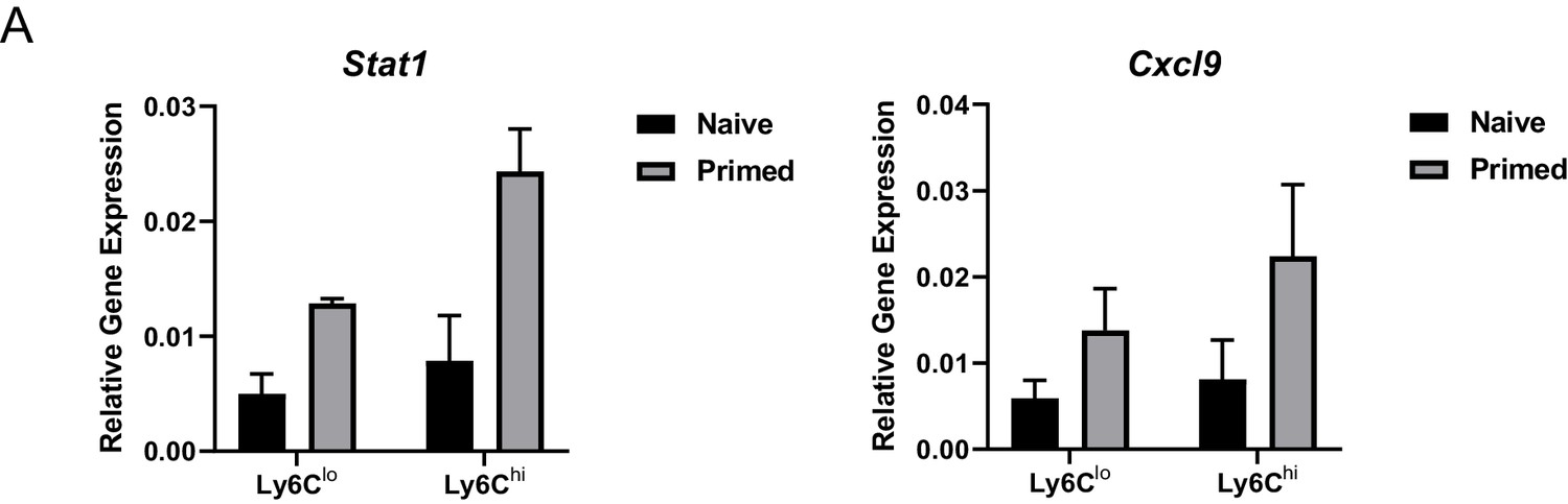
Ly6Chi dermal Mφ can be primed within the infected tissue.
CX3CR1int Ly6Chi and CX3CR1lo Ly6Clo Mφ subsets were sorted from Cx3cr1gfp/+ reporter mice 3 weeks after i.d. infection with S. aureus (107 CFU in 10 µl PBS) and analyzed for expression of Stat1 and Cxcl9 (n = 5 biological replicates representing one independent experiment).
Figure 6 with 2 supplements
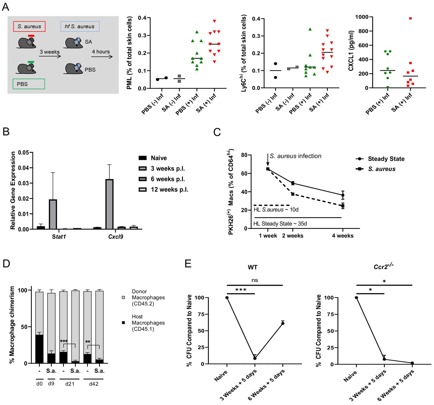
Innate memory is a transient effect and independent of skin microbiota.
(A) C57BL/6J mice harbouring the sDMDMm2 microbiota were colonized with S. aureus under axenic conditions, inoculated 3 weeks later with fixed bacteria (108 CFU in 10 µl PBS) or PBS and analyzed for skin infiltrating PML, Ly6Chi monocytes and skin CXCL1 levels after 4 hr (n: 10–12 biological replicates representing two independent experiments). (B) RNA expression of sorted CD64hi dermal Mφ from S. aureus infected mice or naïve mice analyzed 3, 6 and 12 weeks after infection (n:2–4 biological replicates from two independent experiments). (C) Wt mice were treated i.v. with PKH26 red fluorescent cell linker. One week after labeling, mice were infected i.d. with S. aureus. After another 1 and 3 weeks, the contribution of PHK26 positive to all CD64hi dermal Mφ was quantified (n: two biological replicates per time point from two independent experiments). (D) B6-CD45.1 mice were transplanted with 107 bone marrow cells from B6-CD45.2 congenic mice after fractional lethal irradiation. 8 weeks after transplantation dermal Mφ chimerism was determined by flow cytometry analysis using CD45.1 and CD45.2 antibodies (d0) or infected with S. aureus (107 CFU in 10 µl PBS) intradermally in the left ear. At the indicated time points the mice were sacrificed and dermal Mφ chimerism was determined by flow cytometry in the infected (S.a.) and uninfected ears (-). (n: three biological replicates for d0, and four biological replicates for all other groups). (E) Wt and Ccr2-/- mice were i.d. infected with S. aureus (107 CFU in 10 µl PBSl) and i.d. (re-)infected 3 or 6 weeks later with the same amount of bacteria and analyzed for bacterial load after an additional 5 days (n: three biological replicates from one independent experiment for the 6 week time points). Data were analyzed using two tailed unpaired T test. Error bars are mean ± SEM. *p<0.05, **p<0.01, ***p<0.001.
Figure 6—figure supplement 1
Innate memory is partially Myd88 independent.
(A) Myd88-/- mice were i.d. infected with S. aureus (107/ml) and i.d. re-infected 3 weeks later (primed) or for the first time (naïve) with the same amount of bacteria and analyzed for bacterial load and skin infiltrating PML after an additional 5 days. The dashed red line indicates PML numbers in equivalent treated wt mice (n: 7–8 biological replicates representing two independent experiments). (B) RNA expression of all CD64hi dermal Mφ sorted from S. aureus infected Myd88-/- mice (primed) or naïve mice, analyzed 3 weeks after infection. (C) CD64hi dermal Mφ were sorted from primed or naïve Myd88-/- mice 3 weeks after infection and stimulated ex vivo with fixed S. aureus and analyzed for expression of pro-Il1β (n: 4–8 biological replicates from two independent experiments). Data were analyzed using either two tailed Mann-Whitney test or two tailed unpaired T test (Error bars represent mean ± SEM. *p<0.05, **p<0.01,.
Figure 6—figure supplement 2
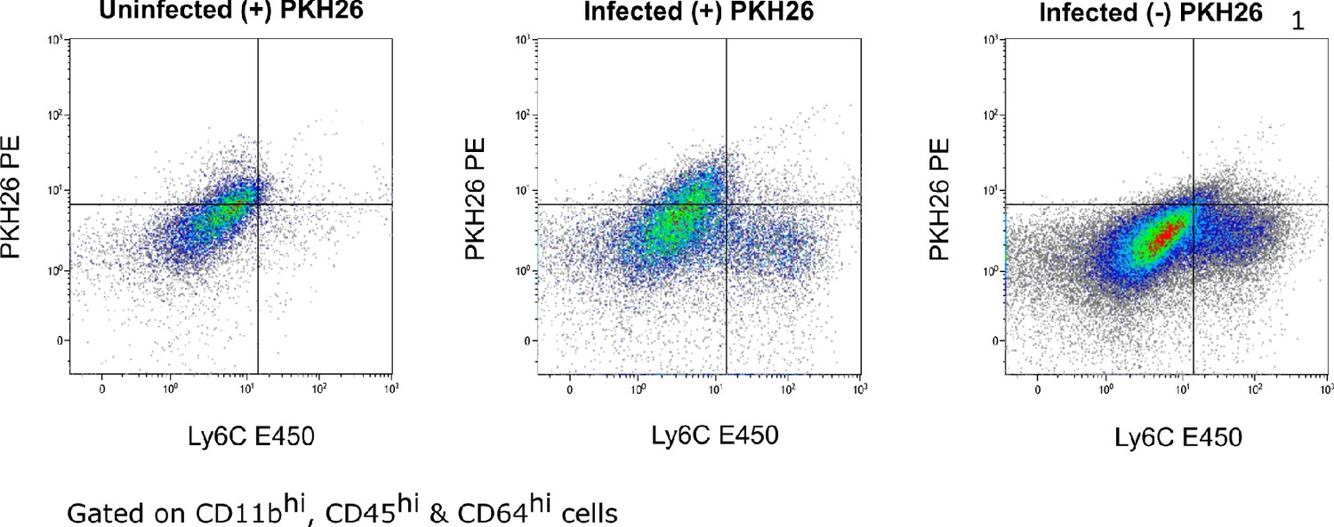
PKH26 labels Ly6Clo but not Ly6Chi dermal Mφ.
Representative FACS plots of PKH26 staining in the skin of wt mice. Left panel shows PKH26 positive CD64hi Mφ in steady state, middle panel shows PKH26 positive CD64hi Mφ and PKH26 negative Ly6Chi macrophages after infection. Right panel shows PKH26 FMO (fluorescence minus one) after infection.
Figure 7
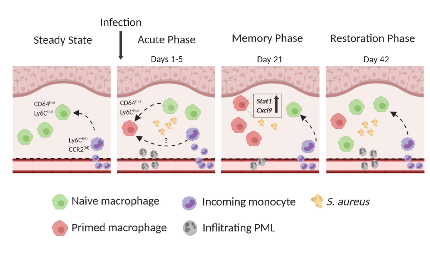
Model of innate immune memory during S. aureus skin infection.
Steady State: CD64hi, Ly6Clo Mφ have a half-life of 5–6 weeks and are replaced by CCR2hi, Ly6Chi incoming monocytes. Within the tissue, monocytes downregulate CCR2 and Ly6C and upregulate CD64. Acute Phase: Resident CD64hi Mφ respond to primary S. aureus infection and adopt a memory phenotype, thus becoming primed for subsequent infection. However, infection results in reduced Mφ half-life and increased influx of CCR2hi, Ly6Chi monocytes which may also undergo priming within the microenvironment. The acute phase is further characterized by high numbers of PMLs infiltrating the tissue. Memory Phase: S. aureus is cleared faster during secondary infection due to primed Mφ which are characterized by increased expression of Stat1 and Cxcl9. Reduced numbers of skin infiltrating PMLs are present in the tissue compared to naïve mice. Restoration Phase: Innate immune memory is lost due to continual influx of Ly6Chi monocytes which give rise to naïve Mφ and replace primed Mφ. Naïve Mφ lack increased expression of Stat1 and Cxcl9 resulting in slower bacterial clearance and increased skin PMLs after secondary infection.
Tables
Key resources table
| Reagent type (species) or resource | Designation | Source or reference | Identifiers | Additional information |
|---|---|---|---|---|
| Genetic reagent (Mus. musculus) | C57BL/6(J) | Jackson lab | Stock No: 000664 | |
| Genetic reagent (Mus. musculus) | C57BL/6(N) | Jackson lab | Stock No: 005304 | |
| Genetic reagent (Mus. musculus) | Ccr2-/- | Gift of Marco Prinz, (University of Freiburg) | ||
| Genetic reagent (Mus. musculus) | Rag2–/–γc–/– | Gift of Tilman Brummer (University of Freiburg) | BALB/c background | |
| Genetic reagent (Mus. musculus) | Cx3cr1gfp/+ | Gift of Steffen Jung (Weizmann Institute of Science) | ||
| Genetic reagent (Mus. musculus) | Myd88-/- | Deshmukh et al., 2012 | ||
| Genetic reagent (Mus. musculus) | sDMDMm2 | Clean Mouse Facility, University of Bern, Switzerland | C57BL/6(J) background | |
| Antibody | anti-mouse CD45 eFluor450 | Invitrogen | Cat: 48-0451-82 Clone: 30-F11 | 1:300 |
| Antibody | anti-mouse CD11b PE-Cy7 | Invitrogen | Cat: 25-0112-82 Clone: M1/70 | 1:3000 |
| Antibody | anti-mouse Ly6G FITC | BD Pharmingen | Cat: 551460 Clone: 1A8 | 1:200 |
| Antibody | anti-mouse Ly6C PerCP-Cy5.5 | BD Pharmingen | Cat: 560525 Clone: AL-21 | 1:500 |
| Antibody | anti-mouse CD64 PerCP/Cy5.5 | Biolegend | Cat: 139301 Clone: X54-5/7.1 | 1:100 |
| Antibody | anti-mouse CD11c APC | BD Pharmingen | Cat:561119 Clone HL3 | 1:100 |
| Antibody | mouse MHC class II eFluor450 | eBioscience | Cat: 48-5320-82 Clone: AF6120.1 | 1:400 |
| Antibody | anti-mouseCD3e | Invitrogen | Cat: 12-0031-82 Clone: 145–2 C11 | 1:200 |
| Antibody | anti-mouse Ly6C PE | Invitrogen | Cat: 12-5932-80 Clone: HK1.4 | 1:1000 |
| Antibody | Anti-mouse pSTAT1 AF647 | BD Biosciences | Cat:612597 Clone: 4a | |
| Chemicalcompound/drug | liposomal clodronate | Clodrosome Encapsula NanoSciences | Cat: CLD-8909 | |
| Chemicalcompound/drug | PKH26 Red | Sigma Aldrich | Cat: PKH26GL | |
| Software | GraphPad Prism | https://www.graphpad.com/scientific-software/prism/ | Version 8 | |
| Software | Kaluza analysis | https://www.beckman.de/flow-cytometry/software/kaluza |
Additional files
Download links
A two-part list of links to download the article, or parts of the article, in various formats.
Downloads (link to download the article as PDF)
Open citations (links to open the citations from this article in various online reference manager services)
Cite this article (links to download the citations from this article in formats compatible with various reference manager tools)
Resident macrophages acquire innate immune memory in staphylococcal skin infection
eLife 9:e55602.
https://doi.org/10.7554/eLife.55602


