Temporal transcription factors determine circuit membership by permanently altering motor neuron-to-muscle synaptic partnerships
- Department of Molecular Genetics and Cell Biology, University of Chicago, United States
- Program in Cell and Molecular Biology, University of Chicago, United States
- Committee on Development, Regeneration, and Stem Cell Biology, University of Chicago, United States
- Grossman Institute for Neuroscience, University of Chicago, United States
Figures
Figure 1 with 2 supplements
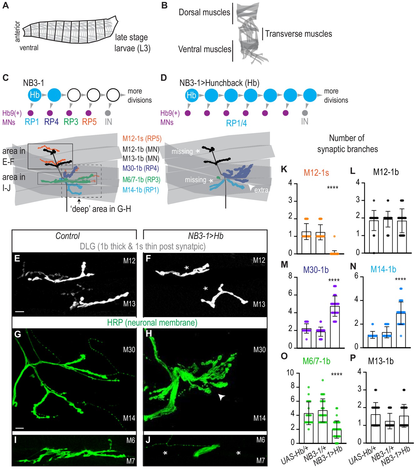
Prolonged expression of Hunchback alters RP motor neuron synapses.
(A) Illustration of the late stage larvae (L3), body is organized into repeated left-right, mirror image hemisegments. Muscles (in gray) have a stereotyped pattern. (B) Illustration of muscles in a single hemisegment of a late stage larvae (L3). (C–D) Illustrations of NB3-1 lineage progression. Each gray arrowhead represents cell division. Each gray arrowhead represents cell division. Large circles are neuroblasts, and smaller circles are neurons. Abbreviations: IN is interneuron. Illustrations of neuromuscular synapses on dorsal muscles in a L3 body wall segment and embryonic molecular identity depicted as circles (blue = RP1/4, green = RP3, orange = RP5). In NB3−1>Hb the number of synaptic branches (in blue) are increased (white arrowhead) onto Muscle 14 and 30 (RP1 and RP4 muscle targets, respectively), 1b synaptic branch number is decreased (asterisk) on Muscle 6 and 7 (RP3 muscle target) and 1s synaptic branch number is lost (asterisk) assessed on Muscle 12 (RP5 muscle target). (E–J) Images of neuronal membrane, both axons and neuromuscular synapses, on ventral muscles in L3 abdominal segments. Arrowhead indicates increased branching onto Muscle 30. An asterisk * indicates missing synapses. Data quantified in (K-P). (K–P) Quantification of the number of 1b or 1s branches on L3 muscles. Color code as in (A). Each dot represents the number of branches onto a single muscle. (K-P) For UAS-Hb/+ n = 22,21,22,22,21,21. For NB3-1/+ n = 30,30,29,29,30,29. For NB3−1>Hb n = 39,38,39,39,38,39. Control is NB3-1/+. NB3−1>Hb is NB3-1 GAL4/UAS Hb; UAS Hb/+. All images are shown dorsal up, anterior left. Scale bars represent 10 microns. For quantifications average and standard deviation are overlaid. ANOVA, corrected for multiple samples ‘****’ p<0.0001.
-
Figure 1—source data 1
Test for difference in the number of synaptic branches on ventral muscles in NB3-1>Hb compared to Control.
- https://cdn.elifesciences.org/articles/56898/elife-56898-fig1-data1-v2.docx
Figure 1—figure supplement 1
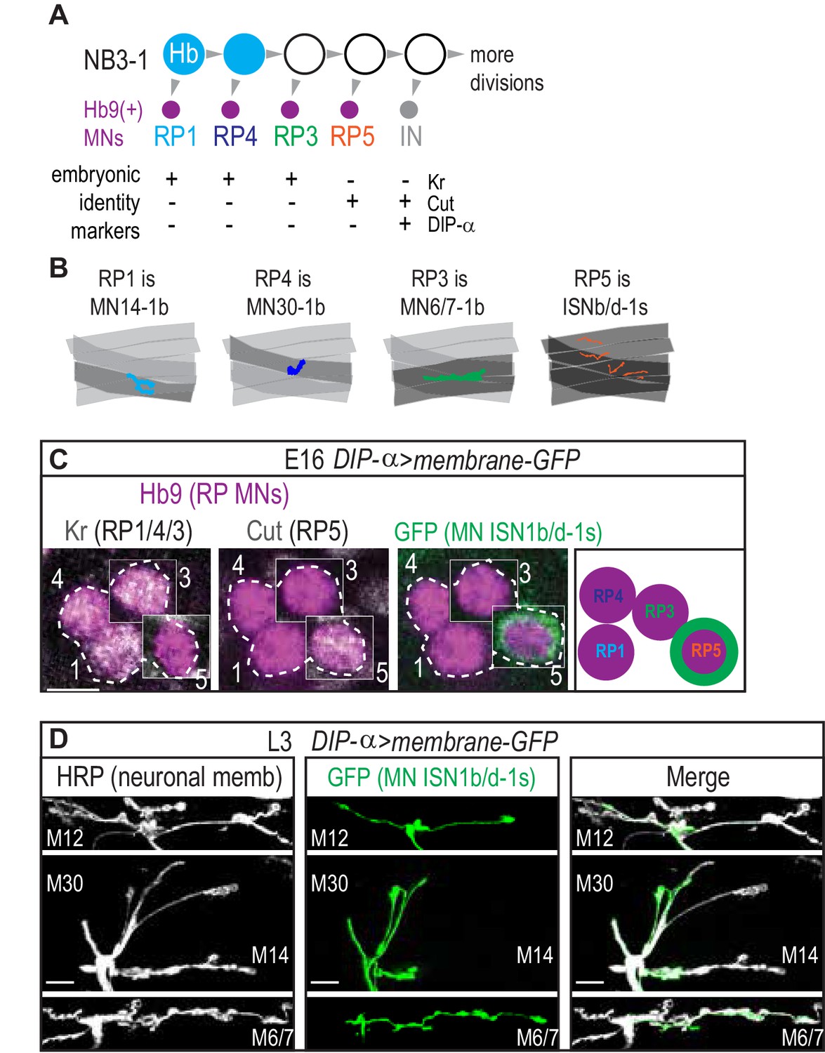
NB3-1 generates RP motor neurons, with known features.
(A) Illustration of NB3-1 lineage progression. Each gray arrowhead represents cell division. Large circles are neuroblasts and smaller circles are neurons. Abbreviations: MN is motor neuron, IN is interneuron, Hb is Hunchback, and Kr is Kruppel. (B) Illustration of individual RP motor neuron neuromuscular synapses onto ventral muscles in third instar larvae. RPs populate the ventral muscle group motor circuit, which is independently recruited during locomotion. (C) Image of embryonic molecular identity marker and GFP expression driven by DIP alpha driver in Hb9(+) cell bodies in late stage embryonic CNSs. RP5 expresses GFP in late stage embryos. Boxes are insets from different z-planes (dotted outline around the RP motor neurons labeled by number). Scale bars represent five microns. Images are shown anterior up, midline to the left. (D) Images of individually labeled DIP alpha(+) motor neuron axon in third instar larvae (L3) fillet dissected larval prep. Scale bars represent 10 microns. Images shown in ventral view, anterior to the right, midline down. DIP-alpha >membrane GFP is DIP-alpha-GAL4/+; UAS-myr-GFP/+.
Figure 1—figure supplement 2
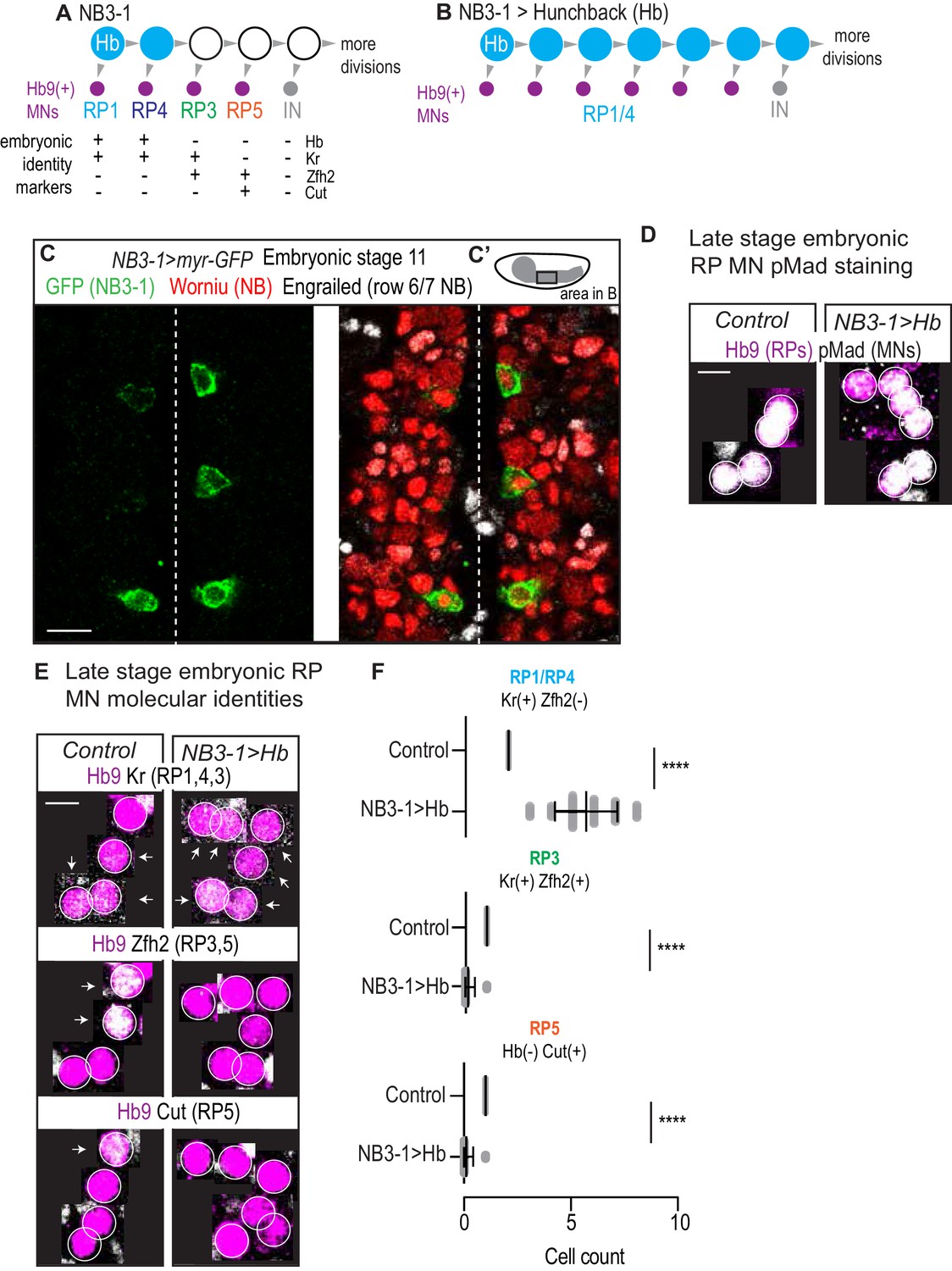
In embryos, prolonged expression of Hunchback generates more RP motor neurons with early born molecular identity.
(A–B) Illustrations of NB3-1 lineage progression. Each gray arrowhead represents cell division. Each gray arrowhead represents cell division. Large circles are neuroblasts, and smaller circles are neurons. Abbreviations: IN is interneuron, Hb is Hunchback, Kr is Kruppel, Zfh2 is Zinc finger homeodomain 2. In NB3−1>Hb, there is an increase in the number of Hb9(+) with RP1/4 embryonic molecular identity. (C) Image of GFP in NB3-1 as a reporter of NB3-1-GAL4 activity. Three complete abdominal segments are shown with anterior up, and midline dotted. C’ Illustration of a Drosophila late stage embryo, CNS in gray. For NB3−1>myr GFP, n = 28 hemisegments. Scale bar represents 10 microns. (D) Images of co-expression of Hb9 and the pan-motor neuron marker pMad in Control and NB3−1>Hb CNS of late stage embryos. For Control, n = 160 hemisegments from four embryos. For NB3−1>Hb, n = 360 from six embryos. Scale bar represents five microns. (E) Images of embryonic molecular identity marker expression in Hb9(+) cells in late stage embryonic CNSs. In NB3−1>Hb, extra Hb9(+) cells with RP1/4 molecular identity are produced. Boxes are neurons from different z-planes. Arrows indicate co-expression. Scale bar represents 5 microns. (F) Quantification of NB3-1 neurons in Control and NB3−1>Hb. Cells with RP1/RP4, RP3, and RP5 molecular markers. Each dot represents a single hemisegment. For Control, n = 31,31,31 hemisegments from top to bottom graph. For NB3−1>Hb, n = 60,90,60 hemisegments from top to bottom graph. NB3−1 > myr GFP is NB3-1 GAL4/UAS-myr GFP. Control is W1118. NB3−1>Hb is NB3-1 GAL4/UAS Hb; UAS Hb/+. Images are shown anterior up, midline to the left. For quantifications average and standard deviation are overlaid. ANOVA, corrected for multiple samples ‘****’ p<0.0001.
-
Figure 1—figure supplement 2—source data 1
Test for difference in the number of embryonic RP MN molecular identities in NB3-1>Hbcompared to Control.
- https://cdn.elifesciences.org/articles/56898/elife-56898-fig1-figsupp2-data1-v2.docx
Figure 2
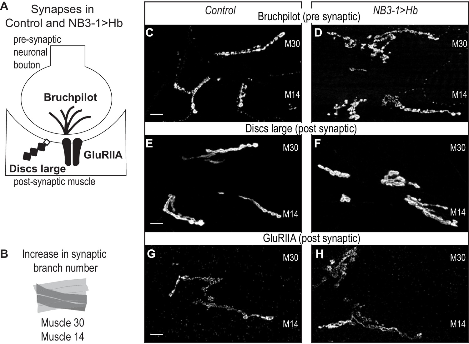
Increased synaptic branches contain pre and postsynaptic markers necessary for function.
(A) Illustration of subcellular localization of neuromuscular synapse markers. Brunchpilot labels active zones, Discs large is a scaffolding protein strongly localized at post-synapse, and GluRIIA is the post-synaptic glutamate receptor IIA. (B) Illustration highlighting (darkened) muscles 14 and 30, which have increased synaptic branching in NB3−1>Hb (see Figure 2). (C–H) Images of neuromuscular synapses on L3 Muscle 14 and 30 (see B). There is no difference in distribution or abundance of synaptic markers between Control and NB3−1>Hb. Control is Hb/+ and NB3−1>Hb is NB3-1-GAL4/UAS-Hb; UAS-Hb/+. All images are shown dorsal up, anterior to the left, scale bars represent 10 microns.
Figure 3
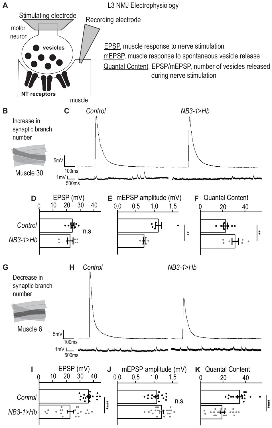
Altered synapses onto Ventral muscles are functional.
(A) Illustration of neuromuscular junction (NMJ) electrophysiology approach on third instar larvae (L3). Abbreviation NT is neurotransmitter. (B) Illustration highlighting (darkened) Muscle 30 whose electrophysiological recordings are presented in C-F. (C) Traces of EPSP and mEPSPs for Muscle 30. (D–F) Quantification of electrophysiological recordings for Muscle 30. (D) Evoked response (EPSP) is not changed in Muscle 30 in NB3−1>Hb vs Control. (E) Spontaneous response (mEPSP) is significantly decreased in Muscle 30 in NB3-1 vs Control. (F) Transmitter release (Quantal Content (EPSP/mEPSP)) is slightly significantly increased in Muscle 30 NB3-1 vs Control. (D–F) For Control, n = 8. For NB3−1>Hb, n = 8. For C-E and H-J, each dot (black represents Control and gray represents NB3−1 > Hb) on the graph represents a single recording from a unique bodywall segment from A2-A4. (G) Illustration highlighting (darkened) Muscle 6 whose electrophysiological recordings are presented in H-K. (H) Traces of EPSP and mEPSPs for Muscle 6. (I–K) Quantification of electrophysiological recordings for Muscle 6. (I) Evoked response (EPSP) is significantly decreased in Muscle 6 in NB3−1>Hb vs Control. (J) Spontaneous response (mEPSP) is not changed in Muscle 6 in NB3-1 vs Control. (K) Transmitter release (Quantal Content (EPSP/mEPSP)) is significantly decreased in Muscle 6, NB3-1 vs Control. (I–K) For Control, n = 17,16,16. For NB3−1>Hb, n = 21. Control is NB3-1/+ and NB3−1>Hb is NB3-1-GAL4/UAS-Hb; UAS-Hb/+. All images are shown dorsal up, anterior to the left. For quantifications, average and standard deviation are overlaid. Unpaired t-test ‘ns’ not significant, ‘**’ p<0.05, ‘****’ p<0.0001.
-
Figure 3—source data 1
Test for the difference in EPSP, mEPSP, and Quantal Content for muscle 14 and 6 in NB3-1>Hb compared to Control.
- https://cdn.elifesciences.org/articles/56898/elife-56898-fig3-data1-v2.docx
Figure 4 with 1 supplement
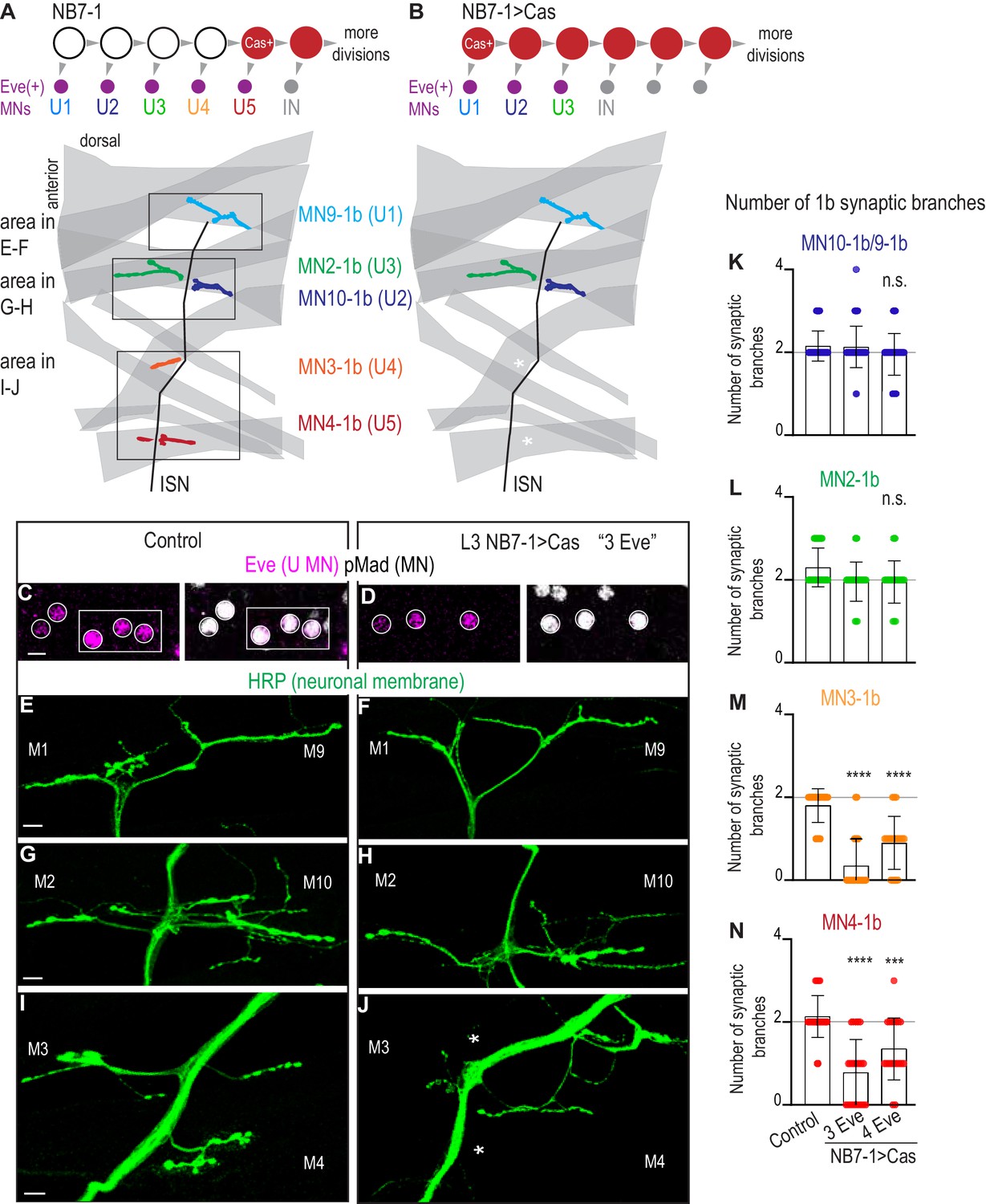
Precocious expression of Castor alters U motor neuron synapses.
(A–B) Illustrations of NB7-1 lineage progression. Each gray arrowhead represents cell division. Each gray arrowhead represents cell division. Large circles are neuroblasts, and smaller circles are neurons. IN is interneuron. In NB7−1>Cas there is a decrease in the number of Eve(+) neurons with U4/U5 embryonic molecular identity. Illustrations of neuromuscular synapses on dorsal muscles in a L3 body wall segment and embryonic molecular identity depicted as circles (light blue = U1, dark blue = U2, green = U3, orange = U4, red = U5). In NB7−1>Cas the number of 1b synaptic branches (in orange and red) are lost (white asterisks) onto Muscle 3 and Muscle 4 (U4 and U5 muscle targets, respectively). (C–D) Images of L3 nerve cord abdominal hemisegment Eve(+) neurons co-expressing the motor neuron marker pMad. For Control, n = 12 hemisegments in three animals. For NB7−1 > Cas, n = 23 hemisegments in seven animals. All images are shown anterior up, midline left, scale bar represents five microns. (E–J) Images of neuronal membrane, both axons and neuromuscular synapses, on ventral muscles in L3 abdominal segments corresponding to hemisegments containing the number of Eve(+) neurons as imaged in (D–E). An asterisk * indicates missing synapses on Muscle 3 and Muscle 4. All images are shown dorsal up, anterior left, scale bar represents 10 microns. Data quantified in (K–N). (K–N) Quantification of the number of 1b branches on L3 muscles. Color code as in (A). Line intersects the y-axis at 2. Each dot represents the number of branches onto a single muscle. Decrease in synaptic branching onto Muscle 4 and Muscle 3 in experimental conditions (3 Eve(+) neurons and 4 Eve(+) neurons) vs Control (K). No change (L). (K–N) For Control n = 59,30,30,30. For NB7−1>Cas hemisegments with 3 Eve(+) neurons, n = 46,23,23,23. For NB7−1>Cas hemisegments with 4 Eve(+) neurons, n = 40,20,20,20. Control is Cas/+ and NB7−1>Cas is NB7-1 GAL4/Cas. For quantifications, average and standard deviation are overlaid. ANOVA, corrected for multiple samples ‘ns’ not significant, ‘***’ p<0.001, ‘****’ p<0.0001.
-
Figure 4—source data 1
Test for difference in the number of synaptic branches on dorsal muscles in NB7-1>Cas compared to Control.
- https://cdn.elifesciences.org/articles/56898/elife-56898-fig4-data1-v2.docx
Figure 4—figure supplement 1

In embryos, precocious expression of Castor generates fewer U motor neurons at the expense of later born neurons.
(A–B) Illustrations of NB7-1 lineage progression. Each gray arrowhead represents cell division. Each gray arrowhead represents cell division. Large circles are neuroblasts, and smaller circles are neurons. Abbreviations: IN is interneuron, MN is motor neuron, IN is interneuron, Eve is Even-skipped, Hb is Hunchback, Kr is Kruppel, and Zfh2 is Zinc finger homeodomain 2. In NB7−1>Cas, there is a decrease in the number of Eve(+) neurons with U4/U5 embryonic molecular identity. (C) Illustration of individual U motor neuron neuromuscular synapses onto dorsal muscles in larvae. Embryonic motor neuron (e.g., U1) and larval motor neuron synapse (e.g. MN9-1b) names are shown. Color code as in (A). (D) Images of embryonic molecular identity marker expression in Eve(+) cells in late stage embryonic CNSs. In NB7−1>Cas, Eve(+) cells with U4/U5 molecular identity are not produced. Boxes are neurons from different z-planes. Arrows indicate co-expression. (E) Quantification of the % of hemisegments in late stage embryonic NB7−1>Cas that give rise to f5 Eve(+) neurons (Untransformed ‘WT’), 3 Eve(+) neurons (Transformed ‘3 Eve(+)”), and 4 Eve(+) neurons (Transformed ‘4 Eve(+)”). n = 44/180, n = 86/180, n = 50/180, respectively for the three phenotypes. (F) Images of co-expression of Eve and the pan-motor neuron marker, pMad in Control and NB7−1>Cas CNS of late stage embryos. All images are shown anterior up, midline to the left, scale bars represent 5 microns.
Figure 5 with 1 supplement
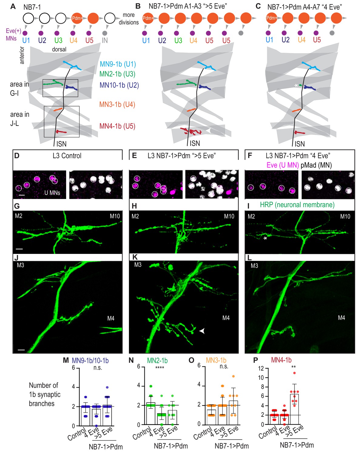
Precocious expression of Pdm alters U motor neuron synapses.
(A–C) Illustration of NB7-1 lineage progression. Each gray arrowhead represents cell division. Each gray arrowhead represents cell division. Large circles are neuroblasts, and smaller circles are neurons. Abbreviations: IN is interneuron. Illustrations of neuromuscular synapses on dorsal muscles in a L3 body wall segment and embryonic molecular identity depicted as circles (light blue = U1, dark blue = U2, green = U3, orange = U4, red = U5). In NB7−1>Pdm in A1-A3 segments where there are more than 5 Eve(+) U motor neurons, the number of 1b synaptic branches (in red) are increased (white arrowhead) onto Muscle 4 (U5 muscle target). In NB7−1>Pdm in A4-A7 segments where there are 4 Eve(+) U motor neurons, the number of 1b synaptic branches (in green) is nearly lost on Muscle 2 (U3 muscle target). (D–F) Images of L3 nerve cord abdominal hemisegment Eve(+) neurons co-expressing the motor neuron marker pMad. All images are shown anterior up, midline left. Scale bars represent 5 microns. (G–L) Images of neuronal membrane, both axons and neuromuscular synapses, on ventral muscles in L3 abdominal segments corresponding to hemisegments containing the number of Eve(+) neurons as imaged in (D–E). An asterisk * indicates missing synapses on Muscle two and an arrowhead indicates increase in synapses on Muscle 4. Data quantified in (M–P). All images are shown dorsal up, anterior left. Scale bars represent 10 microns. (M–P) Quantification of the number of 1b branches on L3 muscles. Color code as in (A). Line intersects the y-axis at 2. Each dot represents the number of branches onto a single muscle. Control is Pdm/+. NB7−1 > Pdm is NB7-1 GAL4/Pdm; Pdm/+. In H, Control is NB7-1 GAL4/UAS myr GFP. (M–P) For Control, n = 33,26,28,28. For NB7−1>Pdm with 4 Eve(+), n = 45,23,22,23. For NB7−1>Pdm with 5 Eve(+), n = 16,8,8,8. For quantifications average and standard deviation are overlaid. ANOVA, corrected for multiple samples ‘ns’ not significant, ‘**’ p<0.05, ‘****’ p<0.0001.
-
Figure 5—source data 1
Test for difference in the number of synaptic branches on dorsal muscles in NB7-1>Pdm compared to Control.
- https://cdn.elifesciences.org/articles/56898/elife-56898-fig5-data1-v2.docx
Figure 5—figure supplement 1
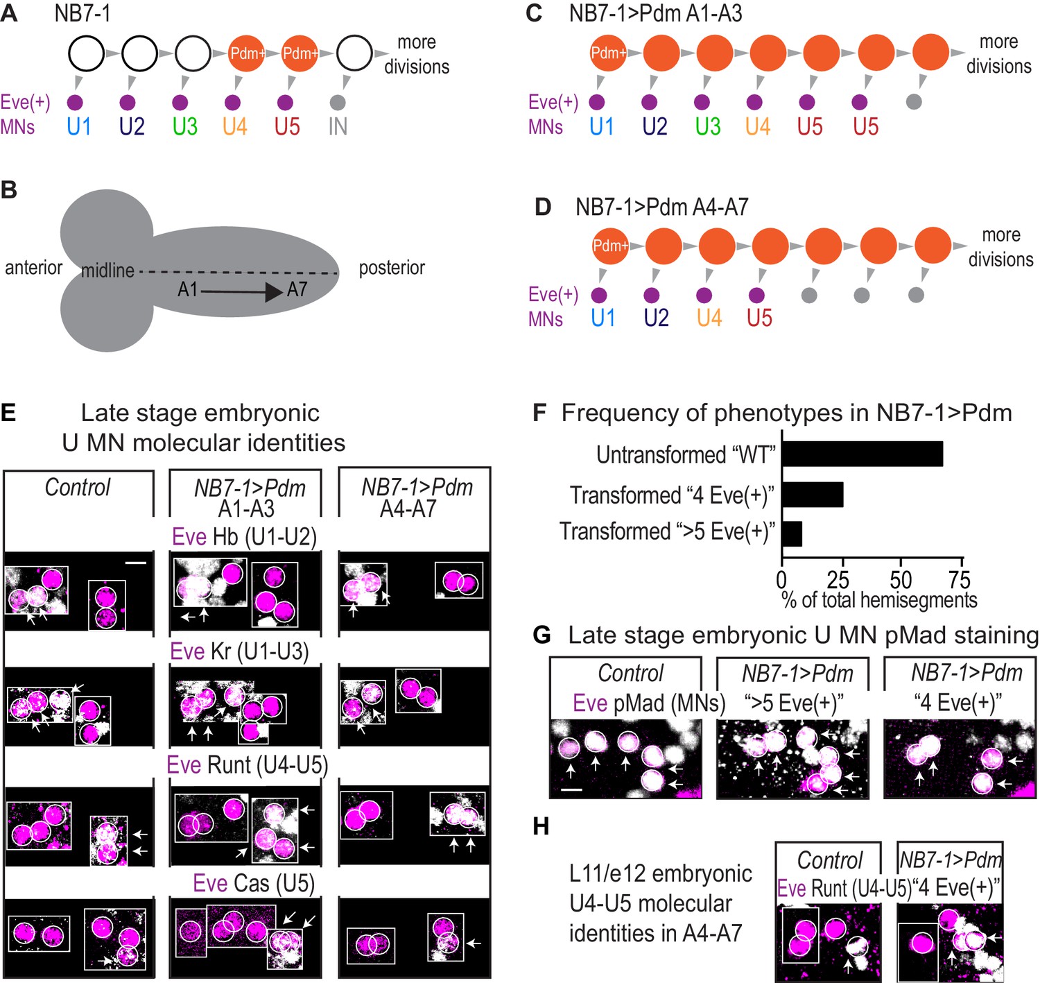
In embryos, precocious expression of Pdm generates either more or fewer U motor neurons depending on A/P positioning.
(A) Illustration of NB7-1 lineage progression. Each gray arrowhead represents cell division. Each gray arrowhead represents cell division. Large circles are neuroblasts, and smaller circles are neurons. Abbreviations: IN is interneuron. (B) Illustration of a Drosophila embryo CNS. Nerve cord abdominal hemisegments are represented as (A). A1 is most anterior and A2-A7 are progressively more posterior. Dotted line represents the midline. (C–D) Illustration follows (A). In NB7−1>Pdm for A1-A3, there is an increase in the number of Eve(+) neurons with U5 embryonic molecular identity (C). In NB7−1>Pdm for A4-A7, there is a decrease in the number of Eve(+) neurons with U3 embryonic molecular identity. (E) Images of embryonic molecular identity marker expression in Eve(+) cells in late stage embryonic CNSs. In NB7−1>Pdm A1-A3, extra Eve(+) cells with U5 molecular identity are produced. In NB7−1>Pdm A4-A7, Eve(+) cells with U3 molecular identity are not produced. Boxes are neurons from different z-planes. Arrows indicate co-expression. (F) Quantification of the % of hemisegments in late stage embryonic NB7−1>Pdm that give rise to 5 Eve(+) neurons (Untransformed ‘WT’), 4 Eve(+) neurons (Transformed ‘4 Eve(+)”), and more than 5 Eve(+) neurons (Transformed ‘>5 Eve(+)”). n = 103/154, n = 39/154, n = 12/154, respectively for the three phenotypes. (G) Images of co-expression of Eve and the pan-motor neuron marker, pMad in Control and NB7−1>Pdm CNS of late stage embryos. (H) Images of embryonic stage late 11 to early 12 (l11/e12) embryonic stage of co-expression of Eve and the U4/U5 marker Runt. In NB7−1>Pdm, cell division rate is not altered because 4 Eve(+) neurons are born, similar to Control. During l11/e12 in NB7−1 > Pdm, two neurons expressing Runt are born, while in Control only one neuron expressing Runt is born. Control is Pdm/+ and NB7−1>Pdm is NB7-1 GAL4/Pdm; Pdm/+. All images are shown anterior up, midline to the left. Scale bars represent 5 microns.
Figure 6 with 1 supplement
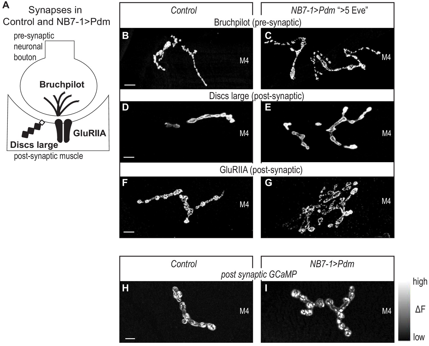
Altered synapses onto Dorsal muscles are functional.
(A) Illustration of subcellular localization of neuromuscular synapse markers. Brunchpilot labels active zones, Discs large is a scaffolding protein strongly localized at post-synapse, GluRIIA is a glutamate receptor IIA. (B–G) Images of neuromuscular synapses on L3 Muscle 4. There is no difference in distribution or abundance of synaptic markers between Control and NB7−1>Pdm. Control is Pdm/+ and NB7−1>Pdm is NB7-1-GAL4/UAS-Pdm; UAS-Pdm/+ (H–I) Images of fluorescence intensity changes in a calcium indicator of synaptic activity. GCaMP was targeted to the post-synaptic density for example (DLG in E-G). When pre-synaptic vesicles are released from active zones (Brp in B-C), post-synaptic neurotransmitter receptors respond (GluRIIA in G-F), increasing GCaMP fluorescence intensity (see Figure 6—figure supplement 1 for details). Images show post-synaptic responses (delta F) in L3 Muscle 4 (M4) in Control and NB7−1>Pdm. Control is Pdm/+; MHC-CD8-GCaMP6f-Sh/Pdm and NB7−1>Pdm is NB7-1-GAL4/Pdm; MHC-CD8-GCaMP6f-Sh/Pdm. All images are shown dorsal up, anterior to the left. Scale bars represent 10 microns.
Figure 6—figure supplement 1
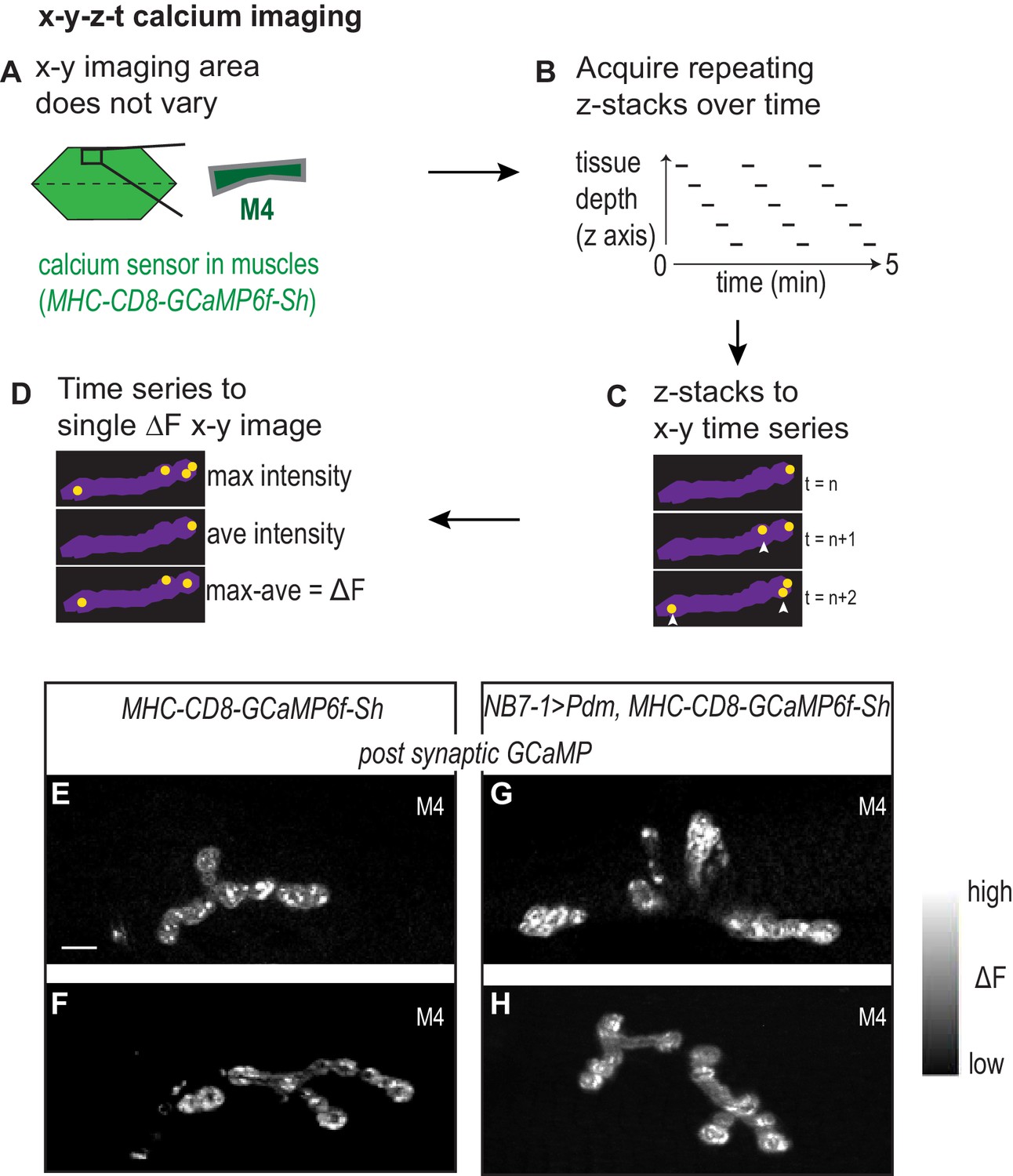
Calcium imaging protocol, analysis, and examples.
(A–D) Illustration of calcium imaging protocol and analysis are shown. See Materials and methods for details. (E–H) Images of calcium signals on L3 Muscle 4. Control is NB7-1-GAL4/+; MHC-CD8-GCaMP6f-Sh/+ and NB7−1>Pdm is NB7-1-GAL4/UAS Pdm; MHC-CD8-GCaMP6f-Sh/UAS Pdm. Images are dorsal up, anterior to the left. Scale bar represents 10 microns.
Figure 7
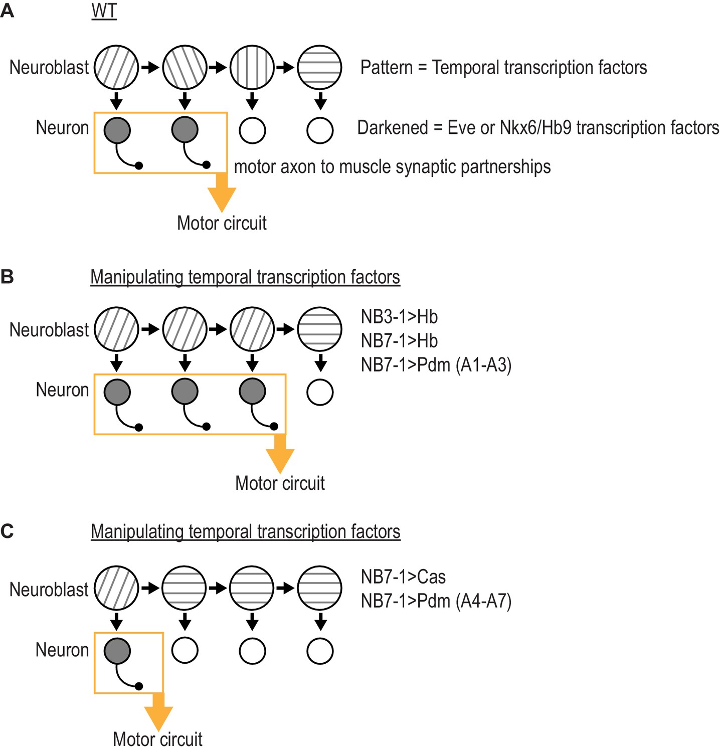
Summary of results in this study.
(A) Illustration of WT (wildtype) neuroblast lineage progression. Different patterns in the neuroblast represent temporal transcription factor expression. Outgrowth projecting from neuron represent motor axon to muscle synaptic partnerships. Yellow box and arrow represent that these neurons are members of the same motor circuit. (A) Illustration of the outcome when expression of Hb (Hunchback) is prolonged in NB7-1 or NB3-1, or expression of Pdm is precocious in NB7-1 (A1-A3) (abdominal segments 1 through 3). Motor axon to muscle synaptic partnerships are altered by increased circuit membership. (C) Illustration of the outcome when expression of Cas (Castor) is precocious in NB7-1 or expression of Pdm is precocious in NB7-1 (A4-A7) (abdominal segments 4 through 7). Motor axon to muscle synaptic partnerships are altered by decreased circuit membership.
Tables
Key resources table
| Reagent type (species) or resource | Designation | Source or reference | Identifiers | Additional information |
|---|---|---|---|---|
| Genetic reagent (D. melanogaster) | DIP-alpha-GAL4 | Robert Carrillo (UChicago) | ||
| Genetic reagent (D. melanogaster) | ac:VP16, gsb:v8v (aka NB7-1-GAL4) | Minoree Kohwi (Columbia) | ||
| Genetic reagent (D. melanogaster) | MHC-CD8-GCaMP6f-Sh | Bloomington stock center [BL] 67739 | BDSC Cat# 67739, RRID:BDSC_67739 | |
| Genetic reagent (D. melanogaster) | UAS-Hb; UAS-Hb/TM2 | Bloomington stock center [BL] 32198 | BDSC Cat# 32198, RRID:BDSC_32198 | |
| Genetic reagent (D. melanogaster) | 5172 J-GAL4 (aka NB3-1-GAL4) | Haluk Lacin (Washington University) | ||
| Genetic reagent (D. melanogaster) | w1118 | Bloomington stock center [BL] 36005 | BDSC Cat# 36005, RRID:BDSC_36005 | |
| Genetic reagent (D. melanogaster) | UAS-Cas | Chris Doe (University of Oregon) | ||
| Genetic reagent (D. melanogaster) | UAS-HA-pdm2/CyO; UAS-HA-pdm2/TM3 | Chris Doe (University of Oregon) | ||
| Genetic reagent (D. melanogaster) | UAS-myr-GFP | Bloomington stock center [BL] 32198 | BDSC Cat# 32198, RRID:BDSC_32198 | |
| Antibody | rabbit anti-Eve (polyclonal) | Heckscher Lab | 1:1000 | |
| Antibody | rabbit anti-Castor (polyclonal) | Chris Doe (Oregon) | 1:1000 | |
| Antibody | guinea pig anti-Hb (polyclonal) | John Rientz (UChicago) | 1:1000 | |
| Antibody | guinea pig anti-Kruppel (polyclonal) | John Rientz (UChicago) | 1:1000 | |
| Antibody | Mouse anti-Eve (monoclonal) | 3C10 | DSHB Cat# 3C10 Anti-even skipped RRID:AB_528229 | 1:50 |
| Antibody | chicken anti-GFP (polyclonal) | Aves #GFP-1020 | Aves Labs Cat# GFP-1020, RRID:AB_10000240 | 1:1000 |
| Antibody | rat anti-Worniu (polyclonal) | Abcam #ab196362 | 1:250 | |
| Antibody | mouse anti-En (monoclonal) | 4D9 | DSHB Cat# 4D9 anti-engrailed/invected, RRID:AB_528224 | 1:5 |
| Antibody | guinea pig anti-Runt (polyclonal) | John Rientz (UChicago) | 1:500 | |
| Antibody | guinea pig anti-HB9 (polyclonal) | Heather Broiher (Case Western) | 1:1000 | |
| Antibody | mouse anti-Cut (monoclonal) | 2B10 | DSHB Cat# 2B10 anti-Cut homeobox RRID:AB_528186 | 1:50 |
| Antibody | rat anti-Zfh2 (polyclonal) | Chris Doe (Oregon) | 1:800 | |
| Antibody | rabbit anti-Smad3 (pMad) (polyclonal) | Abcam #52903 | Abcam Cat# ab52903, RRID:AB_882596 | 1:300 |
| Antibody | mouse anti-Brp (monoclonal) | NC82 | Creative Diagnostics Cat# DMAB9116MD, RRID:AB_2392664 | 1:50 |
| Antibody | mouse anti-DLG (monoclonal) | 4F3 | DSHB Cat# 4F3 anti-discs large, RRID:AB_528203 | 1:500 |
| Antibody | mouse anti-GluRIIA (monoclonal) | 8B4D2 | DSHB Cat# 8B4D2 (MH2B), RRID:AB_52826 | 1:25 |
| Antibody | Cy3-HRP (polyclonal) | Jackson ImmunoResearch 123-025-021 | Jackson ImmunoResearch Labs Cat# 123-025-021, RRID:AB_2338954 | 1:400 |
| Antibody | 647-Phallion (polyclonal) | Thermofisher A22287 | Thermo Fisher Scientific Cat# A22287, RRID:AB_2620155 | 1:600 |
Additional files
Download links
A two-part list of links to download the article, or parts of the article, in various formats.
Downloads (link to download the article as PDF)
Open citations (links to open the citations from this article in various online reference manager services)
Cite this article (links to download the citations from this article in formats compatible with various reference manager tools)
Temporal transcription factors determine circuit membership by permanently altering motor neuron-to-muscle synaptic partnerships
eLife 9:e56898.
https://doi.org/10.7554/eLife.56898


