An ECF-type transporter scavenges heme to overcome iron-limitation in Staphylococcus lugdunensis
- Interfaculty Institute of Microbiology and Infection Medicine, Department of Infection Biology, University of Tübingen, Germany
- Institute of Organic Chemistry, University of Tübingen, Germany
- Groningen Biomolecular Sciences and Biotechnology Institute, University of Groningen, Netherlands
- Department of Microbiology and Immunology, University of Western Ontario, Canada
- Center for Molecular and Vascular Biology, Belgium
- German Centre for Infection Research (DZIF), Partner Site Tübingen, Germany
- (DFG) Cluster of Excellence EXC 2124 Controlling Microbes to Fight Infections, Germany
Figures
Figure 1

LhaSTA represents an iron-regulated heme transporter.
(A) Schematic diagram of the isd operon of S. lugdunensis N920143. Coding sequences, direction of transcription and Fur-binding sites are indicated. ABC membrane-transporters are shown in green. lhaS - SLUG_00900; lhaT - SLUG_00910; lhaA - SLUG_00920 (B) Iron-regulated expression of Lha: S. lugdunensis was grown overnight in TSB, TSB + 200 µM EDDHA or TSB + 200 µM EDDHA + 200 µM FeSO4. Gene expression was quantified by qPCR. Expression was normalized to 5srRNA and to the TSB standard condition using the ΔΔCt method. Fold differences in gene expression are shown. Data represent mean and SD of four independent experiments. Statistical evaluation was performed using students unpaired t-test (lhaS: t = 7,045, df = 6; lhaA: t = 2,979, df = 6) C/D Growth curves of S. lugdunensis N920143 and isogenic mutants. The wild type (WT) S. lugdunensis N920143 strain and the indicated isogenic null mutant strains were grown in the presence of 20 µM FeSO4 (C) or 150 nM heme (D) as a sole source of iron. 500 µl of bacterial cultures were inoculated to an OD600 = 0,05 in 48 well plates and OD600 was monitored every 15 min using an Epoch1 plate reader. For reasons of clarity values taken every 5 hr are displayed. Mean and SD of three experiments are shown. Statistical analysis was performed using one-way ANOVA followed by Dunett’s test for multiple comparisons. * - p<0,05, ****p<0,00001.
Figure 2 with 1 supplement
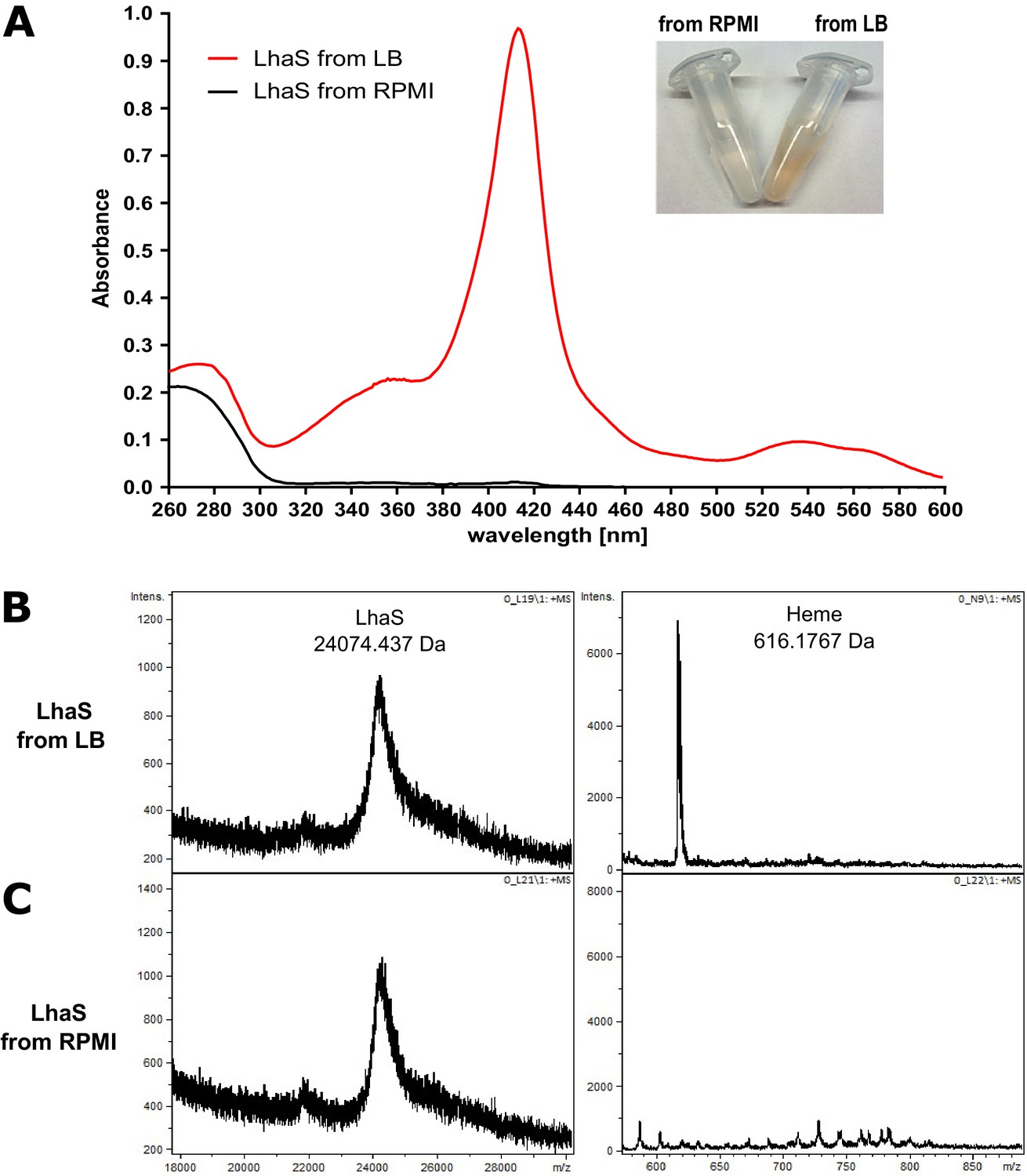
LhaS binds heme.
(A) Ultraviolet-visible (UV-vis) spectrum of recombinant LhaS. C-terminal His-tagged LhaS was heterologously expressed in E. coli and purified from heme-containing LB medium or heme-free RPMI medium. The UV-vis spectrum of the purified LhaS was measured with a BioPhotometer. (B) and (C) MALDI-TOF mass spectra of recombinant LhaS. LhaS (B) was purified out of LB medium and apo-LhaS (C) was purified out of RPMI medium. Mass spectra were recorded with a Reflex IV in reflector mode. All spectra are a sum of 50 shots. Prior to measurements the protein samples were mixed with a 2,5-dihydroxybenzoic acid matrix dissolved in water/acetonitrile/trifluoroacetic acid (50/49.05/0.05) at a concentration of 10 mg ml−1 and spotted onto the MALDI polished steel sample plate.
Figure 2—figure supplement 1
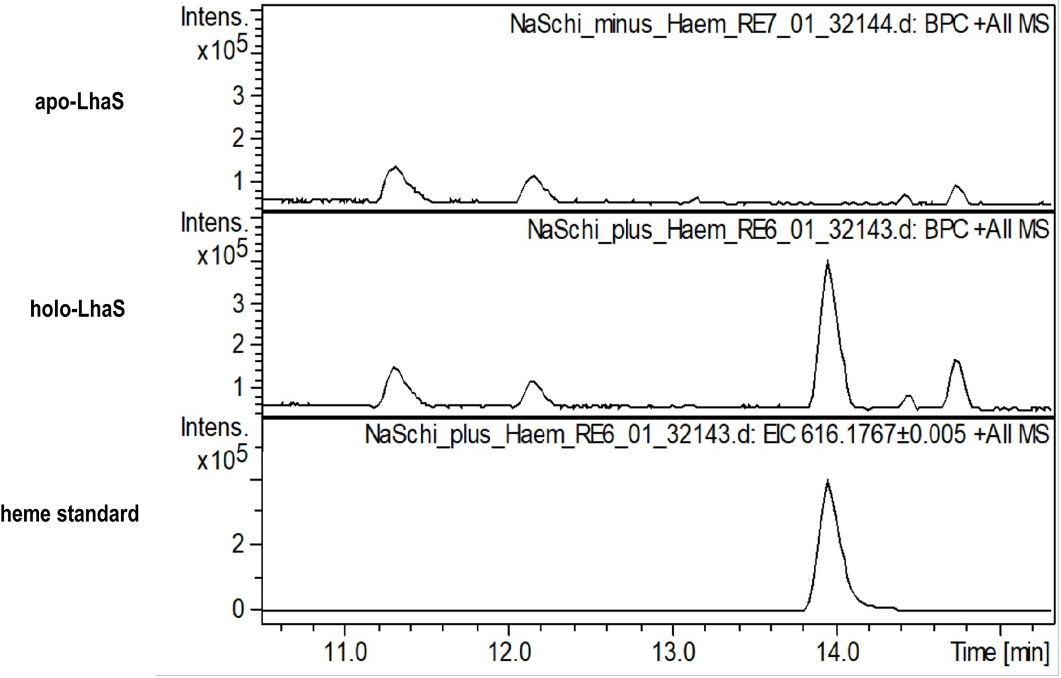
High resolution mass spectra of apo- and holo-LhaS.
Spectra of apo-LhaS isolated from E. coli grown in RPMI medium (upper panel), holo-LhaS isolated from LB-medium (middle panel) and a heme standard (lower panel) were recorded on a HPLC-UV-HR mass spectrometer. The samples were diluted with MilliQ-H2O and applied to a Dionex Ultimate 3000 HPLC system that is coupled to the MaXis 4G ESI-QTOF mass spectrometer.
Figure 3 with 1 supplement

LhaSTA represents a functionally autonomous iron acquisition system.
(A) LhaSTA-dependent proliferation. S. lugdunensis N920143 deletion mutant strains lacking the entire isd operon and expressing LhaSTA (∆isd pRB473:lhaSTA) or not (∆isd pRB473) from the plasmid pRB473 were grown in the presence of 200 nM heme as a sole source of iron. 500 µl of cultures were inoculated to an OD600 = 0,05 in 48 well plates and OD600 was monitored every 15 min using an Epoch1 plate reader. For reasons of clarity values taken every 5 hr are displayed. Mean and SD of three experiments are shown. Statistical analysis was performed using students unpaired t-test. ***p<0,0001 (B) Intracellular accumulation of iron. Strains were grown in iron limited medium to OD600 = 0,6 and 5 µM heme were added for 3 hr. Cell fractionation of 1 ml OD600 = 50 was performed and the iron content of the cytosolic fraction was determined using the ferrozine assay. Data represent the mean and SD of three independent experiments. Statistical analysis was performed using students unpaired t-test (t = 5,12729, df = 4).
Figure 3—figure supplement 1
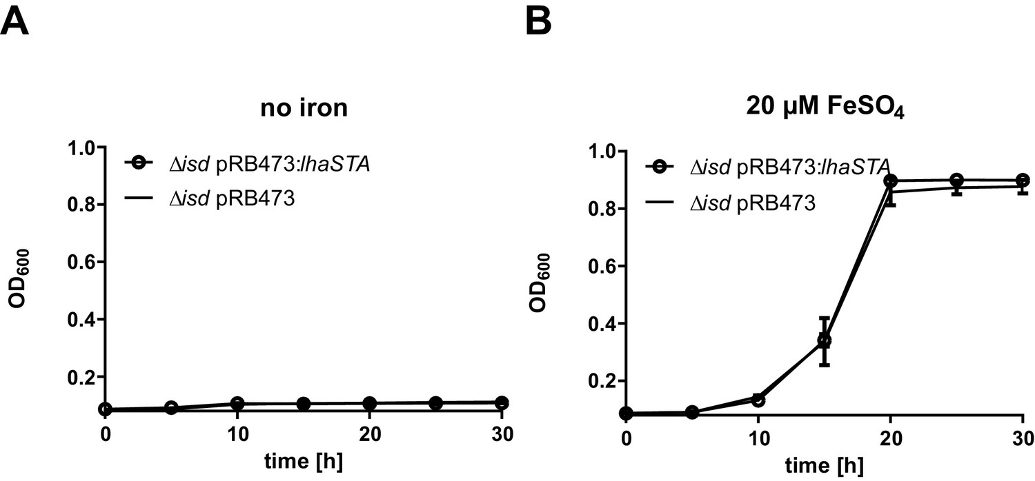
LhaSTA dependent growth.
(A/B) Proliferation of S. lugdunensis N920143 ∆isd pRB473 and ∆isd pRB473:lhaSTA strains. The indicated strains were grown in the absence of nutritional iron (A) or in the presence of 20 µM FeSO4 (B). 500 µl of cultures were inoculated to an OD600 = 0,05 in 48 well plates and OD600 was monitored every 15 min using a Epoch1 plate reader. For reasons of clarity values taken every 5 hr are displayed. Mean and SD of three experiments are shown. Statistical analysis was performed using students unpaired t-test.
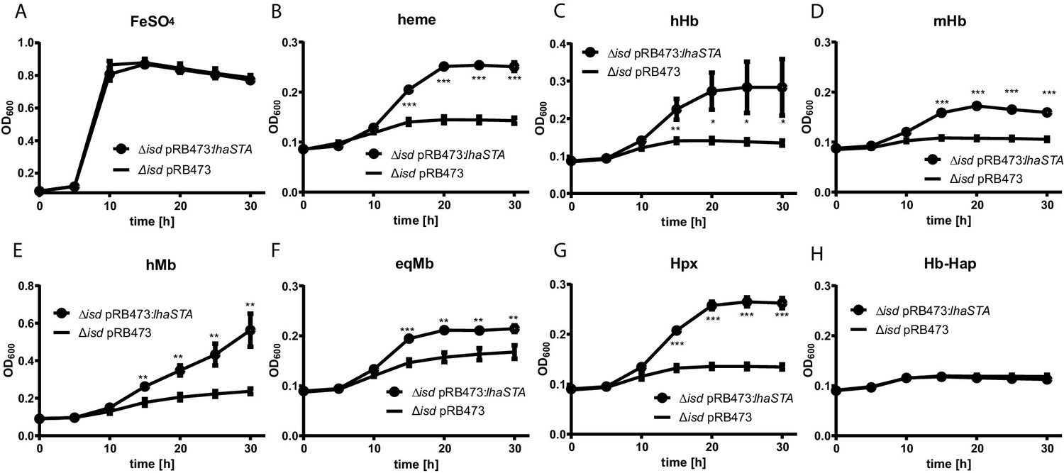
Figure 4 with 1 supplement
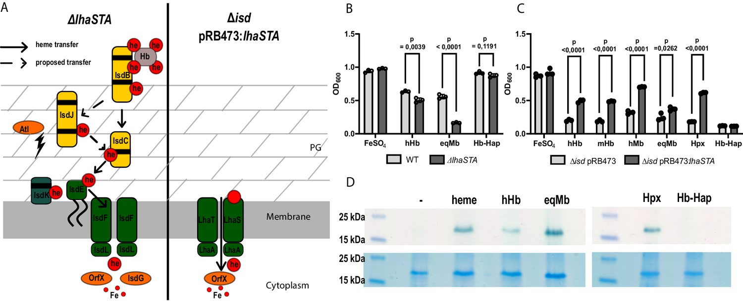
LhaSTA facilitates heme acquisition from a wide range of hemoprotein substrates.
(A) Schematic diagram of known heme acquisition systems in the S. lugdunensis mutant strains lacking either the genes encoding LhaSTA (∆lhaSTA, left) or the entire isd operon and expressing LhaSTA from the plasmid pRB473 (∆isd pRB473:lhaSTA). ABC membrane transporters are shown in green. Cell wall-anchored proteins of the Isd-system are shown in yellow. Heme/hemoglobin-binding NEAT motifs within each protein are indicated as black boxes. Black arrows indicate the transfer of heme. he: heme; hb: hemoglobin; PG: peptidoglycan. (B) Growth of S. lugdunensis N920143 wild type (WT) and ∆lhaSTA. Strains were grown in the presence of 20 µM FeSO4 or 2.5 µg/ml human hemoglobin (hHb) or 10 µg/ml equine myoglobin (eqMb) or 117 nM hemoglobin-haptoglobin complex (Hb-Hap) as a sole source of iron. 500 µl of cultures were inoculated to an OD600 = 0,05 in 48 well plates and OD600 was measured after 30 hr using an Epoch1 plate reader. Mean and SD of three experiments are shown. Statistical analysis was performed using students unpaired t-test. hHb - t = 6,0007, df = 4; eqMb – t = 20,52, df = 4; Hb-Hap – t = 1,978, df = 4. (C) Growth of S. lugdunensis N920143 ∆isd pRB473 and ∆isd pRB473:lhaSTA. Strains were grown in the presence of 20 µM FeSO4 or 2.5 µg/ml hHb or 2.5 µg/ml murine hemoglobin (mHb) or 10 µg/ml human myoglobin (hMb) or 10 µg/ml eqMb or 200 nM human hemopexin (Hpx) or 117 nM Hb-Hap as a sole source of iron. 500 µl of cultures were inoculated to an OD600 = 0,05 in 48 well plates and OD600 was measured after 30 hr using an Epoch1 plate reader. Mean and SD of three experiments are shown. Statistical analysis was performed using students unpaired t-test hHb – t = 18,5, df = 4; mHb – t = 29,03, df = 4; hMb – t = 25,98, df = 4; eqMb – t = 3,442, df = 4; Hpx – t = 77,12 df = 4; Hb-Hap t = 2758 df = 4. (D) TMBZ-H2O2 stain of TGX gels for heme-associated peroxidase activity. Membrane vesicles were saturated with excess of hemoprotein (5.6 µM heme, 476 µg/ml hHb, 437 µg/ml eqMb, 5.6 µM Hpx, 476 µg/ml Hb-Hap) or no hemoprotein (-) for 10 min at RT. LhaS was purified, 15 µg protein was loaded on a TGX gel and stained for peroxidase activity with TMBZ-H2O2 (upper panel). Gels were destained and subsequently stained with BlueSafe (lower panel) to confirm the presence of the protein in all conditions.
Figure 4—figure supplement 1
Growth of S. lugdunensis N940135 ∆isd pRB473 and ∆isd pRB473:lhaSTA.
The indicated strains were grown in the presence of 20 µM FeSO4 or 2.5 µg/ml human hemoglobin (hHb), 2.5 µg/ml or murine hemoglobin (mHb) or 10 µg/ml human myoglobin (hMb) or 10 µg/ml equine myoglobin (eqMb) or 200 nM human hemopexin (Hpx) or 117 nM Hb-Hap as a sole source of iron. 500 µl of cultures were inoculated to an OD600 = 0,05 in 48 well plates and OD600 was measured every 15 min. For reasons of clarity values taken every 5 hr are displayed. Mean and SD of three experiments are shown. Statistical analysis was performed using students unpaired t-test.
Figure 5 with 2 supplements
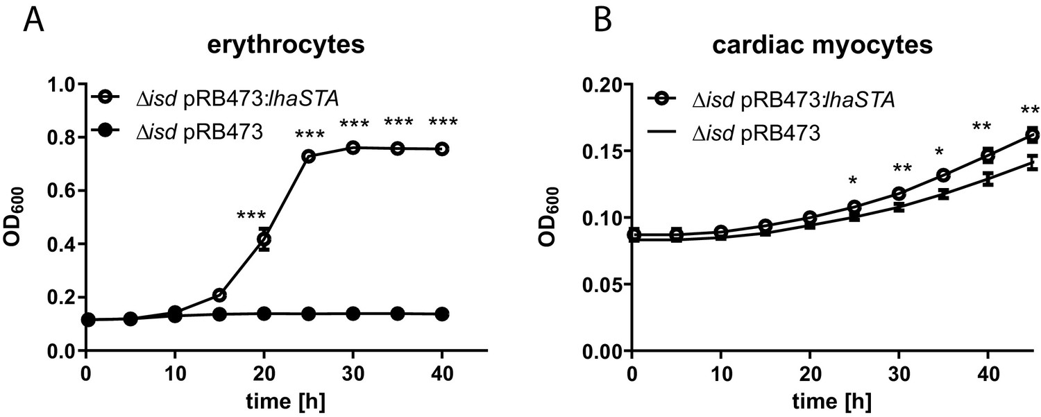
LhaSTA allows usage of host cells as an iron source.
(A) Growth of S. lugdunensis N940135 ∆isd pRB473:lhaSTA and ∆isd pRB473 on human erythrocytes. Strains were grown in the presence of freshly isolated human erythrocytes (105 cells/ml) as a sole source of iron. 500 µl of cultures were inoculated to an OD600 = 0,05 in 48 well plates and OD600 was monitored every 15 min using an Epoch1 plate reader. For reasons of clarity values taken every 5 hr are displayed. Mean and SD of three experiments are shown. Statistical analysis was performed using students unpaired t-test. ***p<0,0001 (B) Growth of S. lugdunensis N940135 ∆isd pRB473 and ∆isd pRB473:lhaSTA on human cardiac myocytes. Strains were grown in the presence of 40000 primary human cardiac myocytes per well as a sole source of iron. Cardiac myocytes were detached and washed once with RPMI+200 µM EDDHA prior addition to the wells. 500 µl of cultures were inoculated to an OD600 = 0,05 in 48 well plates and OD600 was monitored every 15 min using an Epoch1 plate reader. For reasons of clarity values taken every 5 hr are displayed. Mean and SD of three experiments are shown. Statistical analysis was performed using students unpaired t-test. *p<0,05, **p<0,01.
Figure 5—figure supplement 1
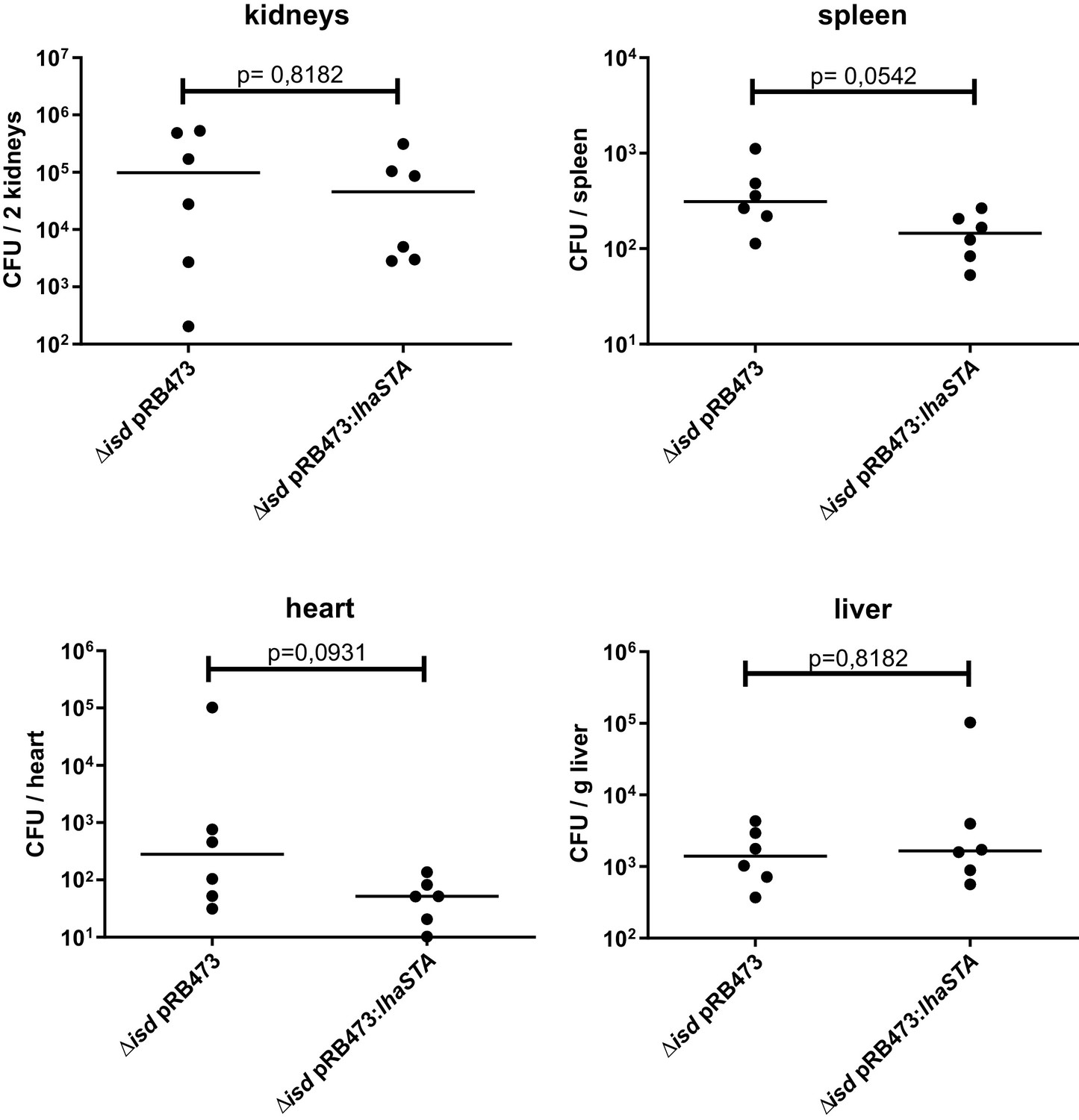
Mouse systemic infection model.
C57BL/6 mice were infected with 3 × 10⁷ CFU per animal. Mice were sacrificed 72 hr post infection and CFUs within the indicated organs were enumerated. Horizontal lines show the median. Statistical analysis was performed using Mann Whitney test.
Figure 5—figure supplement 2
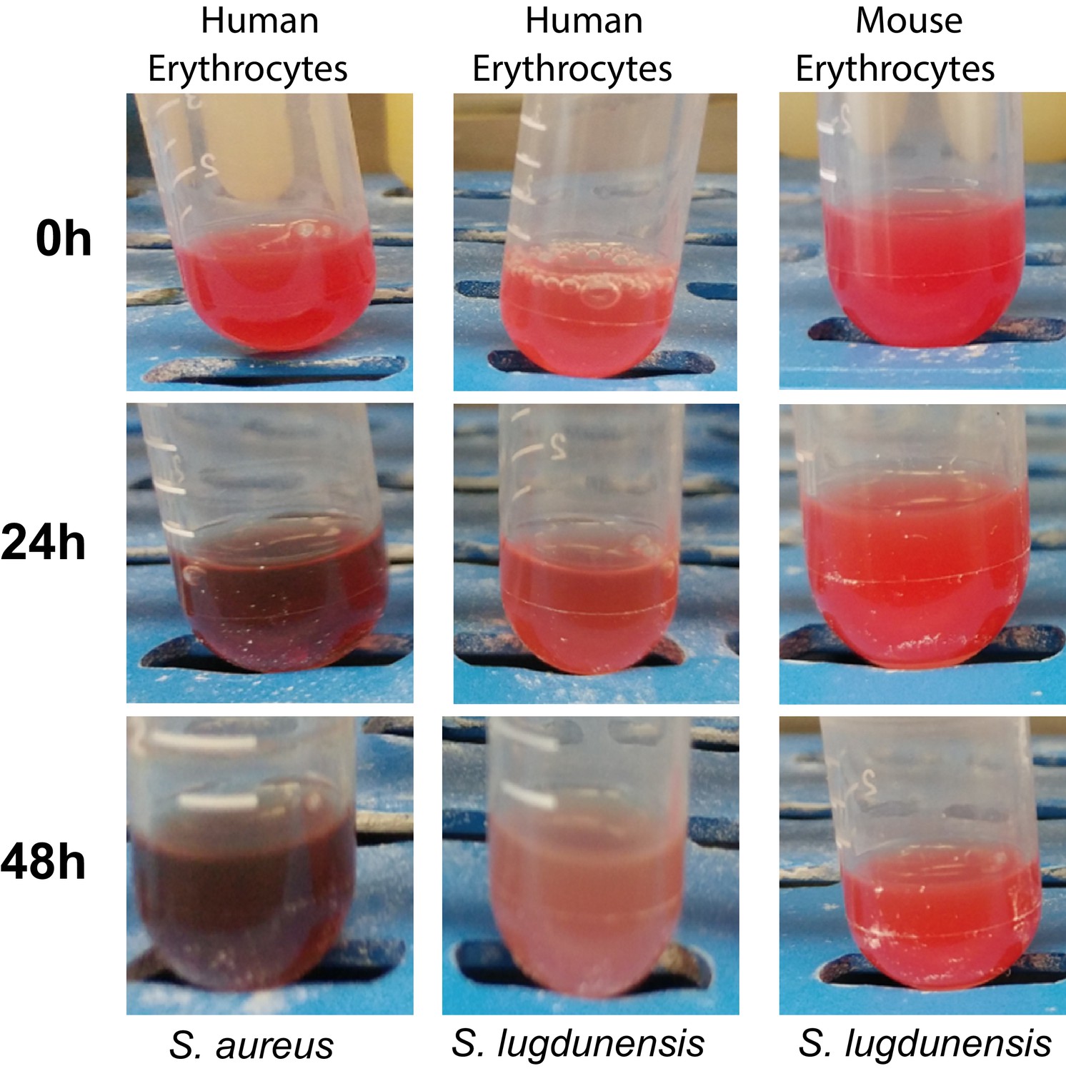
Hemolysis of human and mouse erythrocytes.
Human (left and middle panels) or mouse erythrocytes (right panel) were incubated with the filtrated culture supernatants of S. aureus (left panel) or S. lugdunensis (middle and right panels) for 24 and 48 hr.
Figure 6
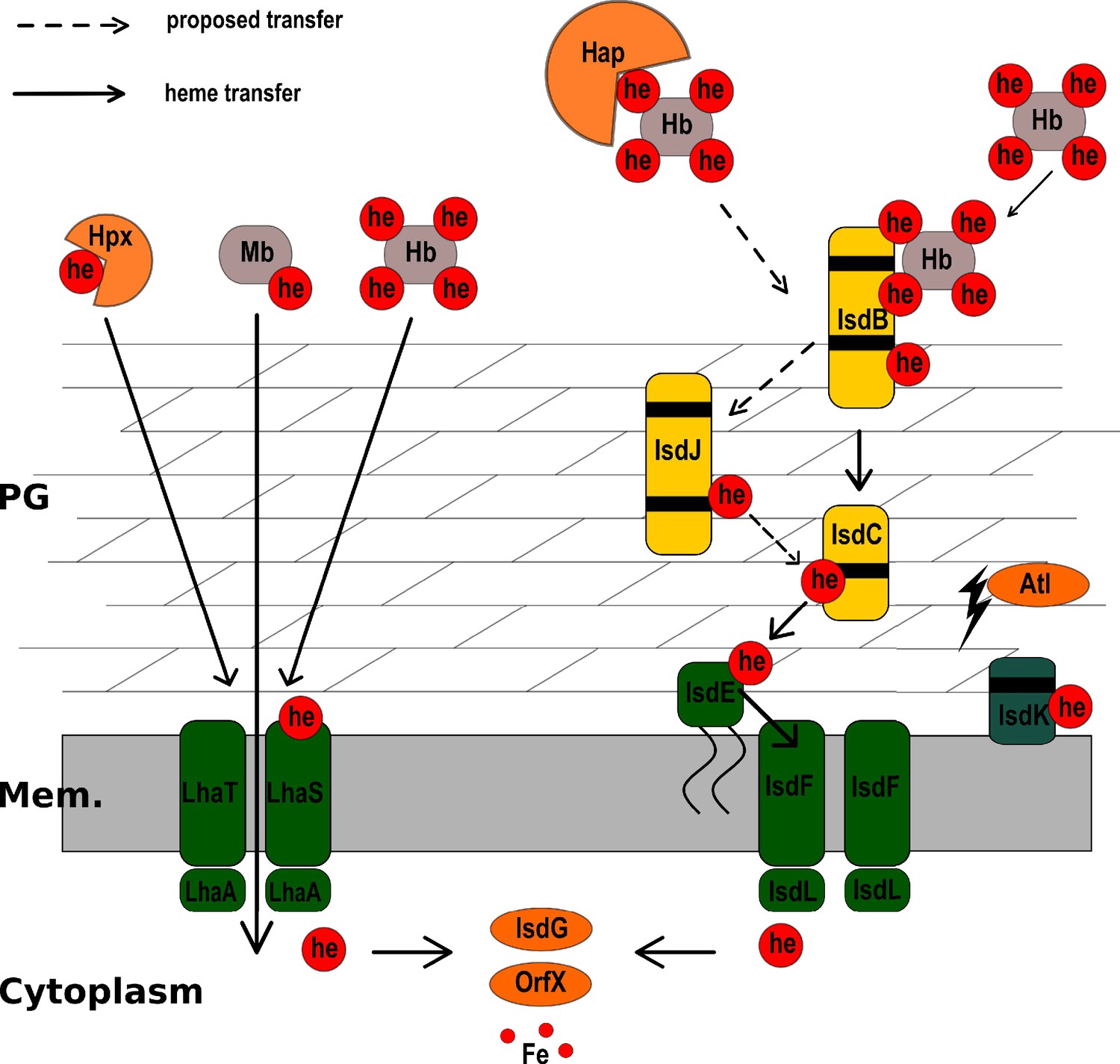
Model of heme acquisition in S. lugdunensis.
ABC membrane transporters are shown in green. Cell wall-anchored proteins of the Isd-system are shown in yellow. Heme/hemoglobin-binding NEAT motifs within each protein are indicated as black boxes. Black arrows indicated the transfer of heme. he: heme; hb: hemoglobin; PG: peptidoglycan; Mem: Membrane; Hap: Haptoglobin; Hpx:Hemopexin; Atl: Autolysin.
Tables
Key resources table
| Reagent type (species) or resource | Designation | Source or reference | Identifiers | Additional information |
|---|---|---|---|---|
| Strain, strain background (Staphylococus lugdunensis) | N940135 | National Reference Center for Staphylococci, Lyon, France (Heilbronner et al., 2011) | ||
| Strain, strain background (S. lugdunensis) | N920143 | National Reference Center for Staphylococci, Lyon, France (Heilbronner et al., 2011) | ||
| Strain, strain background (S. lugdunensis) | N920143 ΔisdEFL | This paper | Markerless deletion mutant of isdEFL | |
| Strain, strain background (S. lugdunensis) | N920143 ΔlhaSTA | This paper | Markerless deletion mutant of lhaSTA | |
| Strain, strain background (S. lugdunensis) | N920143 ΔisdEFLΔlhaSTA | This paper | Markerless double deletion mutant of isdEFL and lhaSTA | |
| Cell line (Human) | Human cardiac myocytes (HCM) | PromoCell | C-12810 | |
| Recombinant DNA reagent | pQE-30 | Qiagen | IPTG inducible expression plasmid | |
| Recombinant DNA reagent | pQE30:lhaS | This paper | LhaS expressing plasmid for protein purification | |
| Recombinant DNA reagent | pRB473: lhaSTA | This paper | LhaSTA expressing plasmid for complementation | |
| Recombinant DNA reagent | pIMAY (plasmid) | Monk et al., 2012 | See Material and methods | Thermosensitive vector for allelic exchange |
| Recombinant DNA reagent | pIMAY:∆isd | Zapotoczna et al., 2012 | Plasmid for the deletion of the entire isd locus | |
| Recombinant DNA reagent | pIMAY:∆isdEFL | This study | Plasmid for the deletion of conventional heme transporter isdEFL | |
| Recombinant DNA reagent | pIMAY:∆lhaSTA | This study | Plasmid for the deletion of heme specific ECF-transporter | |
| Recombinant DNA reagent | pRB473 | Brückner, 1992 | Expression plasmid without promotor region. | |
| Biological sample (Human) | Human hemoglobin | Own preparation | See Material and methods | Sex male |
| Biological sample (Pork) | Porcine hemin | Sigma | 51280 | |
| Biological sample (Human) | Human Myoglobin | Sigma Aldrich | M6036 | |
| Biological sample (Horse) | Equine Myoglobin | Sigma Alrich | M1882 | |
| Biological sample (Human) | Human Haptoglobin (Phenotype 1–1) | Sigma Aldrich | SRP6507 | |
| Biological sample (Human) | Human Hemopexin | Sigma Aldrich | H9291 | |
| Chemical compound, drug | RPMI 1640 Medium | Sigma Aldrich | R6504-10L | |
| Chemical compound, drug | Casamino acids | BACTO | 223050 | |
| Chemical compound, drug | EDDHA | LGC Standarts | TRC-E335100-10MG | |
| Chemical compound, drug | Dodecyl-β-D-maltosid (DDM) | Carl Roth | CN26.1 | |
| Chemical compound, drug | 3,3',5,5'-tetramethylbenzidine (TMBZ) | Sigma Aldrich | 860336 | |
| Chemical compound, drug | Profinity IMAC resin nickel chrged | BIO RAD | 1560135 |
Additional files
-
Supplementary file 1
Key Resources Table PCR primers.
PCR and qPCR primers sequences used in this study.
- https://cdn.elifesciences.org/articles/57322/elife-57322-supp1-v3.docx
-
Transparent reporting form
- https://cdn.elifesciences.org/articles/57322/elife-57322-transrepform-v3.docx
Download links
A two-part list of links to download the article, or parts of the article, in various formats.
Downloads (link to download the article as PDF)
Open citations (links to open the citations from this article in various online reference manager services)
Cite this article (links to download the citations from this article in formats compatible with various reference manager tools)
An ECF-type transporter scavenges heme to overcome iron-limitation in Staphylococcus lugdunensis
eLife 9:e57322.
https://doi.org/10.7554/eLife.57322


