Drosophila seminal sex peptide associates with rival as well as own sperm, providing SP function in polyandrous females
- Department of Molecular Biology and Genetics, Cornell University, United States
Figures
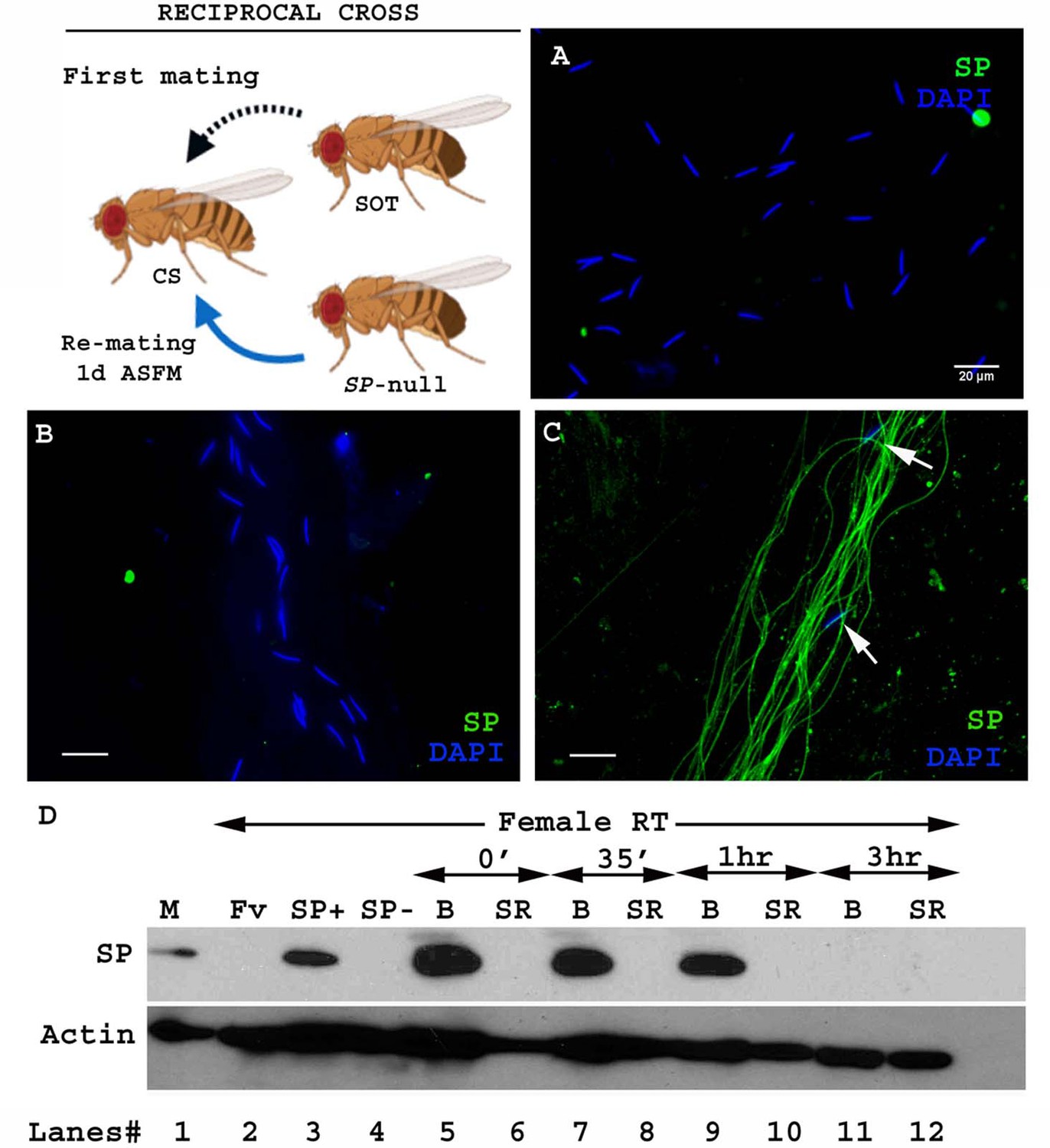
Figure 1

SP from a second male can bind to SP-deficient sperm of previous male stored within a mated female.
Cartoon: Pictorial representation of the crossing scheme (fly images from Biorender). Wild type (CS) females were first mated to an SP-null male and then, at the indicated time, to a spermless (SOT) male. Sperm heads were stained with DAPI (blue) and SP was visualized with Alexa fluor 488, staining the sperm tail (green) and sperm head (cyan; overlapping blue/green). (A) Sperm from females singly mated to SP-null males, 1d ASM. (B) Sperm from females mated to SP-null males, remated to spermless males at 1d ASFM and (C) at 4d ASFM, both frozen 2 hr ASSM. White arrows indicate sperm heads. Bar = 20 µm (D) Western blot lane numbers 1: Fv, reproductive tract (RT) of virgin female (negative control; n = 5), 2: M, a pair of male accessory gland (positive control; n = 1), 3: SP-, reproductive tracts of females mated to SP-null males, 2 hr ASM (n = 5), 4: SOT, reproductive tracts of females mated to spermless males, 1d ASM (n = 5), 5: SOT, SP-, reproductive tract of females mated to spermless males and then remated to SP-null males, 1d ASFM (n = 8 RT), 6: SOT, SP+, reproductive tract of females mated to spermless males and then remated to control (SP+) males at 1d ASFM, frozen 2 hr ASSM (positive control; n = 8 RT), 7: (SP-, SOT), 1d and 8: (SP-, SOT), 4d sperm isolated from the seminal receptacle of females mated to SP-null males and then remated to spermless males at 1d ASFM and 4d ASFM, frozen 2 hr ASSM (n = 15 SS). Actin served as loading control.
Figure 2 with 1 supplement
Sperm from a second male are not bound to SP from a prior spermless male.
(Cartoon): Pictorial representation of the cross (fly images from Biorender); it is reciprocal of that in Figure 1. Females mated first with spermless (SOT) males and then a day later with SP-null males that provided sperm. Sperm heads were stained with DAPI (blue) and SP visualized with Alexa fluor 488, staining the sperm tail (green) and sperm head (cyan; overlapping blue/green). (A) Sperm from females singly mated to SP-null males, 2 hr ASM. (B) Sperm from females mated to spermless males and then remated to SP-null males, 1d ASFM. (C) Sperm from females mated to spermless males and then remated to SP+ males, 1d ASFM, serve as positive controls. Flies were frozen 2 hr ASSM. White arrows indicate sperm heads. Bar = 20 µm (D) Western blot lane numbers 1: M, a pair of male accessory gland (positive control; n = 1), 2: Fv, reproductive tract (RT) of virgin female (negative control; n = 5), 3: SP+, reproductive tract of females mated to control males (TM3 siblings of SP-null males; n = 5; positive control), 4: SP-, reproductive tract of females mated to SP-null males (n = 5; negative control). 5–12: Proteins from Bursa (B) or seminal receptacle (SR) from females mated to spermless males frozen at 0 min immediately after mating, 35 min, 1 hr, and 3 hr ASM, respectively (n = 15). Actin served as loading control.
Figure 2—figure supplement 1
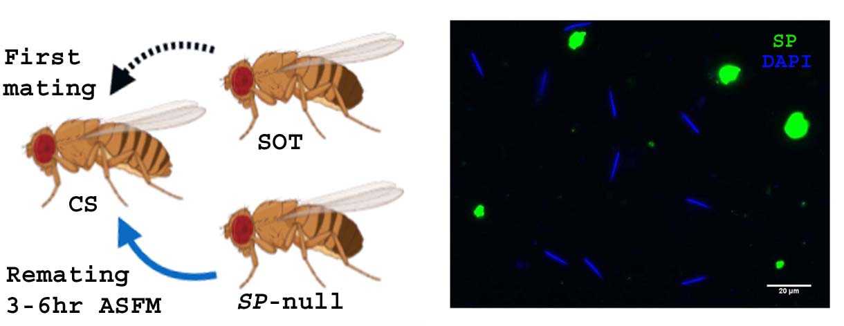
Cartoon: Pictorial representation of cross (fly images from Biorender).
Females mated first with spermless (sot) male and then 3–6 hr ASFM with SP-null male that provided sperm. Panel: Sperm from SR of females mated to spermless males and then remated to SP-null males, 3–6 hr ASFM, frozen at 2 hr ASSM. Sperm heads were stained with DAPI (blue) and SP (green) probed with Alexa fluor 488 (n = 5; Bar = 20 µm).
Figure 3 with 1 supplement

Remating with spermless males restores fertility, delays receptivity and optimizes efficient sperm release in females that previously mated to SP-null males.
(A) Graphical representation of numbers of progeny produced by each female over the span of 10 days, following mating to control (TM3 siblings of SP-null males: SP+; red), SP-null males (SP-null; green), or spermless males (SOT), p***=<0.001; n = 15–20. (B) Fertility of females mated to SP-null males and then remated to spermless males at 1d ASFM (SP-null, SOT; blue, n = 15–20) and (C) Fertility of females mated to SP-null males and then remated to spermless males at 4d ASFM (SP-null, SOT; blue, n = 15–20) compared to females mated to control males and then remated to spermless males (SP+, SOT, red, ns = non significant). (D) Percentage receptivity of females mated to SP-null males and then remated to spermless males (SP-null, SOT) at 1d ASFM, when compared to females singly mated to SP-null males (red arrows), spermless (SOT, blue arrows) or CS males, 1d ASM (p*=<0.05; p***=<0.001; n = 15–20 for each technical replicate). (E) Percentage receptivity of females mated to SP-null males and then remated to spermless males (SP-null, SOT) at 4d ASFM, when compared to females singly mated to SP-null males, spermless (SOT) or CS males (purple arrows), 4d ASM (p***=<0.001; n = 15–20 for each technical replicate). (F) Western blot lane numbers 1: Fv, reproductive tract (RT) of five virgin females (negative control); 2: M, a pair of male accessory gland (positive control); 3, 4, 5: RT of females mated to CS males, flash frozen at 2 hr (n = 5), 1d (n = 15) and 4d (n = 15) ASM, respectively; 6, 7, 8: RT of females mated to SP-null males and then subsequently mated to spermless males at 1d ASFM, flash frozen 2 hr (n = 5), 1d (n = 15) and 4d (n = 15) ASSM, respectively. Actin served as loading control. (G) Sperm in the seminal receptacle (SR) of a typical female mated to a control male (SP+; ProtB-eGFP) at 4d ASM. (H) Sperm in the SR of a typical female mated to SP-null; ProtB-eGFP male at 4d ASM. (I) Sperm in the SR of a typical female, mated to SP-null; ProtB-eGFP and subsequently remated to a spermless male at 1d ASFM, and frozen at 4d ASSM. In (G–I) sperm heads are green due to eGFP. Bar = 50 µm. (J) Graphical representation of sperm counts in SRs of females singly-mated to control (SP+, red, TM3 siblings of SP-null; ProtB-eGFP), SP-null (green) or doubly-mated to SP-null and spermless male (SP-null, SOT, blue) represented in G, H, I panels (p**=<0.01; p*=<0.05; ns = non significant; n = 15–20).
-
Figure 3—source data 1
Source data for Figure 3D, E.
- https://cdn.elifesciences.org/articles/58322/elife-58322-fig3-data1-v2.xlsx
Figure 3—figure supplement 1
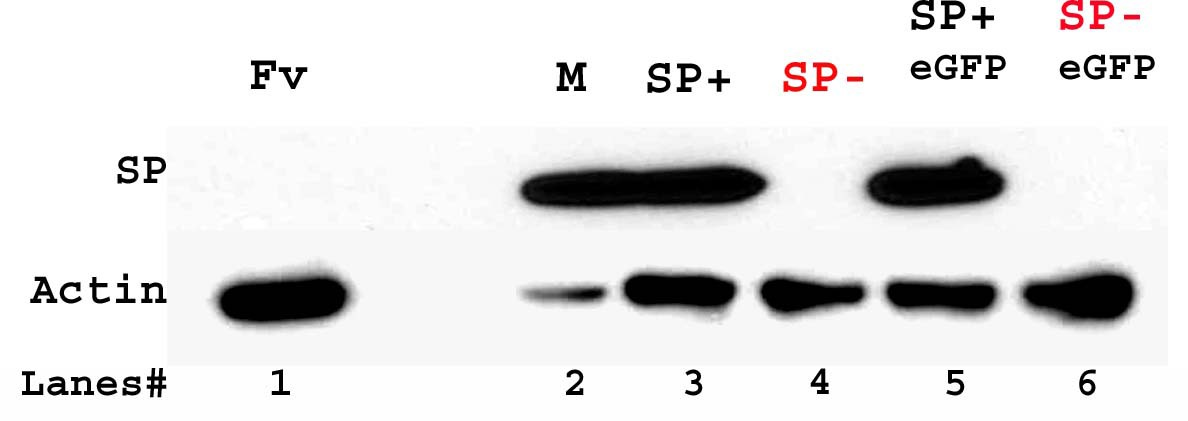
Western blot probed for SP.
Lanes/samples are 1: Fv, reproductive tract (RT) of three virgin females (negative control); 2: M, one pair of male accessory glands (positive control); 3: SP+, RT of three females mated to control (TM3 siblings of SP-null males; positive control) males at 2 hr ASM; 4: SP-, RT of three females mated to SP-null males at 2 hr ASM; 5: SP+ eGFP, RT of three females mated to control (TM3 siblings of SP-null; ProtB-eGFP males; positive control) males at 2 hr ASM; 6: SP- eGFP, RT of three females mated to SP-null; ProtB-eGFP males at 2 hr ASM. Actin served as loading control.
Figure 4 with 1 supplement
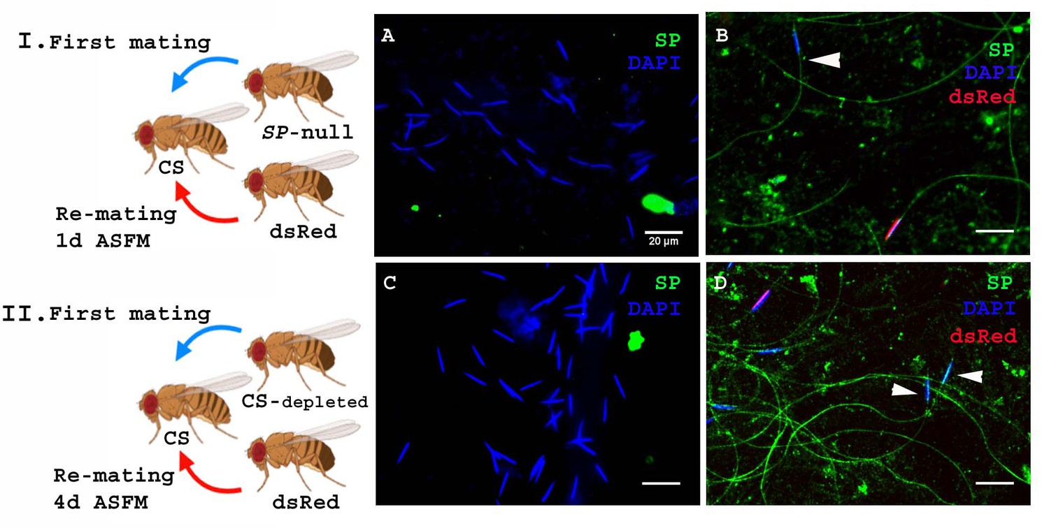
SP from a male who also provides sperm can bind to SP-deficient sperm as well as to the donor’s sperm.
Cartoon (I): Pictorial representation of the experimental cross (fly images from Biorender). Females mated to SP-null males were remated to control (ProtB-dsRed) males at 1d ASFM. (A) Sperm from females singly mated to SP-null males, 2 hr ASM (blue sperm-head). (B) Sperm from females mated to SP-null males (blue sperm-head) remated to ProtB-dsRed (red+ blue sperm-head) males at 1d ASFM. SP was visualized with Alexa fluor 488, staining the sperm (head+ tail) green. Flies were frozen 2 hr ASSM. White arrows indicate sperm heads (n = 10; Bar = 20 µm). Cartoon (II): Pictorial representation of the substitute cross (fly images from Biorender). Females mated to SFP depleted control (CS) males were remated to control (Prot B-dsRed) males at 4d ASFM. (C) Sperm from females singly-mated to SFP depleted CS males at 4d ASM (blue sperm-head). (D) Sperm from females mated to SFP depleted CS males (blue sperm-head), remated to ProtB-dsRed (red+ blue sperm-head) males at 4d ASFM. SP was visualized with Alexa fluor 488, staining the sperm (head+ tail)green. Flies were frozen 2 hr ASSM. White arrows indicate sperm heads (n = 10; Bar = 20 µm).
Figure 4—figure supplement 1
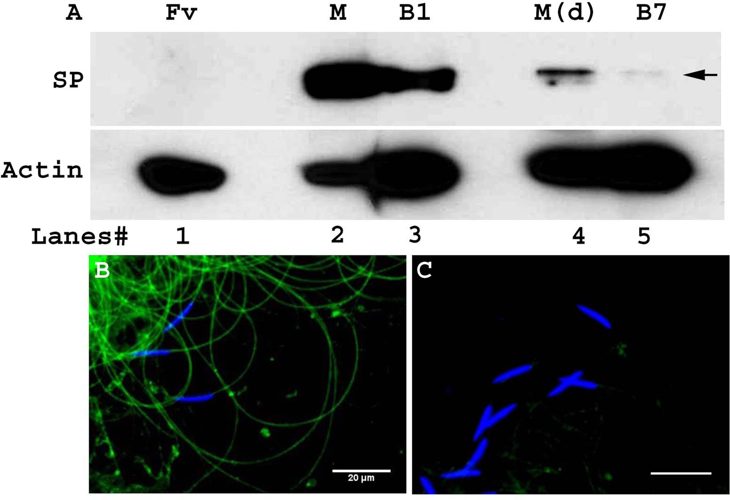
Little to no SP is transferred by multiply-mated males.
(A) Western blot probed for SP. Lanes/samples are 1: Fv, reproductive tract (RT) of two virgin females (negative control); 2: M, one pair of male accessory glands from a 3-day-old unmated virgin male; 3: B1, RT of four females mated to control unmated virgin males, frozen at 2 hr ASM; 4: M(d), one pair of male accessory glands dissected from a multiply mated male (previously mated with six virgin females); 5: B7, RT of four females mated to multiply-mated males, frozen at 2 hr ASM. Actin served as loading control. (B) Sperm dissected from females mated to unmated males, frozen at 2 hr ASM. (C) Sperm dissected from females mated to multiply mated males, frozen at 2 hr ASM. Sperm heads were stained with DAPI (blue) and presence of SP (green) was detected with Alexa fluor 488 (n = 5; Bar = 20 µm).
Figure 5
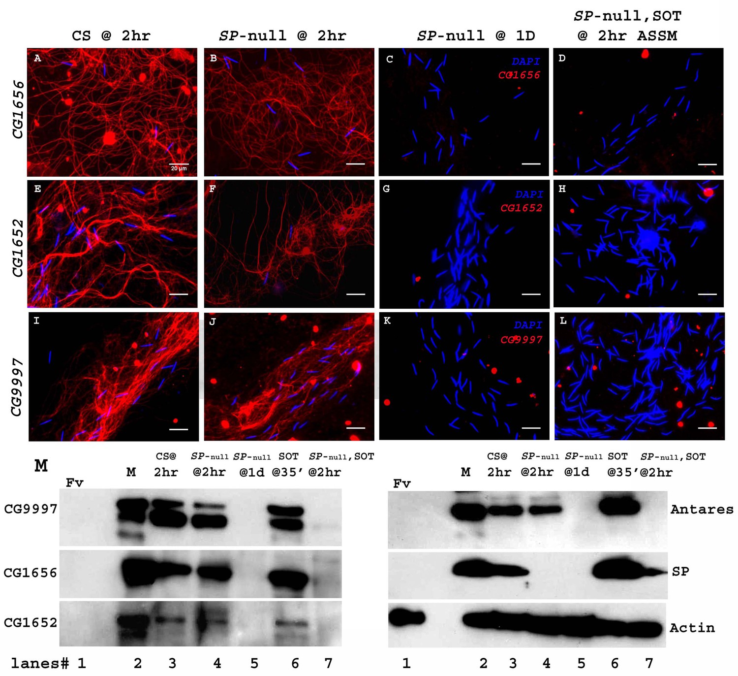
Sperm do not bind detectable LTR-SFPs from a second male.
Females mated to wild type (CS) males at 2 hr ASM show LTR-SFPs bound to sperm, CG1656 (A), CG1652 (E), CG9997 (I). Females mated to SP-null males show the same (B,F,J) but by 1d postmating LTR-SFPs’ signal were no longer detected on sperm (C,G,K) confirming previous reports (Singh et al., 2018). Females mated to SP-null males and then remated to spermless males also do not show detectable signal for sperm-LTR-SFP binding for CG1656 (D), CG1652 (H) and CG9997 (L), 2 hr ASSM, although they have SP bound (Figure 1). Sperm stained for the indicated LTR-SFP detected with Alexa fluor 594 (red) and sperm-head stained with DAPI (blue). Bar = 20 µm (M) Western blot probed for indicated LTR-SFPs. Lanes/samples are 1: Fv, reproductive tract (RT) of three virgin females (negative control); 2: M, one pair of male accessory glands (positive control); 3: CS @ 2 hr, sperm dissected from SR of 20 females mated to wild type (CS) males at 2 hr ASM; 4: SP-null @ 2 hr, sperm dissected from SR of 20 females mated to SP-null males at 2 hr ASM; 5: SP-null @1d, sperm dissected from SR of 20 females mated to SP-null males at 1d ASM; 6: SOT@35’, reproductive tract of three females mated to spermless males at 35’ASM (positive control); 7: SP-null, SOT @ 2 hr, sperm dissected from SR of 20 females mated to SP-null males and then remated to spermless males at 1d ASFM, and frozen at 2 hr ASSM. Lanes were probed for LTR-SFPs CG9997, CG1656, antares and CG1652 and SP as described in the text. Actin served as loading control.
Figure 6 with 1 supplement
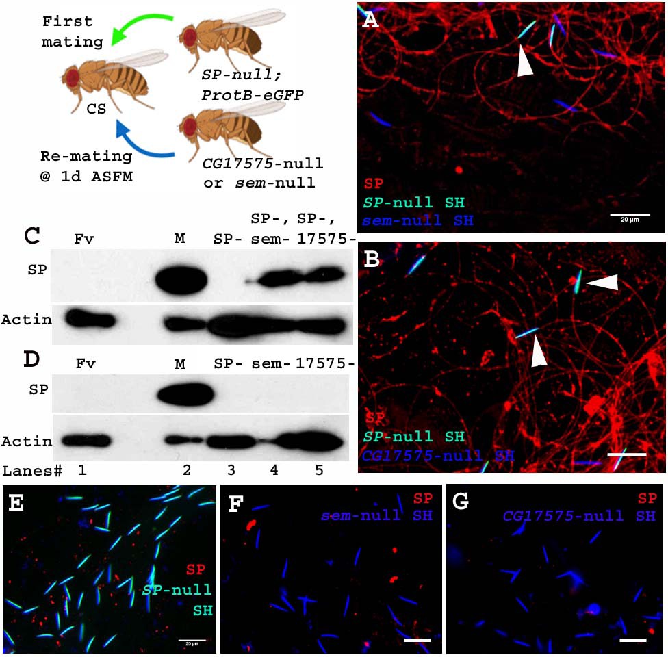
Sperm received from SP-null males do not require CG17575 or seminase from a second male to bind SP from that male.
Cartoon: Pictorial representation of the experimental cross (fly images from Biorender). Females mated first with SP-null; ProtB-eGFP males [cyan sperm-head; DAPI(blue)+eGFP(green)] and then a day later with CG17575-null or seminase-null males (blue sperm-head; DAPI stained) and frozen, 2 hr ASSM. SP was visualized with Alexa fluor 594, staining the sperm (head+ tail) red. (A) Sperm from females mated to SP-null; ProtB-eGFP males and then remated to seminase-null males, 1d ASFM. (B) Sperm from females mated to SP-null; ProtB-eGFP males and then remated to CG17575-null males, 1d ASFM. (C) Western blot probed for SP. Lanes/samples are 1: Fv, reproductive tract (RT) of three virgin females (negative control); 2: M, one pair of male accessory glands (positive control); 3: SP-, sperm dissected from 20 females mated to SP-null; ProtB-eGFP males at 2 hr ASM; 4: SP-, sem-, sperm dissected from 20 females mated to SP-null; ProtB-eGFP males and subsequently to seminase-null males at 1d ASFM, frozen at 2 hr ASSM; 5: SP-, 17575-, sperm dissected from 20 females mated to SP-null; ProtB-eGFP males and subsequently to CG17575-null males at 1d ASFM, frozen at 2 hr ASSM. (D) Western blot probed for SP. Lanes/samples are 1: Fv, reproductive tract (RT) of three virgin females (negative control); 2: M, one pair of male accessory glands (positive control); 3: SP-, sperm dissected from 20 females mated to SP-null; ProtB-eGFP males at 2 hr ASM; 4: sem-, sperm dissected from 20 females mated to seminase-null males at 2 hr ASM 5: 17575-, sperm dissected from 20 females mated to CG17575-null males at 2 hr ASM. Actin served as loading control. (E) Sperm isolated from females singly mated to SP-null; ProtB-eGFP males, 2 hr ASM. (F) Sperm isolated from females singly mated to seminase-null male, 2 hr ASM. (G) Sperm isolated from females singly mated to CG17575-null male, 2 hr ASM. White arrows indicate sperm heads (represented as SH, n = 10; Bar = 20 µm).
Figure 6—figure supplement 1
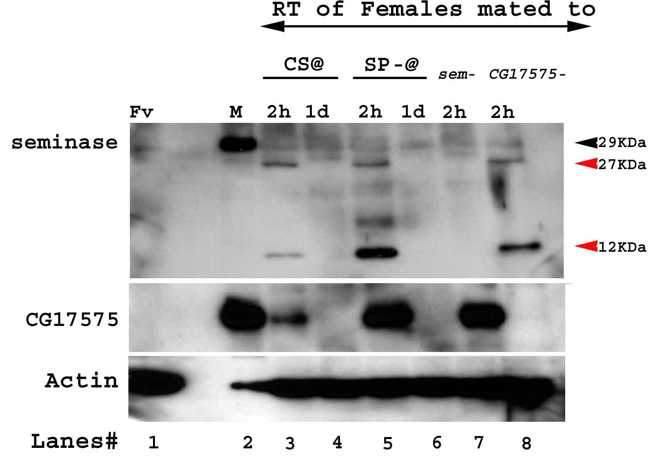
Western blot probed for seminase and CG17575.
Lanes/samples are 1: Fv, reproductive tract (RT) of three virgin females (negative control); 2: M, one pair of male accessory glands (positive control); 3–4: RT of five females mated to wild type (CS) males at 2 hr and 1d ASM, respectively; 5–6: RT of five females mated to SP-null; ProtB-eGFP males at 2 hr and 1d ASM, respectively; 7: RT of five females mated to seminase-null males at 2 hr ASM; 8: RT of five females mated to CG17575-null males at 2 hr ASM. Black arrows indicate full length seminase, red arrows indicate the cleavage products of seminase, post-mating in the female RT. Actin served as loading control.
Tables
Key resources table
| Reagent type (species) or resource | Designation | Source or reference | Identifiers | Additional information |
|---|---|---|---|---|
| Genetic reagent (D. melanogaster) | Tudor | R. Boswell; similar stock now available from BloomingtonDrosophila Stock Center | BDSC:1735; FBst0001735;RRID:BDSC_1735 | FlyBase Genotype:tud1 bw1 sp1/CyO |
| Genetic reagent (D. melanogaster) | Δ325/TM3; Sb ry (SP-knockout line) | Gift from Eric Kubli | ||
| Genetic reagent (D. melanogaster) | Δ130/TM3; Sb ry (deficiency line) | Gift from Eric Kubli | ||
| Genetic reagent (D. melanogaster) | ProtB-eGFP(X); TM3/TM6 | Gift from Scott Pitnick | ||
| Genetic reagent (D. melanogaster) | ProtB-DsRed | Gift from Scott Pitnick | ||
| Antibody | anti-SP (rabbit polyclonal) | Wolfner lab | IF (1:200), WB (1:2000) | |
| Antibody | anti-CG1656 (rabbit polyclonal) | Wolfner lab | IF (1:100), WB (1:1000) | |
| Antibody | anti-CG1652 (rabbit polyclonal) | Wolfner lab | IF (1:50), WB (1:500) | |
| Antibody | anti-CG9997 (rabbit polyclonal) | Wolfner lab | IF (1:50), WB (1:1000) | |
| Antibody | IgG (H+L) Goat anti-Rabbit, Alexa Fluor 488 (goat anti-rabbit polyclonal) | Invitrogen | Cat. # A11008 RRID:AB_143165 | IF (1:300) |
| Antibody | IgG (H+L) Goat anti-Rabbit, Alexa Fluor 594 (goat anti-rabbit polyclonal) | Invitrogen | Cat. # A11012 RRID:AB_2534079 | IF (1:300) |
| Antibody | anti-Antares (rabbit polyclonal) | Wolfner lab | WB (1:500) | |
| Antibody | anti-seminase (rabbit polyclonal) | Wolfner lab | WB (1:1000) | |
| Antibody | anti-CG17575 (rabbit polyclonal) | Wolfner lab | WB (1:1000) | |
| Antibody | Anti-actin (mouse monoclonal) | Millipore Corp | Cat# MAB1501 RRID:AB_2223041 | WB (1:3000) |
| Antibody | Peroxidase AffiniPure Goat Anti-Rabbit IgG (goat anti-rabbit polyclonal) | Jackson Research | Code#111-035-003 RRID:AB_2313567 | WB (1:2000) |
| Antibody | Peroxidase AffiniPure Goat Anti-Mouse IgG (goat anti-mouse polyclonal) | Jackson Research | Code#115-035-003 RRID:AB_10015289 | WB (1:2000) |
| Other | DAPI stain | Invitrogen | Cat. # PI62247 | (1 µg/mL) |
| Other | Poly-L-Lysine (0.1 % w/v in H2O) | Sigma | P8920-100ML | 0.01% w/v in H2O |
| Other | Albumin from Bovine Serum (BSA) | Sigma | A9418-50G | 5% in 1X PBS |
| Other | CitiFluor Mountant Solution | Electron Microscopy Sciences | Cat. #17970–100 | |
| Software, Algorithm | Graph Pad Prism | RRID:SCR_002798 | Version 6.01 |
Additional files
Download links
A two-part list of links to download the article, or parts of the article, in various formats.
Downloads (link to download the article as PDF)
Open citations (links to open the citations from this article in various online reference manager services)
Cite this article (links to download the citations from this article in formats compatible with various reference manager tools)
Drosophila seminal sex peptide associates with rival as well as own sperm, providing SP function in polyandrous females
eLife 9:e58322.
https://doi.org/10.7554/eLife.58322


