Lichen mimesis in mid-Mesozoic lacewings
- College of Life Sciences and Academy for Multidisciplinary Studies, Capital Normal University, China
- Department of Paleobiology, National Museum of Natural History, Smithsonian Institution, United States
- Department of Entomology, University of Maryland, United States
- State Key Laboratory of Mycology, Institute of Microbiology, Chinese Academy of Sciences, China
Figures
Figure 1
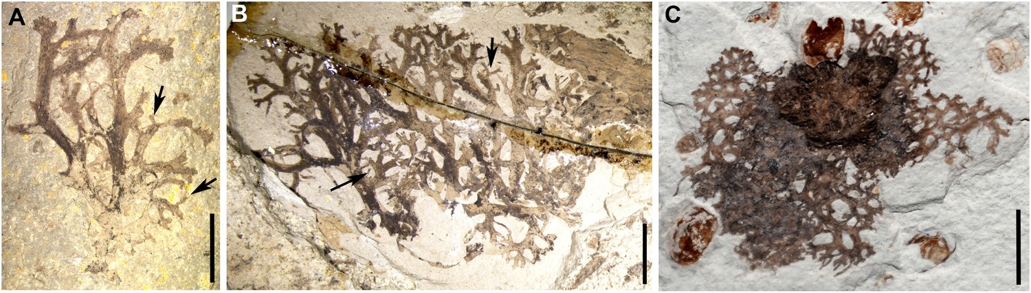
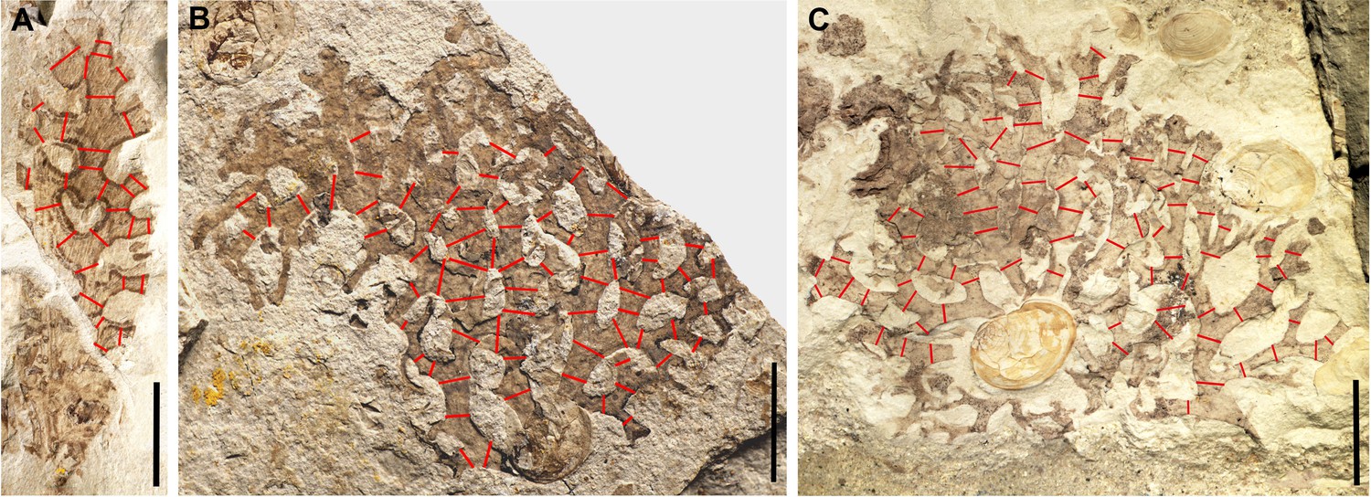
Photos of the lichen Daohugouthallus ciliiferus Wang, Krings et Taylor, 2010.
(A) Specimen B0476P, with arrows indicating the lobules. (B) Specimen CNU-LICHEN-NN2019001, with arrows indicating the lobules. (C) Specimen CNU-LICHEN-NN2019002P. Scale bars: 5 mm in A–C.
Figure 2
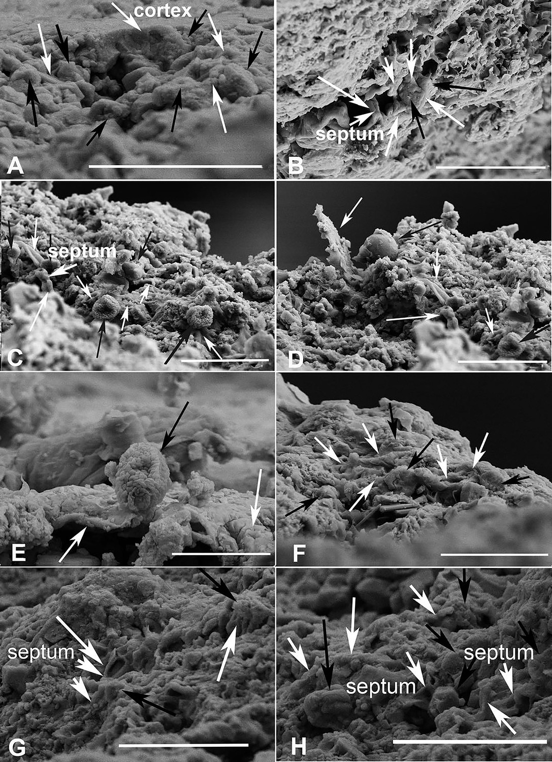
Scanning electron microscopy (SEM) micrographs of lichen fossil (CNU-LICHEN-NN2019001).
(A) Thallus longitudinal section containing the cortex, with white arrows pointing to the fungal hyphae, and black ones to the algal cells. The fungal hyphae are interweaved with algal cells. (B–D, F–H) Fungal hyphae indicated by white arrows; algal cells are indicated by black arrows showing entanglement and encirclement by fungal hyphae; septa shown in B, C, G, H. (E) One algal cell indicated by the black arrow, displaying adherence to other fungal hyphae indicated by the white arrow. Scale bars: 5 μm in A, C, D, G, H; 10 μm in B; 3 μm in E; 4 μm in F.
Figure 3
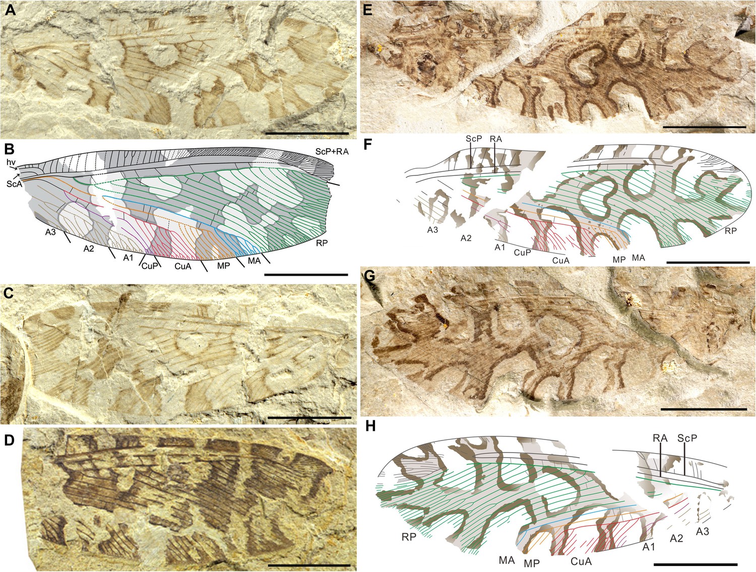
Photos and line drawings of Lichenipolystoechotes angustimaculatus gen. et sp. nov., and L. ramimaculatus gen. et sp. nov.
(A–C) Holotype CNU-NEU-NN2016040P/C of L. angustimaculatus, photo of part in (A). Accompanying overlay drawing in (B). Photo of counterpart in (C). (D) Photo of the paratype CNU-NEU-NN2016041 of L. angustimaculatus. (E–H) The holotype CNU-NEU-NN2019006P/C of L. ramimaculatus, with a lichen mimicking forewing pattern. Photo of part in (E); accompanying overlay drawing in (F); photo of counterpart in (G); and accompanying overlay drawing in (H). Scale bars: 5 mm in A–H.
Figure 4 with 1 supplement
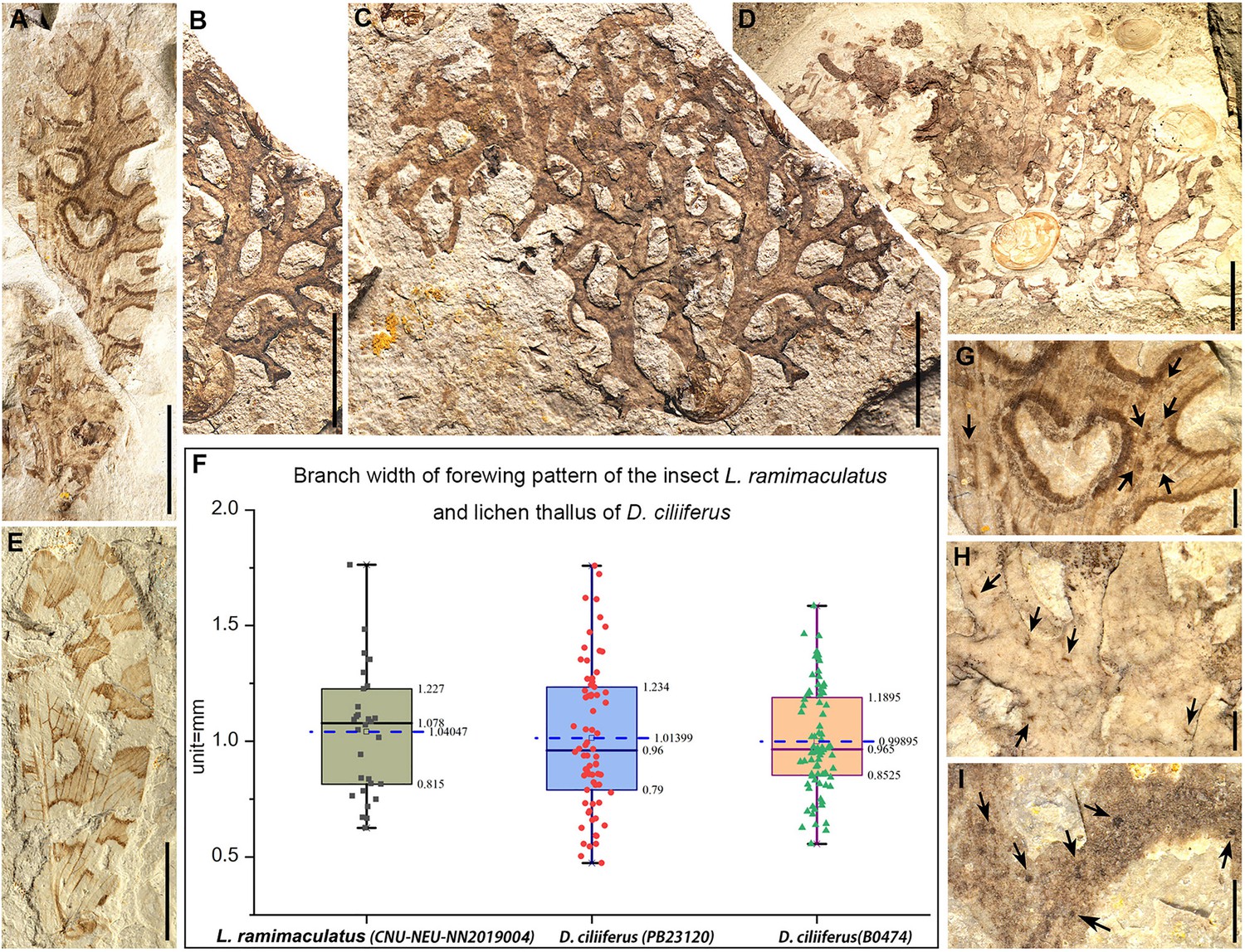
The lichen mimicking lacewing Lichenipolystoechotes ramimaculatus gen. et sp. nov. and L. angustimaculatus gen. et sp. nov., and fossils of the contemporaneous lichen Daohugouthallus ciliiferus Wang, Krings et Taylor, 2010.
(A) Photo of part of L. ramimaculatus, with a lichen mimicking forewing pattern, CNU-NEU-NN2019004P. (B–C) Photos of the lichen thallus D. ciliiferus, PB23120; thallus segment in (B); and entire thallus in (C). Photos A–C are at the same scale. (D) Photo of a nearly intact lichen thallus of D. ciliiferus, B0474. (E) Photo of L. angustimaculatus with a lichen mimicking wing pattern; CNU-NEU-NN2016040P. (F) Box scatter plots of measurement data displaying lower and upper extremes, lower and upper quartile, median and average (in the blue dotted line) of branch widths of L. ramimaculatus’s forewing pattern (CNU-NEU-NN2019004C) and thallus branch widths of lichen D. ciliiferus (PB23120, B0474) separately. (Black, red and green dots represent measurement results of branch pattern widths of lichen-mimicking lacewing and thallus widths of the two lichen specimens, respectively.) (G) Part of the wing pattern of L. ramimaculatus, with irregular wing spots. (H, I) Portion of the thallus of D. ciliiferus, with irregular spot-like punctiform pycnidia, B0474 (H), B0476P (I) The dark arrows indicate the spots on wing of L. ramimaculatus and thallus of D. ciliiferus. Scale bars: 5 mm in A–E, 1 mm in G–I.
Figure 4—figure supplement 1
Measuring lines on lichen-mimicking L. ramimaculatus and lichen D. ciliiferus.
(A) Measuring lines on the forewing of lichen-mimicking L. ramimaculatus, CNU-NEU-NN2019004P. (B) Measuring lines on lichen specimen PB23120. (C) Measuring lines on lichen specimen B0474. Measuring lines are indicated by their red color. Scale bars: 5 mm in A–C.
Figure 5
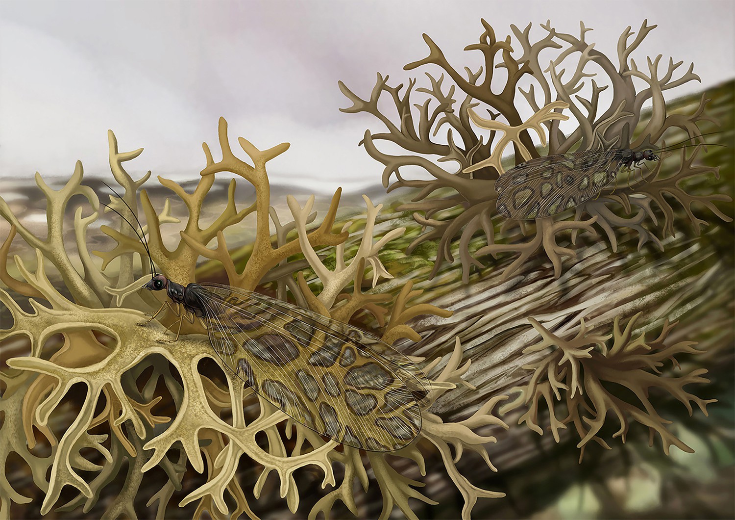
Habitus reconstruction of the lichen mimicking lacewing Lichenipolystoechotes ramimaculatus gen. et sp. nov. on the lichen Daohugouthallus ciliiferus Wang, Krings et Taylor, 2010.
The colors used in the drawing of D. ciliiferus is Taupe, referring to the color of extant lichen Everniastrum cirrhatum. The body of the L. ramimaculatus is reconstructed based on living ithonid species, and the wing is based on the fossil of holotype CNU-NEU-NN2019006P/C. The color of insect is yellowish-brown based on the general coloration of extant polystoechotids. Xiaoran Zuo did the reconstruction drawing.
Figure 6
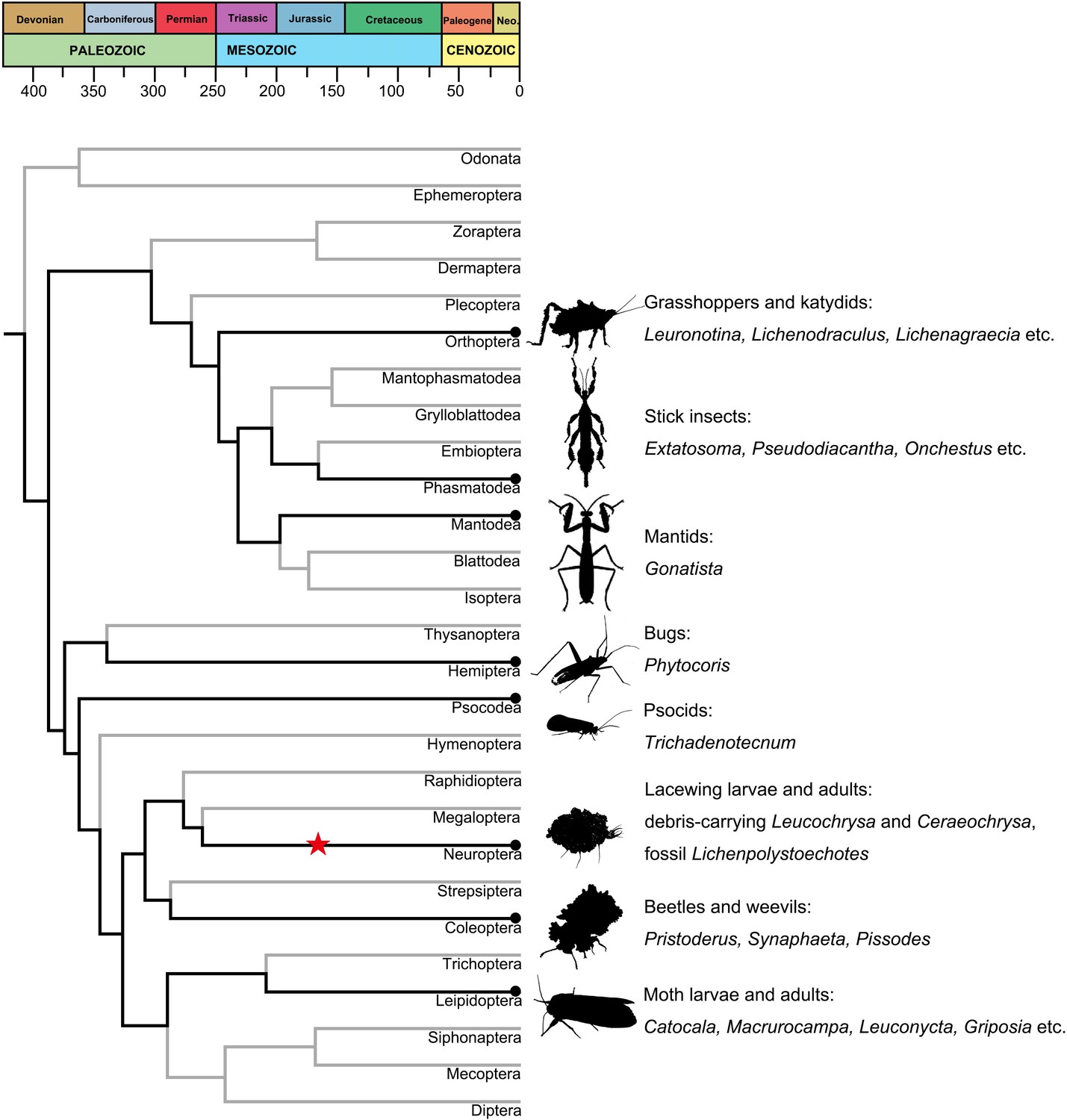
Lichen mimicry and camouflage by insects across major insect lineages.
Time-dated chronogram based on Misof et al., 2014. Specific examples of fossil and modern lichen mimesis by various insect taxa are provided at right. Black dots represent modern insect–lichen-mimetic associations; the star represents the fossil Lichenipolystoechotes–lichen mimicry of this study.
Additional files
-
Supplementary file 1
Table S1 Branch width of forewing pattern of Lichenipolystoechotes ramimaculatus and lichen thallus of Daohugouthallus ciliiferus.
- https://cdn.elifesciences.org/articles/59007/elife-59007-supp1-v2.docx
-
Transparent reporting form
- https://cdn.elifesciences.org/articles/59007/elife-59007-transrepform-v2.pdf
Download links
A two-part list of links to download the article, or parts of the article, in various formats.
Downloads (link to download the article as PDF)
Open citations (links to open the citations from this article in various online reference manager services)
Cite this article (links to download the citations from this article in formats compatible with various reference manager tools)
Lichen mimesis in mid-Mesozoic lacewings
eLife 9:e59007.
https://doi.org/10.7554/eLife.59007


