Chimpanzee brain morphometry utilizing standardized MRI preprocessing and macroanatomical annotations
- Institute of Systems Neuroscience, Medical Faculty, Heinrich-Heine-University, Germany
- Institute of Neuroscience and Medicine (INM-7) Research Centre Jülich, Germany
- Keeling Center for Comparative Medicine and Research, The University of Texas MD Anderson Cancer Center, United States
- Department of Anthropology and Center for the Advanced Study of Human Paleobiology, The George Washington University, United States
- Department of Experimental Medicine, University of Copenhagen, Denmark
- Department of Psychology, Georgia State University, United States
- Institute of Neuroscience and Medicine (INM-1), Research Centre Jülich, Germany
- Institute for Anatomy I, Medical Faculty, Heinrich-Heine-University, Germany
- JARA-BRAIN, Jülich-Aachen Research Alliance, Germany
- Structural Brain Mapping Group, Department of Neurology, Jena University Hospital, Germany
- Structural Brain Mapping Group, Department of Psychiatry and Psychotherapy, Jena University Hospital, Germany
- Center of Functionally Integrative Neuroscience, Department of Clinical Medicine, Aarhus University, Denmark
Figures
Figure 1
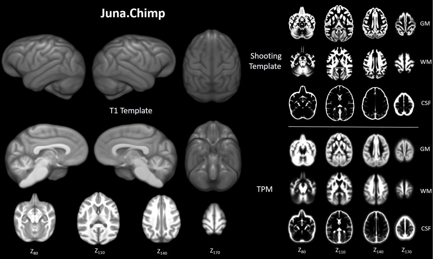
Juna.Chimp templates including the average.
T1- template, tissue probability maps (TPM), and Geodesic Shooting template. For Shooting templates and TPM axial slices are shown of gray matter (GM), white matter (WM), and cerebrospinal fluid (CSF). All templates are presented at 0.5 mm resolution.
Figure 2
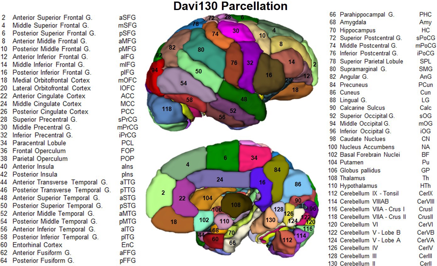
Lateral and medial aspect of the Davi130 parcellation right hemisphere.
Visible regions are numbered with Davi130 parcellation region numbers and correspond to names in the figure. Even numbers correspond to regions in the right hemisphere (as shown in the figure), while left hemisphere regions are odd numbers. A list of all Davi130 labels can be found at Figure 2—source data 1.
-
Figure 2—source data 1
Source file for Complete List of Davi130 Labels.
- https://cdn.elifesciences.org/articles/60136/elife-60136-fig2-data1-v2.docx
Figure 3 with 2 supplements
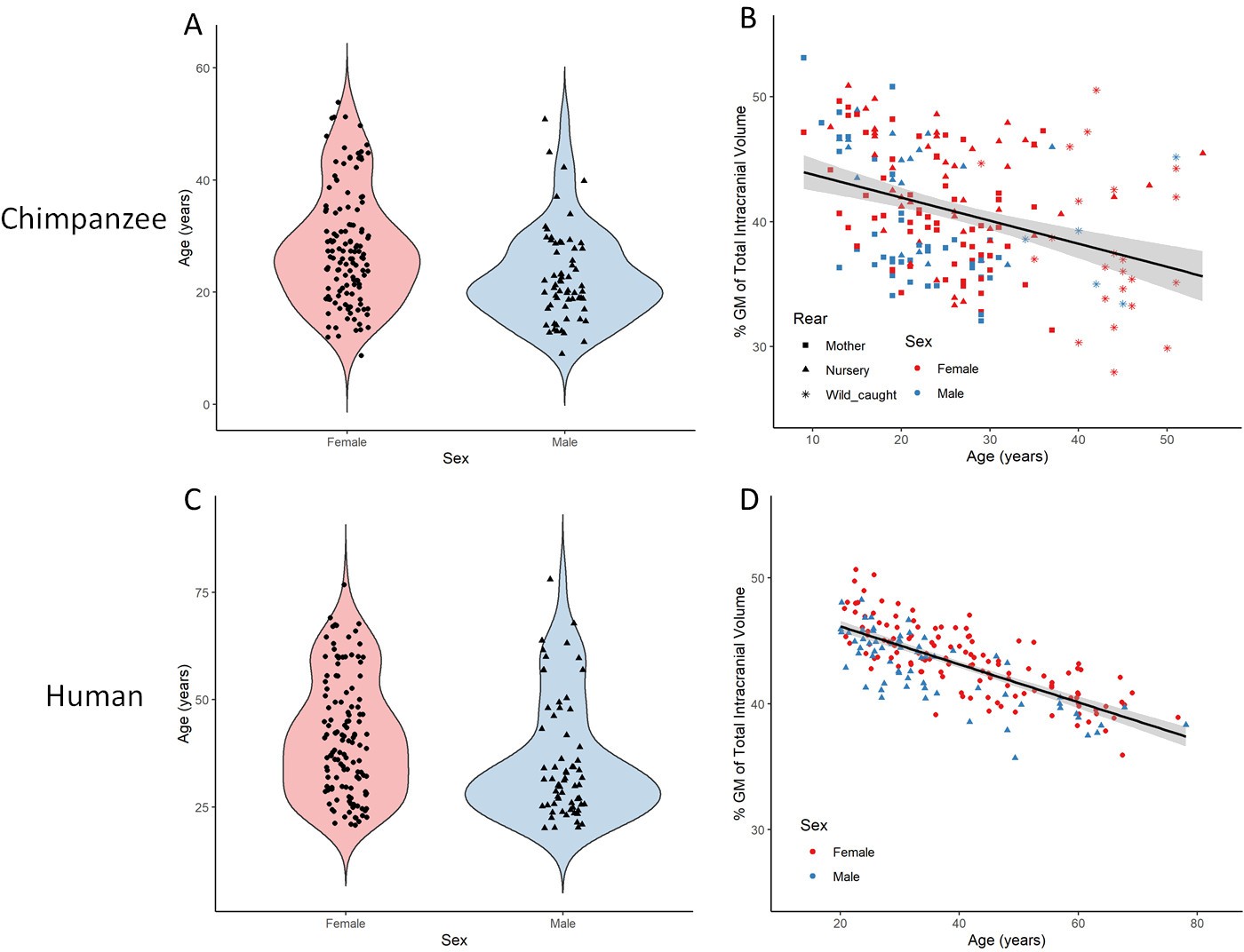
Total gray matter volume decline during aging in chimpanzees and matched human sample.
(A) Distribution of age and sex in the final sample of 194 chimpanzees. (B) Linear relationship between GM and age with standard error for chimpanzee sample. (C) Distribution of age and sex in the human (IXI) matched sample of 194 humans. (D) Linear relationship between GM and age with standard error for human sample. Figure 3—figure supplement 1 presents the age and sex distribution of the whole sample (n = 223). Figure 3—figure supplement 2 presents the age and sex distribution of the whole IXI sample (n = 496).
Figure 3—figure supplement 1
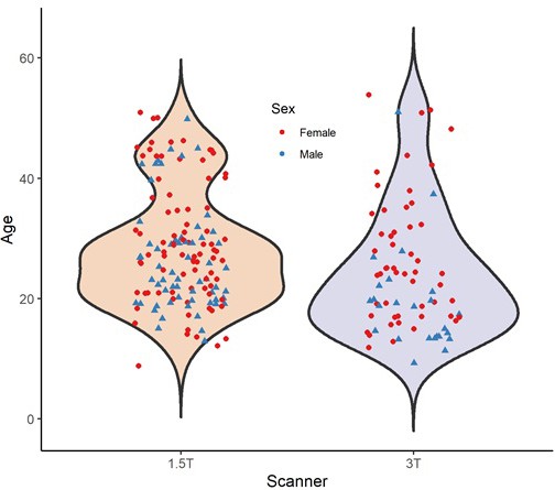
Age and sex distribution of complete chimpanzee (n = 223) sample separated by scanner field strength.
Figure 3—figure supplement 2

Age and sex distribution of complete IXI human sample (n = 496) separated by scanner field strength.
Figure 4
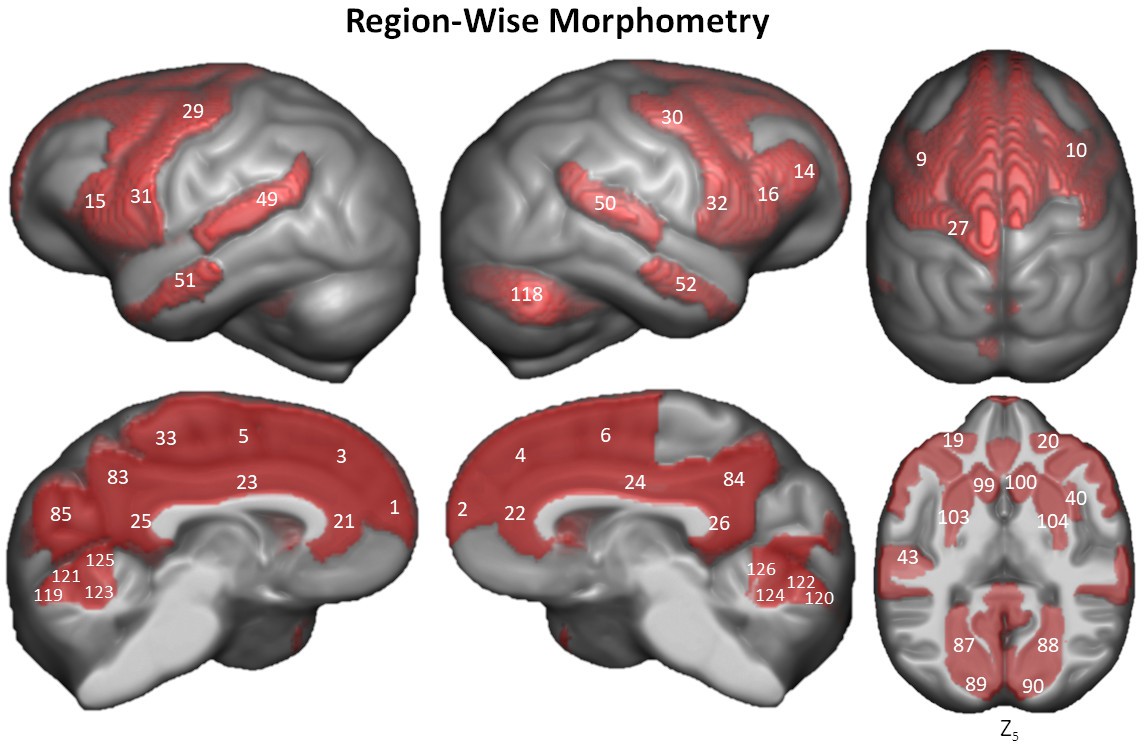
Region-wise morphometry in the Davi130 parcellation age regression.
Red regions represent Davi130 regions that remained significant at p≤0.05 following FWE correction (Holm method). The T-statistic and p-value for all Davi130 labels can be found in Figure 4—source data 1. 1 and 2 – aSFG, 3 and 4 – mSFG, 5 and 6 – pSFG, 9 and 10 – pMFG, 14 – mIFG, 15 and 16 – pIFG, 19 and 20 – lOFC, 21 and 22 – ACC, 23 and 24 – MCC, 25 and 26 – PCC, 27 – sPrCG, 29 and 30 – mPrCG, 31 and 32 – iPrCG, 33 – PCL, 40 – aIns, 43 – aTTG, 49 and 50 – pSTG, 51 and 52 – aMTG, 83 and 84 – PCun, 85 – Cun, 87 and 88 – LG, 89 and 90 – Calc, 97 and 98 – CN, 99 and 100 – NA, 103 and 104 – Pu, 118 – CrusII, 119 and 120 – CerVI, 121 and 122 – CerVB, 123 and 124 – CerVB, 125 and 126 – CerIV.
-
Figure 4—source data 1
Aging effect on gray matter in complete Davi130 Labels.
- https://cdn.elifesciences.org/articles/60136/elife-60136-fig4-data1-v2.docx
Figure 5 with 1 supplement
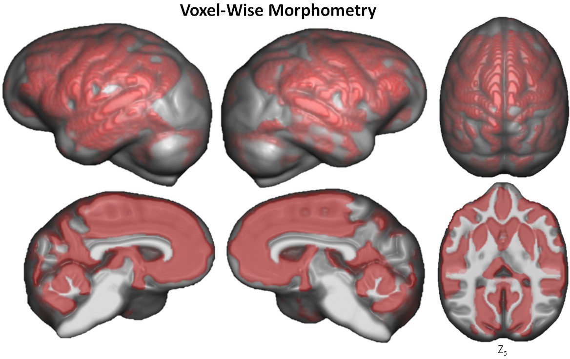
Voxel-based morphometry of aging on GM volume.
The significant clusters are found using TFCE with FWE correction at p≤0.05.
Figure 5—figure supplement 1
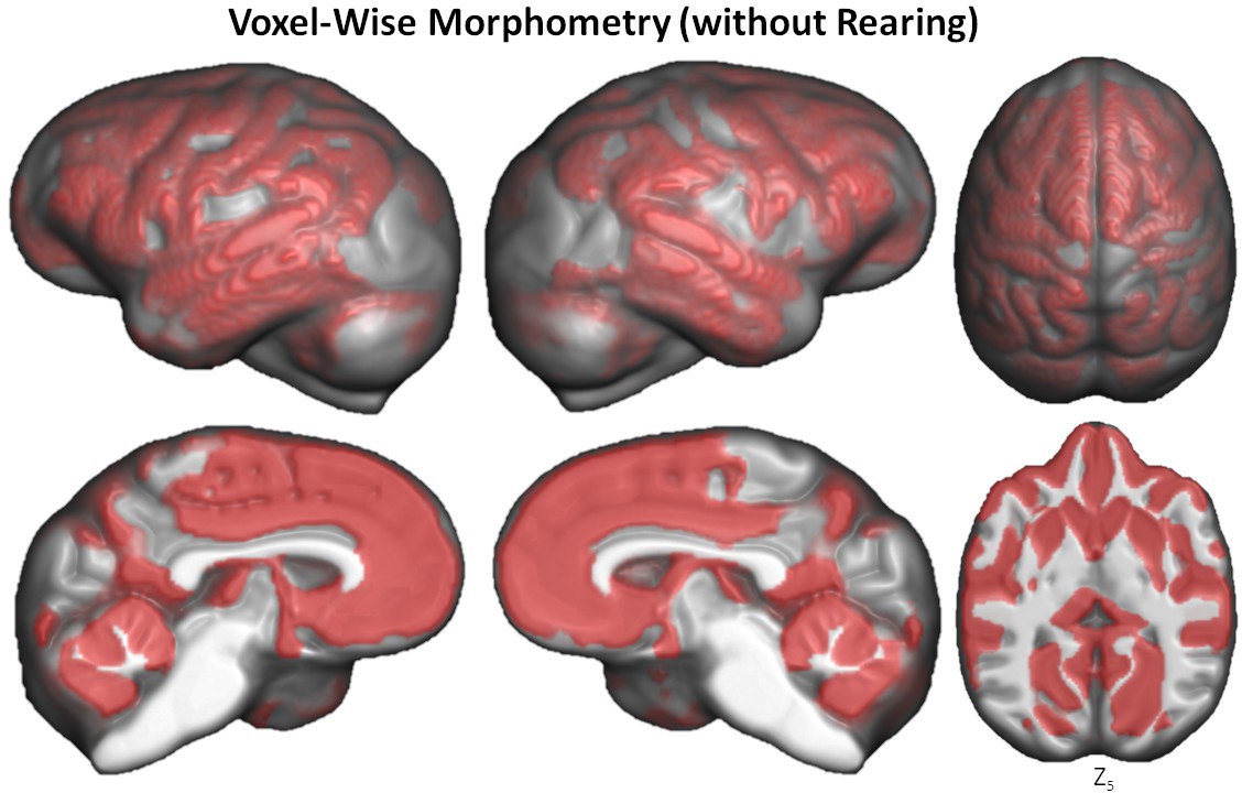
Voxel-based morphometry of aging on GM volume using TFCE with FWE correction at p≤0.05 without rearing as a covariate.
Figure 6
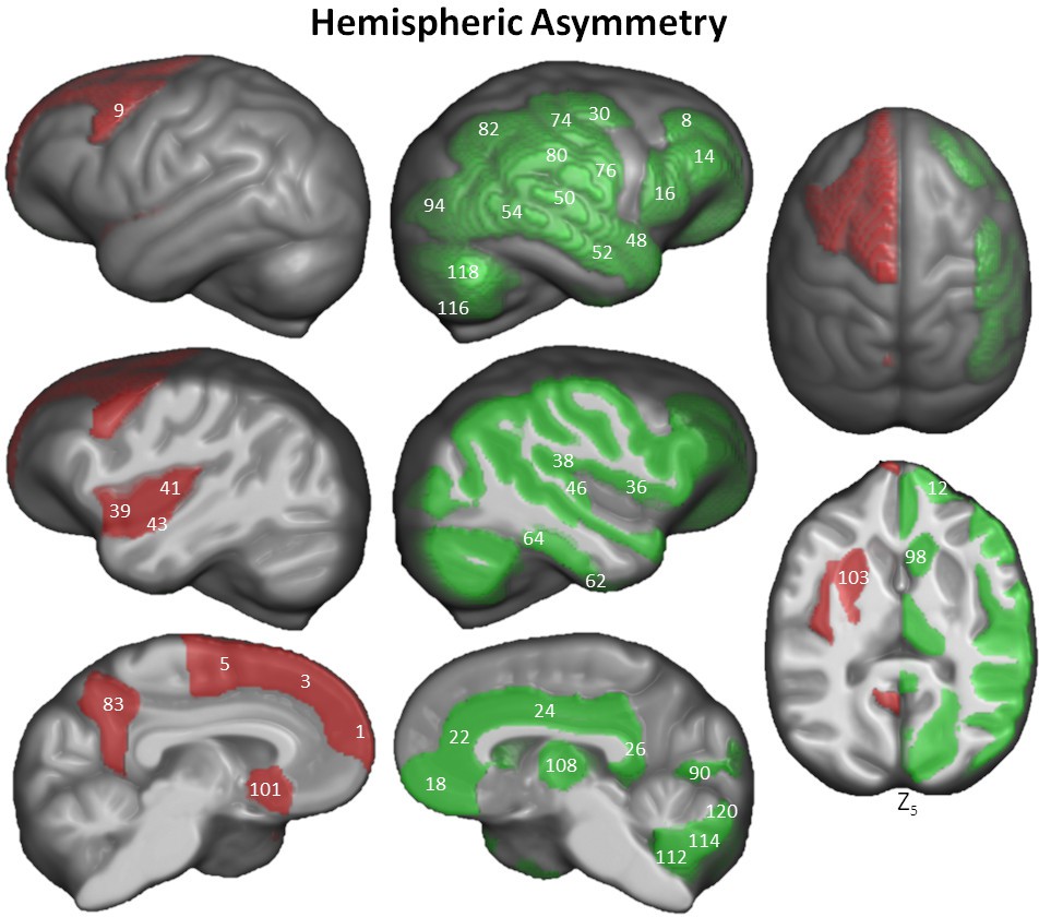
Hemispheric asymmetry of Davi130 regions within the chimpanzee sample.
Significant leftward (red) and rightward (green) asymmetrical regions are those with a p≤0.05 after FWE correction. The T-statistic and p-value for all Davi130 labels can be found in Figure 6—source data 1. 1 – aSFG, 3 – mSFG, 5 – pSFG, 8 – aMFG, 9 – pMFG, 12 – aIFG, 14 – mIFG, 16 – pIFG, 18 – mOFC, 22 – ACC, 24 – MCC, 26 – PCC, 30 – mPrCG, 36 – FOP, 38 – POP, 39 – aIns, 41 – pIns, 43 – aTTG, 46 – pTTG, 48 – aSTG, 50 – pSTG, 52 – aMTG, 54 – pMTG, 62 – aFFG, 64 – pFFG, 74 – mPoCG, 76 – iPoCG, 80 – SMG, 82, AnG, 83 – PCun, 90 – Calc, 94 – mOG, 98 – CN, 101 – BF, 103 – Pu, 108 – Th, 112 – CerIX, 114 – CerVIII, 115 –CrusI, 118 –CrusII, 120 – CerVI.
-
Figure 6—source data 1
Complete Davi130 labels hemispheric asymmetry.
- https://cdn.elifesciences.org/articles/60136/elife-60136-fig6-data1-v2.docx
Figure 7
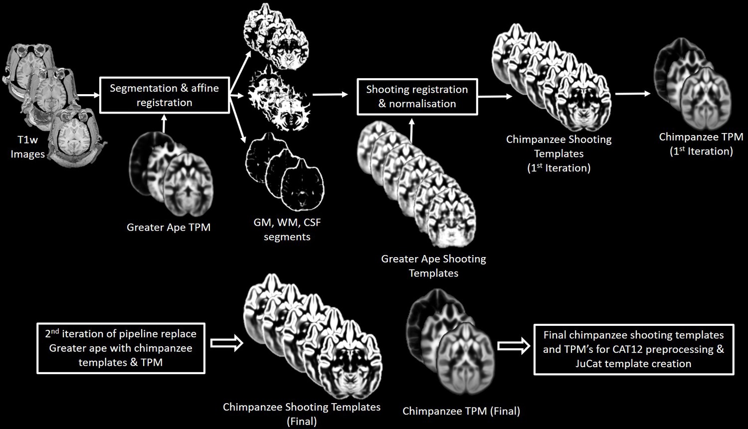
Workflow for creation of chimpanzee-specific shooting template and TPM, which can then be used in CAT12 structural preprocessing pipeline to create the Juna.Chimp template.
The resulting chimpanzee-shooting template, TPM and CAT atlas establishes the robust and reliable base to segment and spatially normalize the T1w images utilizing CAT12’s processing pipeline (Dahnke and Gaser, 2017; Gaser et al., 2020).
Tables
Key resources table
| Reagent type (species) or resource | Designation | Source or reference | Identifiers | Additional information |
|---|---|---|---|---|
| Software, algorithm | CAT12 | http://www.neuro.uni-jena.de/cat/ | RRID:SCR_019184 | |
| Software, algorithm | NCBR | http://www.chimpanzeebrain.org/ | RRID:SCR_019183 | |
| Software, algorithm | MATLAB | http://www.mathworks.com/products/matlab/ | RRID:SCR_001622 | |
| Software, algorithm | SPM | http://www.fil.ion.ucl.ac.uk/spm/ | RRID:SCR_007037 | |
| Software, algorithm | RStudio | http://www.rstudio.com/ | RRID:SCR_000432 | |
| Software, algorithm | 3D Slicer | http://slicer.org/ | RRID:SCR_005619 |
Additional files
Download links
A two-part list of links to download the article, or parts of the article, in various formats.
Downloads (link to download the article as PDF)
Open citations (links to open the citations from this article in various online reference manager services)
Cite this article (links to download the citations from this article in formats compatible with various reference manager tools)
Chimpanzee brain morphometry utilizing standardized MRI preprocessing and macroanatomical annotations
eLife 9:e60136.
https://doi.org/10.7554/eLife.60136


