Embryonic origin and serial homology of gill arches and paired fins in the skate, Leucoraja erinacea
- Department of Zoology, University of Cambridge, United Kingdom
- Marine Biological Laboratory, United Kingdom
Figures
Figure 1 with 1 supplement
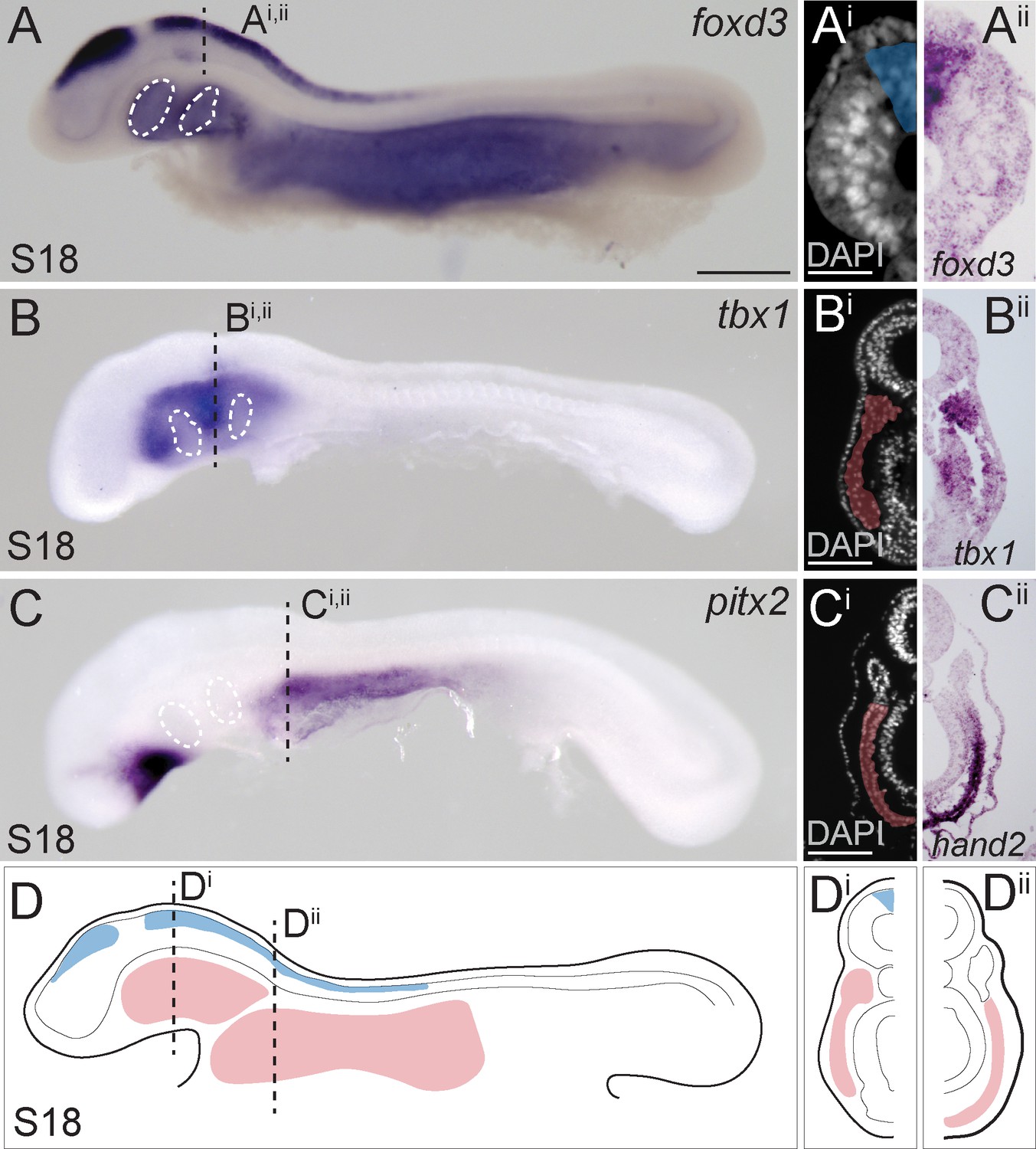
Neural crest and mesoderm after skate neurulation.
(A) Wholemount mRNA in situ hybridization for foxD3 reveals expression in (Ai, Aii) pre-migratory neural crest cells within the dorsal neural tube of the skate embryo at S18. (B, Bi, Bii) tbx1-expressing head mesoderm grades into (C) pitx2- and (Ci, Cii) hand2-expressing lateral plate mesoderm in the skate embryo at S18. (D) Schematic representation of neural crest, head mesoderm and lateral plate mesoderm tissues targeted for cell lineage tracing in this study. White dashed lines indicate the location of developing pharyngeal endodermal pouches. Scale bars: (A, B), C = 700 µm; Ai = 65 µm; (Bi), Ci = 120 µm.
Figure 1—figure supplement 1
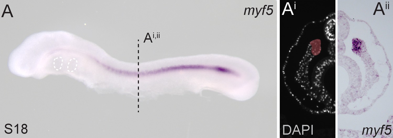
Paraxial mesoderm in the little skate.
(A) Wholemount mRNA in situ hybridization for myf5 reveals expression in (Ai, Aii) somitic and presomitic mesoderm of the skate embryo at S18.
Figure 2 with 1 supplement
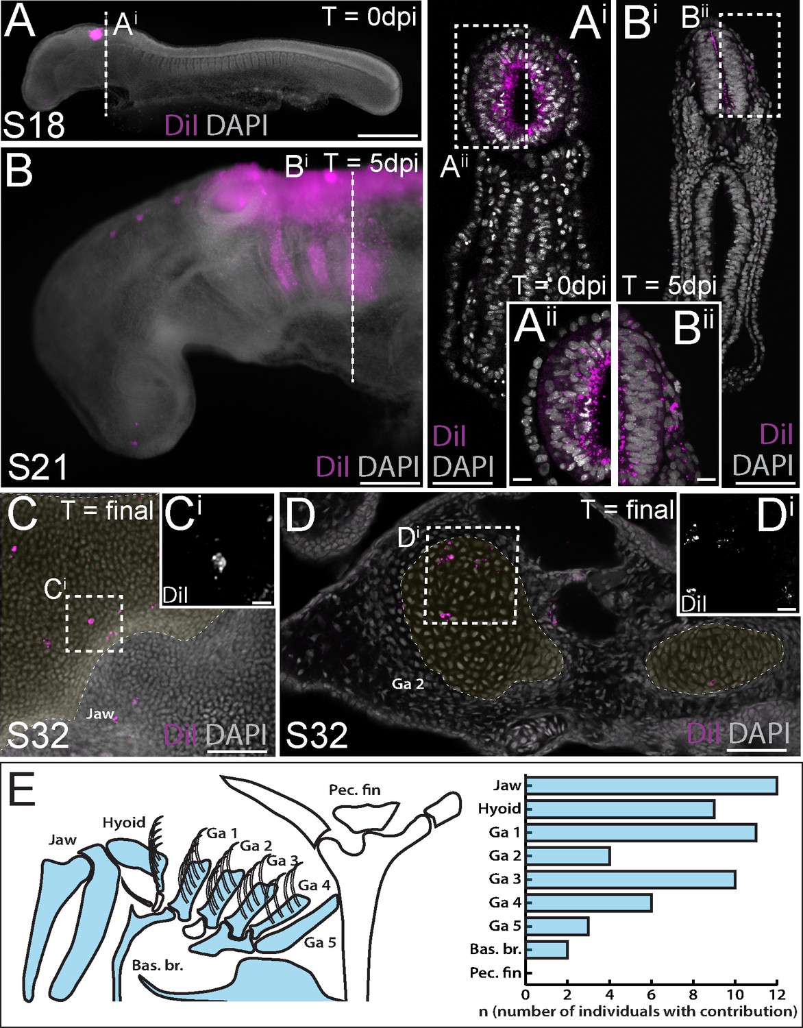
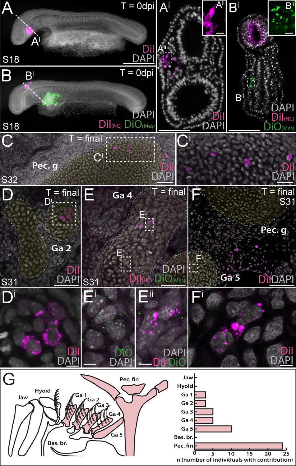
Neural crest contributes to the jaw, hyoid and gill arch skeleton in the skate.
(A) Microinjection of CM-DiI into the lumen of the neural tube at S18 results in (Ai, Aii) labelling of cells throughout the hindbrain neural tube, including premigratory neural crest cells. (B) At 5 days post-injection (dpi), CM-DiI-labelled cranial neural crest cells can be seen streaming from the hindbrain neural tube into the pharyngeal arches (see also Bi, Bii). At S32, CM-DiI (i.e. neural crest-derived) chondrocytes are recovered within pharyngeal arch skeletal elements, including (C, Ci) the palatoquadrate of the jaw and (D, Di) the epibranchial of gill arch 2. (E) Schematic representation of pharyngeal and pectoral fin skeletal elements in the S32 skate embryo, with elements receiving contribution from neural crest coloured blue, and a plot showing the number of embryos observed with neural crest contributions to the pharyngeal arch skeleton. In (C and D), cartilaginous elements are false-coloured yellow. Scale bars: A = 700 µm; Ai = 250 µm; Aii = 50 µm; B = 340 µm; Bi = 250 µm; Bii = 50 µm; C = 165 µm; Ci = 15 µm; D = 70 µm; Di = 20 µm.
Figure 2—figure supplement 1
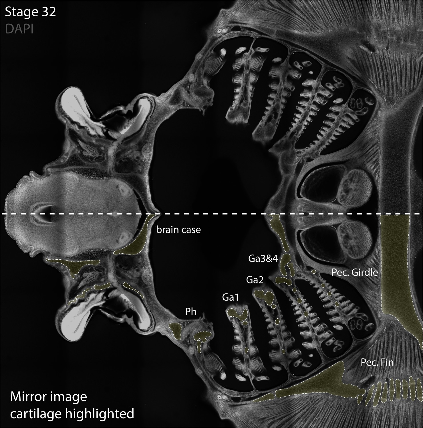
Identification of skate pharyngeal arch skeletal elements in section.
Cartilaginous elements of the pharyngeal skeleton of a S32 skate embryos are readily visible in DAPI-stained sections, as shown here without (top) and with false-colouring of cartilages in yellow (bottom).
Figure 3 with 2 supplements
Mesoderm contributes to the gill arch and pectoral fin skeleton in the skate.
(A, Ai) Microinjection of CM-DiI into the head mesoderm (HM) of a skate embryo at S18. (B, Bi) Simultaneous labelling of the hindbrain neural tube (including premigratory cranial neural crest cells) with CM-DiI and lateral plate mesoderm (LPM) with SpDiOC18 in a S18 skate embryo. (C, Ci) LPM gives rise to chondrocytes within the skeleton of the pectoral fin and girdle, while mesoderm at the HM-LPM boundary and LPM give rise to chondrocytes within the gill arch skeleton – e.g. (D, Di) in the branchial rays of gill arch 2. (E) After double labelling of the LPM with SpDiOC18 and the neural tube with CM-DiI, as in (B) above, both (Ei) SpDiOC18- and (Eii) CM-DiI-labelled chondrocytes are recovered within the gill arch skeleton – for example, in the ceratobranchial of gill arch 4 – demonstrating the dual mesodermal and neural crest origin of these elements. (F, Fi) Mesodermally-derived chondrocytes were also recovered in the ceratobranchial of gill arch 5, in close proximity to the label-retaining pectoral girdle and surrounding connective tissue. (G) Schematic summary of pharyngeal and paired fin skeletal elements in the S32 skate embryo, with elements receiving any mesodermal contributions (HM, HM-LPM or LPM) coloured red, and a plot showing the number of embryos observed with mesoderm contributions to the pharyngeal arch and pectoral fin skeleton. In (D), (E) and (F), cartilaginous elements are false-coloured yellow. Scale bars: A, B = 700 µm; Ai = 125 µm; Aii = 15 µm; Bi = 50 µm; Bii = 15 µm; C = 60 µm; Ci = 20 µm; D = 50 µm; Di = 5 µm; E = 30 µm; Ei = 5 µm; F = 60 µm; Fi = 7 µm.
Figure 3—figure supplement 1
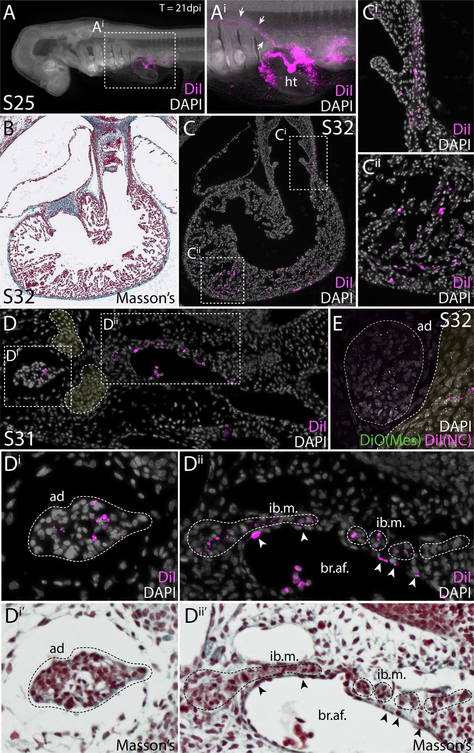
Cardiopharyngeal mesodermal derivatives in the skate.
(A) A skate embryo 21 days after CM-DiI labelling of lateral mesoderm at the HM-LPM boundary. Abundant label is recovered within the heart (ht) and major blood vessels (white arrows in Ai). (B) Histological section through the heart of a S32 skate embryo, stained with Masson’s trichrome. (C) Adjacent section to (B), showing labelling of the heart after CM-DiI injection in lateral mesoderm of the HM-LPM boundary. (D) Example of branchiomeric musculature (branchial adductor muscle, ad, and interbranchial musculature, ib.m.) and vascular endothelium (white arrowheads) derived from lateral mesoderm (Di’ and Dii’) are the same sections as (Di and Dii), respectively, imaged after Masson’s trichrome counterstaining. Condensing gill arch cartilage is false coloured in yellow. (E) SpDiOC18 labelling of branchiomeric musculature (branchial adductor muscle, ad) reveals its mesodermal origin, from an embryo in which lateral mesoderm was labelled with SpDiOC18 and the neural tube was labelled with CM-DiI at S18.
Figure 3—figure supplement 2
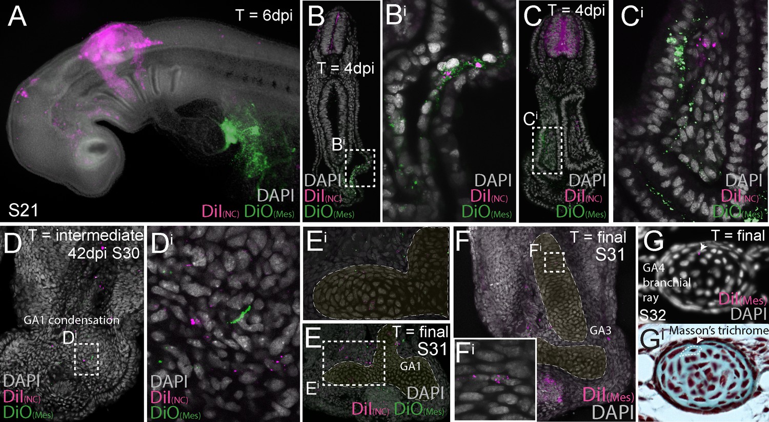
Dual embryonic neural crest and mesodermal origin of gill arch cartilages in the skate.
(A) A skate embryos 6 days after neural tube labelling with CM-DiI and lateral plate mesoderm (LPM) labelling with SpDiOC18. (B–C) Transverse sections reveal that 4 days post neural tube labelling with CM-DiI and LPM labelling with SpDiOC18, CM-DiI-labelled neural crest cells can be seen migrating adjacent to SpDiOC18-labelled LPM. (D) Forty-two days post-dual labelling, both CM-DiI-retaining (i.e. neural crest-derived) and SpDiOC18-retaining (i.e. LPM-derived) cells are recovered within the condensing ceratobranchial cartilage of gill arch 1, while (E) at S32, CM-DiI-retaining and SpDiOC18-retaining chondrocytes are recovered within the ceratobranchial cartilage of gill arch 1. (F) Mesodermal contribution to the ceratobranchial cartilage of gill arch 3. (G) Mesodermal contribution to a branchial ray on gill arch 4. In (E) and (F), cartilaginous elements are false-coloured yellow.
Figure 4
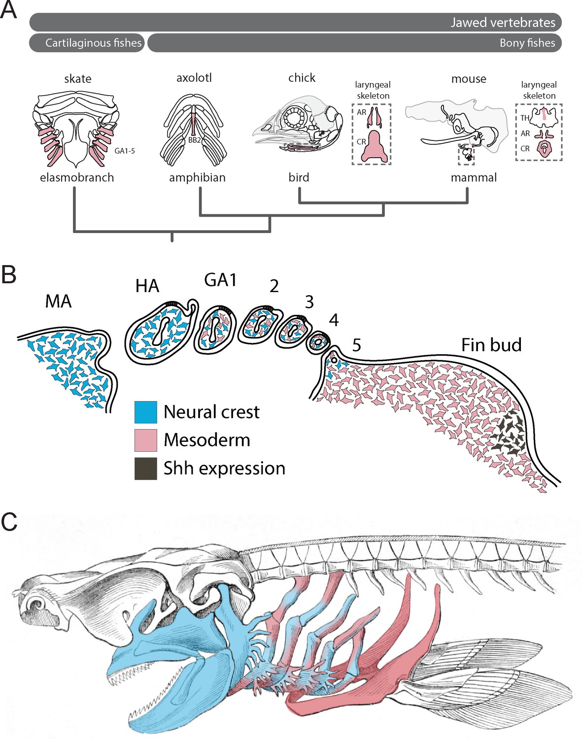
Mesodermal contributions to the pharyngeal endoskeleton in jawed vertebrates.
(A) Mesodermal contributions (red) to the gill arch skeleton in skate, the basibranchial skeleton in axolotl and the laryngeal skeleton of chick and mouse points to an ancestral mesodermal contribution to the pharyngeal arch skeleton of jawed vertebrates. (B) Schematic representation of neural crest- (blue) and mesoderm-derived (red) skeletogenic mesenchyme in the skate pharyngeal arches and pectoral fin bud, in relation to epithelial and mesenchymal Shh expression, respectively. (C) We propose that the mandibular and hyoid arch skeleton are neural crest-derived and the pectoral fin skeleton mesodermal derived, while the gill arch skeletal elements are of dual neural crest and mesodermal origin.
Additional files
-
Supplementary file 1
Fate mapping data for neural crest and mesodermal lineage tracing experiments.
Raw scoring data showing contributions from the neural crest and mesoderm to the skate pharyngeal skeleton, and schematic of the skate pharyngeal skeleton. Skeletal elements abbreviated as follows: BC, basibranchial copula; BH, basihyal; CB1-5, ceratobranchials 1–5; C-ph; ceratopseudohyal; EB1-5, epibranchials 1–5; E-ph, epipseudohyal; HB2; hypobranchial 2; HB3/4, hypobranchial 3/4; HM, hyomandibula; H-ph, hypopseudohyal; HT, heart; MK, Meckel’s cartilage; PB1-4, pharyngobranchials 1–4; PG, pectoral girdle; PQ, palatoquadrate; SC, spiracular cartilage.
- https://cdn.elifesciences.org/articles/60635/elife-60635-supp1-v1.xlsx
-
Transparent reporting form
- https://cdn.elifesciences.org/articles/60635/elife-60635-transrepform-v1.docx
Download links
A two-part list of links to download the article, or parts of the article, in various formats.
Downloads (link to download the article as PDF)
Open citations (links to open the citations from this article in various online reference manager services)
Cite this article (links to download the citations from this article in formats compatible with various reference manager tools)
Embryonic origin and serial homology of gill arches and paired fins in the skate, Leucoraja erinacea
eLife 9:e60635.
https://doi.org/10.7554/eLife.60635


