A versatile system to record cell-cell interactions
- Department of Genetics, Stanford University School of Medicine, United States
- Cancer Biology Program, Stanford University School of Medicine, United States
- Immunology Program, Stanford University School of Medicine, United States
- Neuroscience Program, Stanford University School of Medicine, United States
- Department of Neurosurgery, Stanford University School of Medicine, United States
- Department of Pathology, Stanford University School of Medicine, United States
Figures
Figure 1 with 2 supplements
GFP-based Touching Nexus (G-baToN) leads to cell-cell interaction-dependent receiver cell labeling.
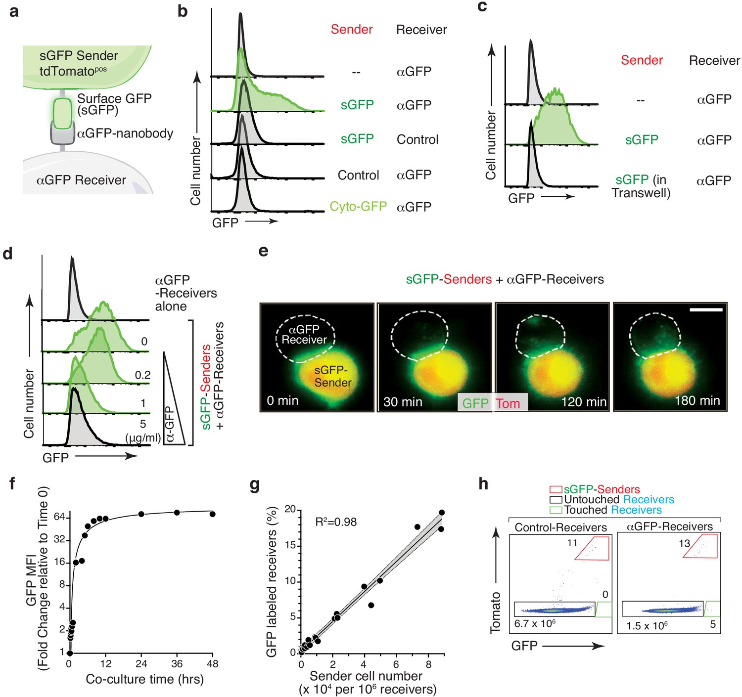
(a) Schematic of the G-baToN system. Surface GFP (sGFP) on a sender cell is transferred to a receiver cell expressing a cell surface anti-GFP nanobody (αGFP) leading to GFP labeling of the ‘touched’ receiver cell. (b) GFP transfer from sGFP-expressing KPT lung cancer sender cells (marked by intracellular tdTomato) to αGFP-expressing 293 receiver cells. Receiver cell labeling is sGFP- and αGFP- dependent. Control sender cells do not express sGFP. Control receiver cells do not express αGFP. Cytoplasmic GFP (Cyto-GFP) is not transferred to receiver cells. Sender and receiver cells were seeded at a 1:1 ratio and co-cultured for 24 hr. Receiver cells were defined as TomatonegPIneg cells. (c) GFP transfer to 293 receiver cells requires direct cell-cell contact. Receiver cells separated from sender cells by a transwell chamber are not labeled. Sender and receiver cells were seeded in upper and lower chambers respectively at a 1:1 ratio and cultured for 24 hr. Receiver cells were defined as TomatonegPIneg cells. (d) GFP transfer to 293 receiver cells requires sGFP-αGFP interaction and is blocked by an anti-GFP antibody in a dose-dependent manner. sGFP sender cells were pre-incubated with the indicated concentration of anti-GFP antibody for 2 hr, washed with PBS, and then co-cultured with receiver cells at a 1:1 ratio for 24 hr. Receiver cells were defined as TomatonegPIneg cells. (e) Time-lapse imaging of GFP transfer from a sGFP-expressing sender cell to an αGFP-expressing receiver cell. Time after contact is indicated. Receiver cell is outlined with white dashed line. Scale bar: 10 μm. (f) Analysis of GFP Mean Fluorescence Intensity (MFI) of αGFP receiver cells (marked by intracellular BFP) co-cultured with sGFP sender cells (marked by intracellular tdTomato) co-cultured for the indicated amount of time. Sender and receiver cells were seeded at a 1:1 ratio. Receiver cells were defined as TomatonegPInegBFPpos cells. (g) Percentage of labeled αGFP receiver cells after co-culture with different numbers of sender cells for 24 hr. Receiver cells were defined as TomatonegPInegBFPpos cells. (h) Detection of rare labeled αGFP receiver cells after co-culture with sGFP sender cells at approximately a 1:105 ratio for 24 hr. Receiver cells were defined as TomatonegPInegBFPpos cells.
Figure 1—figure supplement 1
GFP transfer requires direct GFP-αGFP interaction.
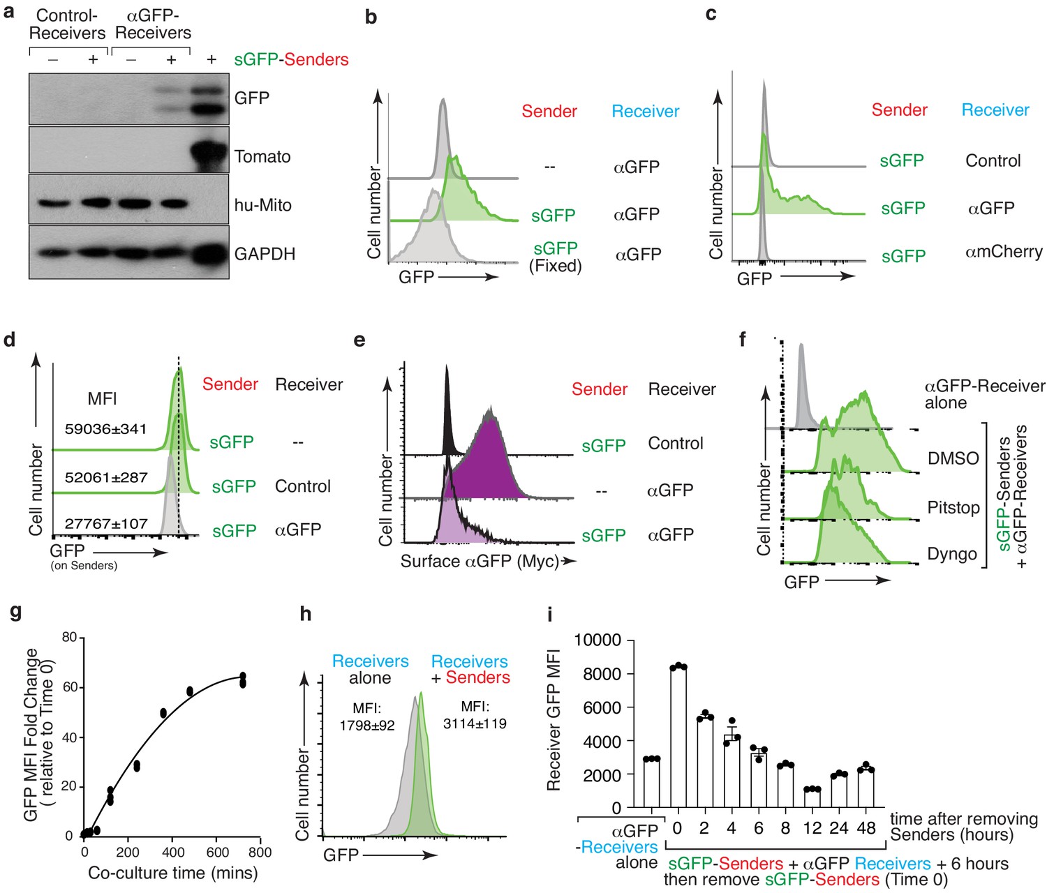
(a) GFP protein is transferred to receiver cells. Western blot analysis of FACS purified Control and αGFP receiver 293 cells cultured in isolation or co-culture with sGFP Tomato-positive sender cells (mouse KPT lung cancer cells) for 24 hr. sGFP but not Tomato is present in touched αGFP receiver cells. Human mitochondrial antigen (hu-Mito) is a marker for receiver cells. GAPDH shows loading. Rightmost lane is sGFP sender cells. (b) GFP cannot be transferred from fixed sGFP sender cells to live αGFP receiver cells. sGFP sender cells were fixed in 1% PFA for 5 min and washed with PBS before co-cultured with αGFP receiver cells at a 1:1 ratio for 24 hr. Receiver cells were defined as TomatonegPInegBFPpos cells. (c) GFP transfer to 293 receiver cells required sGFP-αGFP recognition. GFP is transferred from sGFP sender cells to αGFP-receiver cells but not from sGFP sender cells to αmCherry-receiver cells. Control receiver cells do not express any nanobody. Sender and receiver cells were co-cultured at a 1:1 ratio for 24 hr. Receiver cells were defined as TomatonegPInegBFPpos cells. (d) GFP transfer to receiver cells is accompanied by a reduction of GFP on the sender cells. GFP expression on sender cells after 24 hr co-culture with control or αGFP-expressing 293 receiver cells at a 1:1 ratio. Co-culture with αGFP expressing but not control receiver cells reduced GFP on sGFP sender cells. Sender cells were defined as TomatoposDAPInegBFPneg cells. (e) GFP transfer is accompanied with αGFP internalization on receiver cells. Analysis of surface αGFP (Myc-tag) on 293 receiver cell co-cultured for 24 hr with sGFP sender cells. Receiver cells were defined as TomatonegPIneg cells. (f) GFP transfer to 293 receiver cells is partially dependent on membrane dynamics of endocytosis. Both a clathrin inhibitor (Pitstop, 20 μM) and a dynamin inhibitor (Dyngo 4a, 10 μM) partially inhibit GFP transfer from sGFP sender cells to αGFP receiver cells. Inhibitors were present during the 24 hr co-culture. Receiver cells were defined as TomatonegPIneg cells. (g) Analysis of GFP Mean Fluorescence Intensity (MFI) of αGFP receiver cells (marked by intracellular BFP) co-cultured with sGFP sender cells (marked by intracellular tdTomato) co-cultured for the indicated amount of time in the first 12 hr. Sender and receiver cells were seeded at a 1:1 ratio. Receiver cells were defined as TomatonegPInegBFPpos cells. (h) GFP transfer to αGFP 293 receiver cells can be very rapid. A shift in GFP MFI was detected 5 min after mixing sGFP sender cells with αGFP receiver cell. Receiver cells were defined as TomatonegPInegBFPpos cells. MFI mean +/- SD of triplicate cultures is shown. (i) Rapid GFP degradation in touched receiver cells after removal of the sGFP sender cells. After 6 hr co-culture, GFP-positive receiver cells were purified by FACS followed by culture without sender cells. Receiver cells were defined as mCherrynegPInegBFPpos cells. GFP MFI in receiver cells reduced rapidly (T1/2 approximately 2 hr). MFI mean +/- SD of triplicate cultures is shown.
Figure 1—figure supplement 2
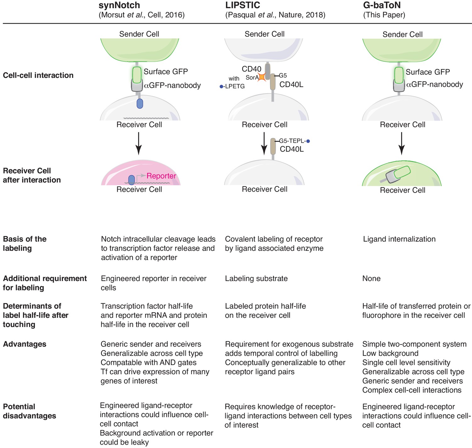
Features of the SynNotch, LIPSTIC, and G-baToN cell-cell interaction reporter systems.
Overview of systems that enable labeling of cells after cell-cell contact.
Figure 2
Transmembrane domains and the nanobody affinity impact sGFP transfer and receiver cell labeling.
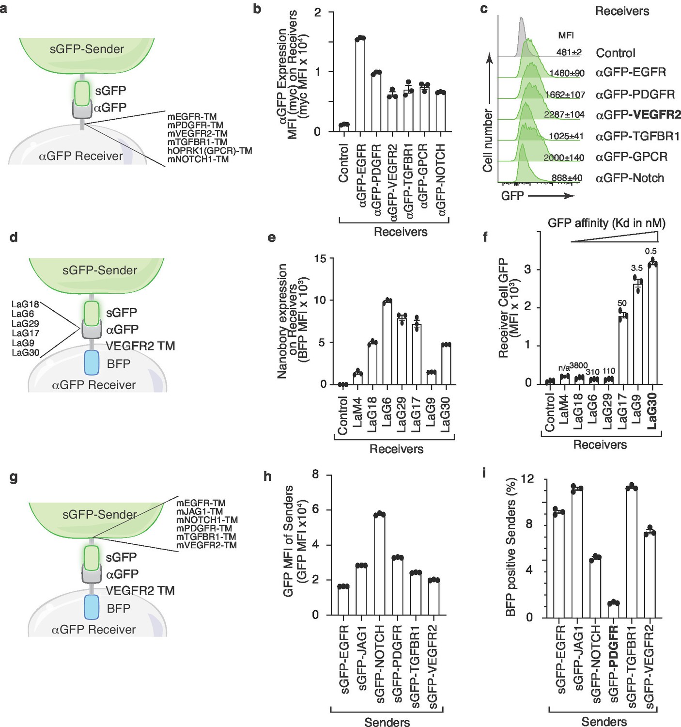
(a) Schematic of the sender and receiver cells used to determine the impact of different αGFP transmembrane (TM) domains. TM domains contain the TM domain itself as well as membrane proximal regions from the indicated mouse (m) and human (h) proteins. (b) Different TM domains impact cell surface αGFP expression on 293 receiver cells. Membrane αGFP was quantified by anti-Myc staining. Control receiver cells do not express any nanobody. Mean +/- SD of Myc MFI of triplicate cultures is shown. (c) VEGFR2 TM domain on αGFP receiver cells enable highest GFP transfer efficiency. Receiver cells expressing αGFP linked to different TM domains were co-cultured with sGFP sender cells at a 1:1 ratio for 6 hr. Receiver cells were defined as TomatonegPIneg cells. (d) Schematic of the sender and receiver cells used to determine the impact of different αGFP nanobodies on G-baToN-based labeling. (e) Different nanobodies exhibit different levels of expression on 293 receiver cells. Total αGFP expression was assessed by BFP intensity. Mean +/- SD of GFP MFI of triplicate cultures is shown. (f) αGFP affinity influences transfer of GFP to touched 293 receiver cells. Receiver cells expressing different αGFP nanobodies were co-cultured with sGFP sender cells at a 1:1 ratio for 6 hr. GFP transfer was assessed by flow cytometry. GFP intensity on TomatonegPInegBFPpos receiver cells is shown as mean +/- SD of triplicate cultures. (g) Schematic of the sender and receiver cells used to determine the impact of different sGFP TM domains on G-baToN-based labeling. TM domains contain the TM domain itself as well as membrane proximal regions from the indicated mouse (m) proteins. (h) Different TM domains on sGFP impact its expression in 293 sender cells. sGFP expression in sender cells was assessed by flow cytometry for GFP. Mean +/- SD of GFP MFI of triplicate cultures is shown. (i) PDGFR TM domain on sGFP minimized retrograde transfer of αGFP from receiver cells to 293 sGFP sender cells. αGFP transfer to sGFP sender cells was determined as the percentage of mCherryposGFPpos sender cells that were also BFPpos. Cells were co-cultured for 6 hr at a 1:1 ratio. Mean +/- SD of triplicate cultures is shown.
Figure 3

G-baToN can be detect cancer cell-endothelial cell and endothelial cell-smooth muscle cell interactions.
(a, b) G-baToN can detect cancer cell-endothelial cell (EC) interactions. HUVECs expressing αGFP were co-cultured with or without Tomatopos sGFP-expressing lung cancer sender cells at a 1:1 ratio for 24 hr. (a) Representative images of Tomatopos sGFP-expressing lung cancer sender cells co-cultured with either control HUVEC receiver cells (HUVECs expressing BFP) or αGFP HUVEC receiver cells at a 1:1 ratio for 24 hr. Scale bars = 50 μm. (b) MFI of GFP on PInegTomatonegBFPposCD31pos Receiver cells was assessed by flow cytometry and is shown as mean +/- SD of triplicate cultures. **p<0.01, n = 3. (c,d) G-baToN can detect endothelial cell (EC)-smooth muscle cell (SMC) interactions. Primary human umbilical artery smooth muscle cells (HUASMC) expressing αGFP were co-cultured with or without sGFP-expressing HUVEC sender cells at a 1:1 ratio for 24 hr. (c) Representative images of sGFP-expressing HUVEC sender cells co-cultured with either control HUASMC receiver cells (expressing BFP) or αGFP HUASMC receiver cells at a 1:1 ratio for 24 hr. Scale bars = 50 μm. (d) MFI of GFP on PInegBFPpos receiver cells was assessed by flow cytometry and is shown as mean +/- SD of triplicate cultures. **p<0.01, n = 3. (e,f,g) G-baToN can detect cancer cell-endothelial cell interactions in 3D-microfluidic culture. (e) Details on design of 3D-microfluidic devices for cancer cell-endothelial cell co-culture. (f) Representative images of Tomatopos sGFP-expressing lung cancer sender cells co-cultured with either control HUVEC receiver cells (HUVECs expressing BFP) or αGFP HUVEC receiver cells at a 1:10 ratio for 24 hr. Scale bars = 200 μm. (g) Average number of GFPpos HUVEC after co-culture with cancer cells for 24 hr. 10 areas from three chips with 200X magnification were used for the quantification. **p<0.01, n = 10.
Figure 4
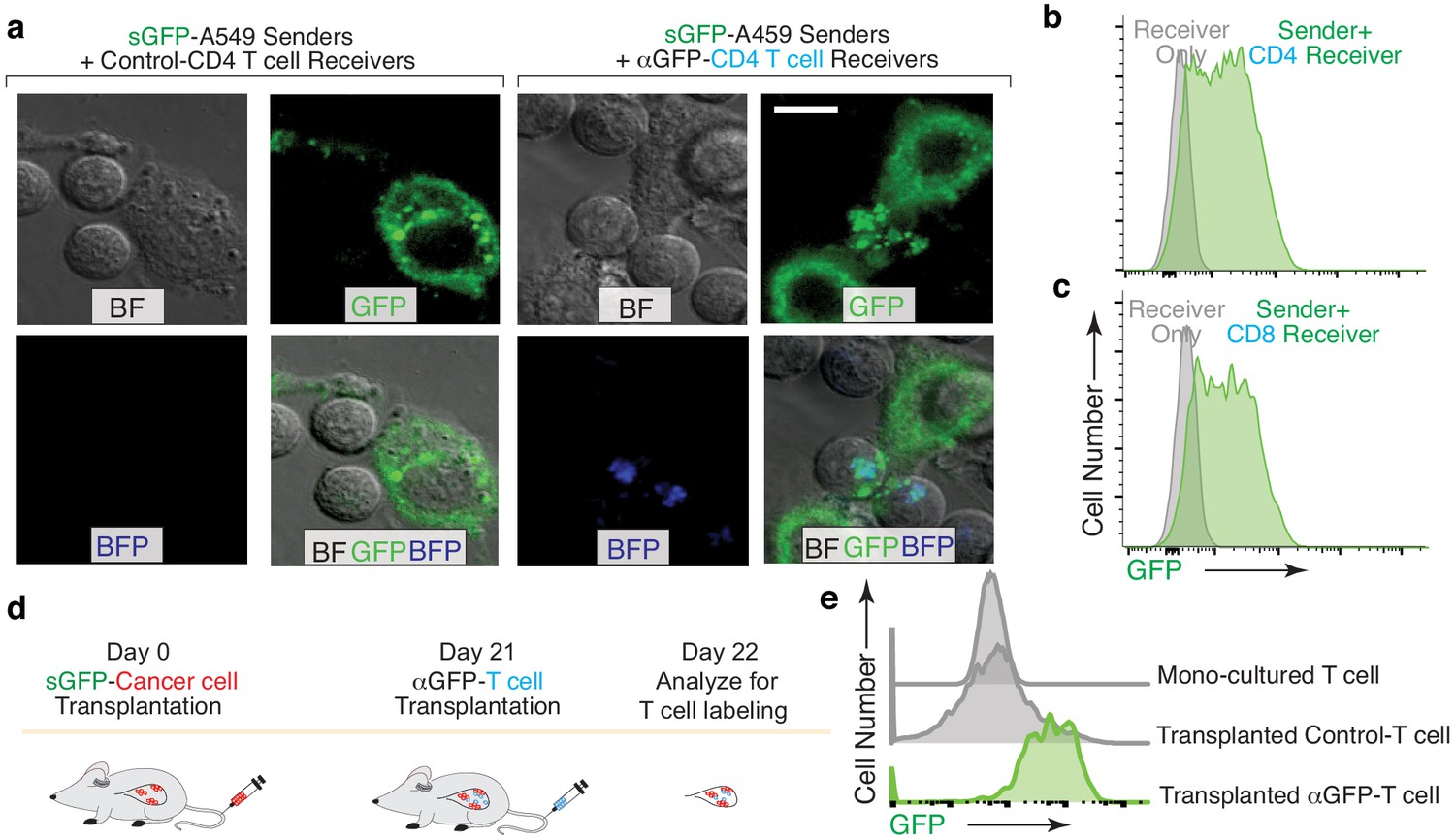
G-baToN can detect cancer cells – T cells interactions.
(a,b,c) G-baToN can detect cancer cell-T cell interactions in vitro. (a) Primary human CD4pos T cells were co-cultured with sGFP-expressing lung cancer sender cells (A549 cells) at a 2:1 ratio for 24 hr. Representative image of A549 cell and CD4 T cell interactions. Scale bars = 10 μm. BF = bright field (b,c) A549 cells expressing sGFP can transfer GFP to αGFP primary human CD4pos (b) or CD8pos (c) T cells after co-culture at a 1:1 ratio for 24 hr. Receiver cells were defined as Near-IRnegBFPposCD4pos or CD8pos T cells. (d,e) G-baToN can detect cancer cell-T cell interactions in vivo. (d) Experiment design for cancer cell-T cell interactions in vivo. 1 × 10⁶ sGFP-expressing lung cancer sender cells were transplanted into NSG mice at day 0. 4 × 10⁶ αGFP primary human CD4pos T cell were transplanted into tumor-bearing mice at day 21. One day after T cell transplantation (day 22), T cells in the mouse lung were analyzed by FACS. (e) sGFP-expressing cancer cell can transfer GFP to αGFP-expressing primary human CD4pos T cells. Receive cells were defined as PInegBFPposCD4posT cells.
Figure 5 with 1 supplement
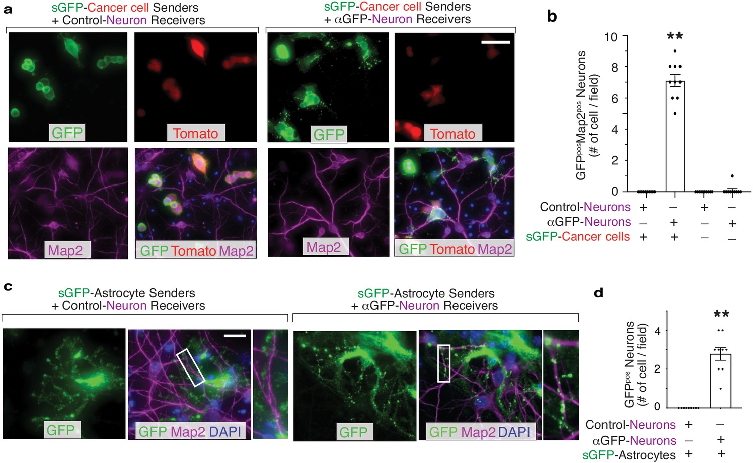
G-baToN can detect cancer cell–neuron and astrocyte-neuron interactions.
(a) Representative image of sGFP-expressing cancer sender cells co-cultured with either control neuron receiver cells or αGFP neuron receiver cells at a 1:1 ratio for 24 hr. Neurons were stained with Microtubule Associated Protein 2 (Map2). Scale bars = 50 μm. (b) Quantification of a using images from 10 different fields. Each dot represents a field. The bar indicates the mean +/- SD. GFPpos neurons were defined as Map2posTomatoneg cells with GFP. **p<0.01, n = 10. (c) Representative images of sGFP-expressing astrocyte sender cells co-cultured with either control neuron receiver cells or αGFP neuron receiver cells at a 1:2 ratio for 24 hr. Neurons were stained with Map2. Scale bars = 50 μm. Higher magnification of the boxed areas are shown on the right. (d) Quantification of c using images from 10 different fields. Each dot represents a field. The bar indicates the mean +/- SD. GFPpos neurons were defined as Map2pos cells with GFP. **p<0.01, n = 10.
Figure 5—figure supplement 1
G-baToN is a generalized system that can be used for touching-based labeling between various cell types.
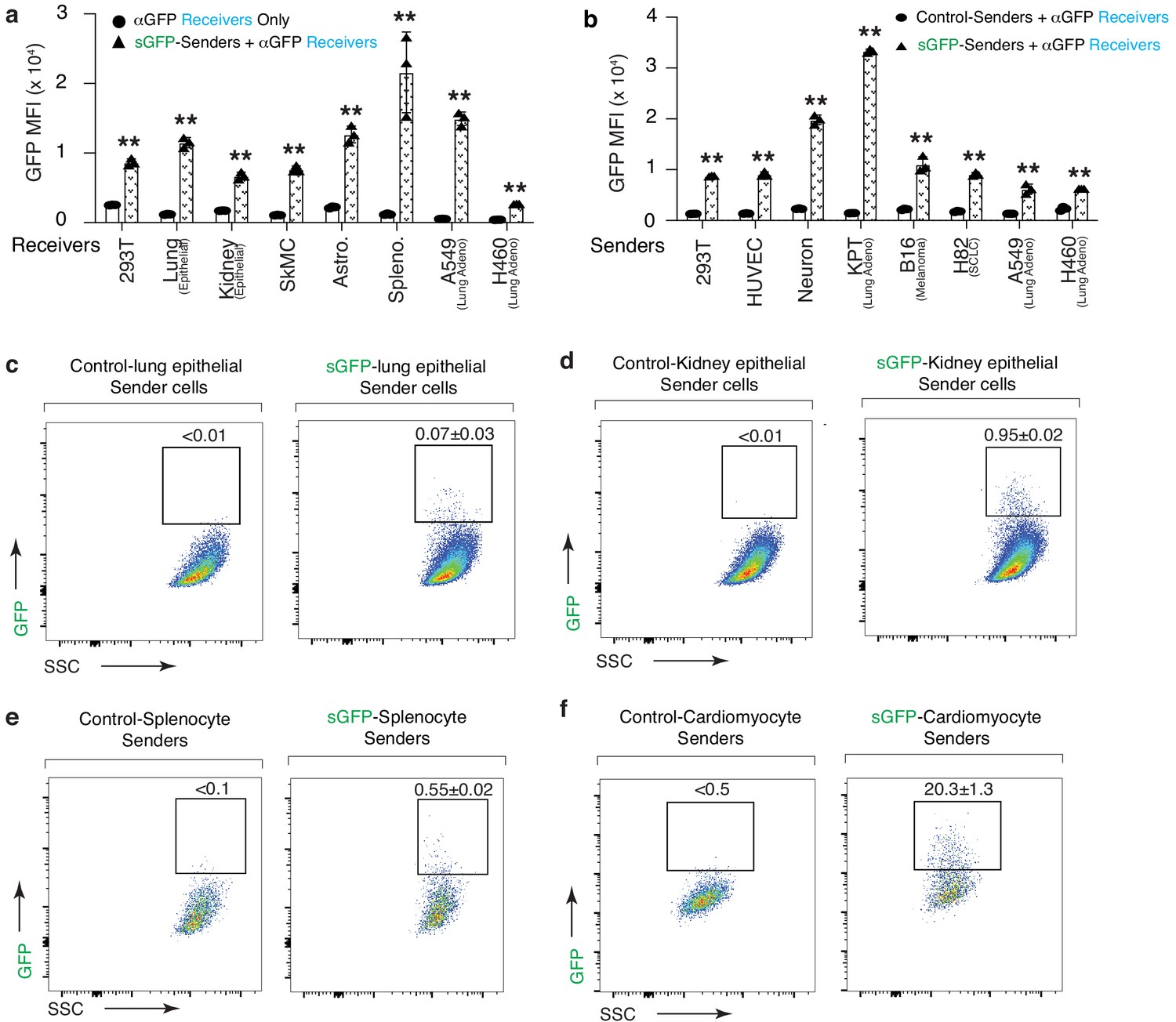
(a) Many different cell lines and primary cell types can serve as receiver cells. Receiver cells expressing αGFP were co-cultured with sGFP-expressing KPT sender cells for 24 hr. MFI of GFP on TomatonegPInegBFPpos receiver cells was assessed by FACS analysis and is shown as fold change relative to monocultured receiver cells. For lung epithelial cells and kidney epithelial cells MFI of GFP on TomatonegPInegBFPposEpCAMpos cells are shown as mean +/- SD of triplicate cultures. SkMC: skeletal muscle cells. Astro: Astocytes. Hepato: hepatocytes. Spleno: splenocytes. (b) Many different cell lines and primary cell types can serve as sender cells. Sender cells expressing sGFP were co-cultured with αGFP 293 receiver cells for 24 hr. MFI of GFP on mCherrynegPInegBFPpos receiver cells was assessed by FACS and is shown as fold change relative to monocultured 293 receiver cells. Mean +/- SD of triplicate cultures is shown. (c,d,e,f) Diverse primary mouse cells expressing sGFP can transfer GFP to αGFP 293 receiver cells. Different primary sender cells were first sorted based on their cell surface markers, then transduced with lentiviral vectors expressing sGFP before being co-cultured with αGFP 293 receiver cells for 24 hr. Percentage of mCherrynegPInegBFPposGFPpos receiver cells was assessed by flow cytometry. For each primary sender, approximate sGFPpos sender: αGFPpos receiver ratio is indicated: (c) Lung epithelial cells (1:180) (sorted EpCampos cells from dissociated adult lung); (d) Kidney epithelial cells (1:20) (sorted EpCampos cells from dissociated adult kidney); (e) Splenocyte (1:20); (f) Cardiomyocyte (1:5) (sorted Sirpapos cells from dissociated adult heart). Percent of labeled cells is indicated as mean +/- SD of triplicate cultures.
Figure 6 with 2 supplements
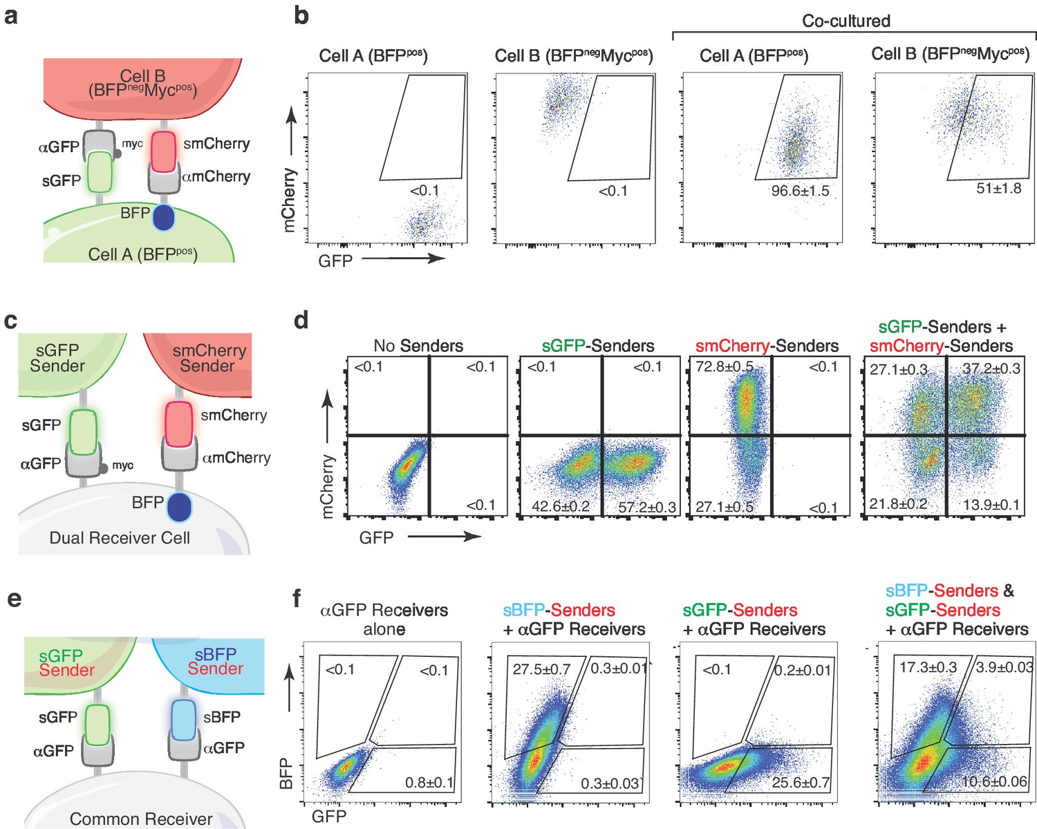
Multicolor-baToN systems enable recording of higher-order interactions.
(a) Diagram of the reciprocal baToN system. Cell A expresses sGFP and αmCherry (tagged by intracellular BFP), Cell B expresses smCherry and αGFP (tagged by Myc-tag). (b) Representative FACS plots of cell A and cell B monocultures (left two panels) and after co-culture at a 5:1 ratio for 24 hr. Percent of labeled cells is indicated as mean +/- SD of triplicate cultures. (c) Schematic of the AND gate-baToN system. sGFP and smCherry sender cells express either sGFP or mCherry. Dual receiver cells express both αGFP (LaG17, tagged by Myc-tag) and αmCherry (LaM4, tagged by intracellular BFP). (d) Representative FACS plots of dual receiver 293 cells cultured with the indicated 293 sender cells at 1:1 (for single sender cell) or 1:1:1 (for dual sender cells) ratios. Percent of labeled receiver cells (defined as BFPpos) after 24 hr of co-culture is indicated as mean +/- SD of triplicate cultures. (e) Diagram of the BFP/GFP AND gate baToN system. sBFP sender cells express intracellular Tomato and surface BFP, sGFP sender cells express intracellular Tomato and surface GFP. Common receiver cells expressed αGFP. (f) Representative FACS plots of common receiver 293 cells cultured with the indicated Tomatopos sender cells at 1:1 (for single sender cell) or 1:1:1 (for dual sender cells) ratios. Receiver cells were defined as TomatonegPIneg. Percent of labeled common receiver cells after 24 hr of co-culture is indicated as mean +/- SD of triplicate cultures.
Figure 6—figure supplement 1
X-baToN systems enable fluorescent labeling via various antigen-nanobody/scFV pairs.
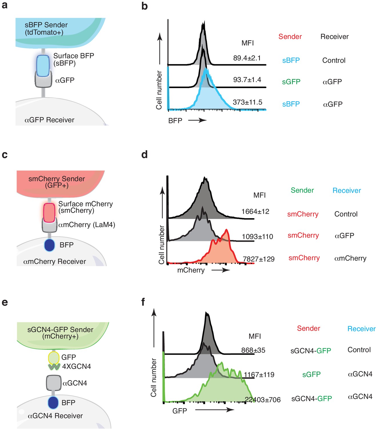
(a) Schematic of the surface BFP (sBFP)-baToN system. Sender cells (marked by intracellular tdTomato) express sBFP and receiver cells express αGFP. (b) The cross reactivity of αGFP with BFP allows BFP to be transferred from sBFP sender cells to αGFP receiver cells. sBFP sender cells and αGFP receiver cells were co-cultured at 1:1 ratio for 24 hr. Receiver cells were defined as toTomatonegPIneg cells. BFP MFI of receiver cells is shown as mean +/- SD of triplicate cultures. (c) Schematic of the surface mCherry (smCherry)-baToN system. Sender cells (marked by intracellular GFP) express smCherry and receiver cells express αmCherry (tagged by intracellular BFP) (d) The pairing between mCherry and αmCherry enable mCherry transfer from smCherry 293 sender cells to αmCherry-expressing 293 receiver cells. smCherry sender cells are not able to transfer mCherry to αGFP receiver cells. smCherry sender cells were co-cultured with αGFP or αmCherry receiver cells at a 1:1 ratio for 24 hr. Receiver cells were defined as GFPnegBFPpos cells. mCherry MFI of receiver cell is shown as mean +/- SD of triplicate cultures. smCherry 293 sender cells are not able to transfer mCherry to αGFP 293 receiver cells. (e) Schematic of the surface GCN4 (sGCN4)-baToN system. Sender cells (marked by intracellular 2A-mCherry) express cell surface 4X GCN4 peptide fused with GFP. Receiver cells express a cell surface anti-GCN4 single-chain variable fragment (scFV; αGCN4, tagged by intracellular BFP). (f) The pairing between GCN4 and αGCN4 enables co-transfer of GFP from 4X sGCN4-GFP sender cells to αGCN4 receiver cells 24 hr after co-culture at a 1:1 ratio. Receiver cells were defined as mCherrynegPInegBFPpos cells. GFP MFI of receiver cells is shown as mean +/- SD of triplicate cultures. sGFP sender cells are not able to transfer GFP to αGCN4 receiver cells.
Figure 6—figure supplement 2
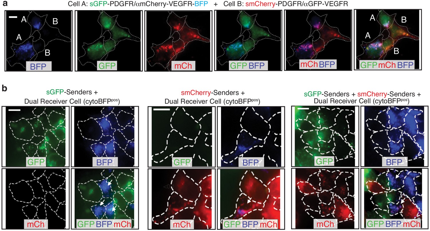
Dual color-baToN systems enable labeling in complex cell-cell interaction systems.
(a) Representative image of reciprocal labeling of cell A and cell B co-cultured for 24 hr. Cells are outlined with a white dashed line. Cell A is BFP and GFP double positive and Cell B is mCherry positive. Touched cell A is BFP, GFP and mCherry triple positive and touched cell B is GFP and mCherry double positive (pointed out by white letter). Scale bar: 10 μm. (b) Representative image of dual receiver cells co-cultured with indicated sender cells. Cells are outlined with a white dashed line. Dual receiver cells are BFP positive and sender touched dual receiver cells are pointed out by white arrows. Scale bar: 10 μm.
Figure 7
The HaloTag-baToN system enables quantitative and sensitive cell-cell interaction-dependent receiver cell labeling.
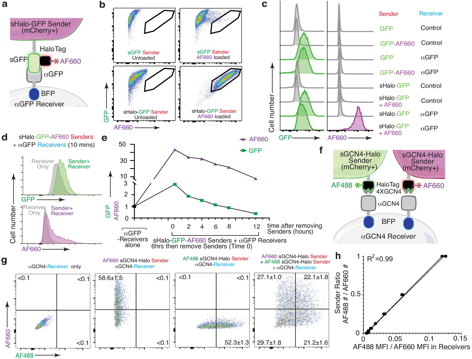
(a) Diagram of HaloTag-baToN system. Sender cells (marked by intracellular 2A-mCherry) express surface HaloTag-GFP fusion which can be loaded with HaloTag ligands (in this example AF660). Receiver cells express αGFP (LaG17, tagged by intracellular BFP). (b) Labeling of HaloTag-expressing sender cells with AF660 fluorophore. Representative FACS plots of KP (lung adenocarcinoma) sender cells expressing either sGFP or sGFP-sHaloTag incubated with AF660-conjugated HaloTag ligand for 5 min on ice. AF660 specifically labeled sHaloTag-GFP sender cells but not sGFP sender cells. (c) Representative plot of GFP and AF660 intensity in αGFP 293 receiver cells co-cultured with HaloTag-GFP KP sender cells at a 1:1 ratio for 6 hr. Receiver cells were defined as mCherrynegPInegBFPpos cells. (d) AF660 transfer to αGFP 293 receiver cells is rapid after cell-cell interaction. AF660 MFI shift was detected after mixing sHalo-GFP sender cells and αGFP receiver cells and co-culture for 10 min. AF660 MFI shift was more dramatic than GFP. Receiver cells were defined as mCherrynegPInegBFPpos cells. (e) Slower AF660 quenching in touched receiver cells after removing sHalo-GFP sender cells. After 6 hr co-culture, GFP/AF660-positive receiver cells were purified via FACS. Analysis of GFP/AF660 MFI in purified receiver cells showed rapid GFP degradation but slower AF660 quenching. Receiver cells were defined as mCherrynegPInegBFPpos cells. (f) Diagram of dual color GCN4-HaloTag-baToN system. Sender cells (marked by intracellular 2A-mCherry) express surface 4XGCN4 associated with HaloTag, loaded with either AF488- or AF660- conjugated HaloTag ligand. Receiver cells express αGCN4 (tagged by intracellular BFP). (g) Representative FACS plots of αGCN4 receiver cells co-cultured with the indicated sender cells at 1:1 (for single sender cell) or 1:1:1 (for dual sender cells) ratios. Percent of labeled receiver cells (gated as mCherrynegPInegBFPpos) after 6 hr of co-culture is indicated as mean +/- SD of triplicate cultures. (h) AF488/AF660 GCN4-HaloTag sender ratio in the co-culture directly proportional to AF488/AF660 intensity (MFI) of αGCN4 receiver after 6 hr of co-culture. Receiver cells were defined as mCherrynegPInegBFPpos cells.
Figure 8
The G-baToN system can co-transfer cargo molecules to touched receiver cells.
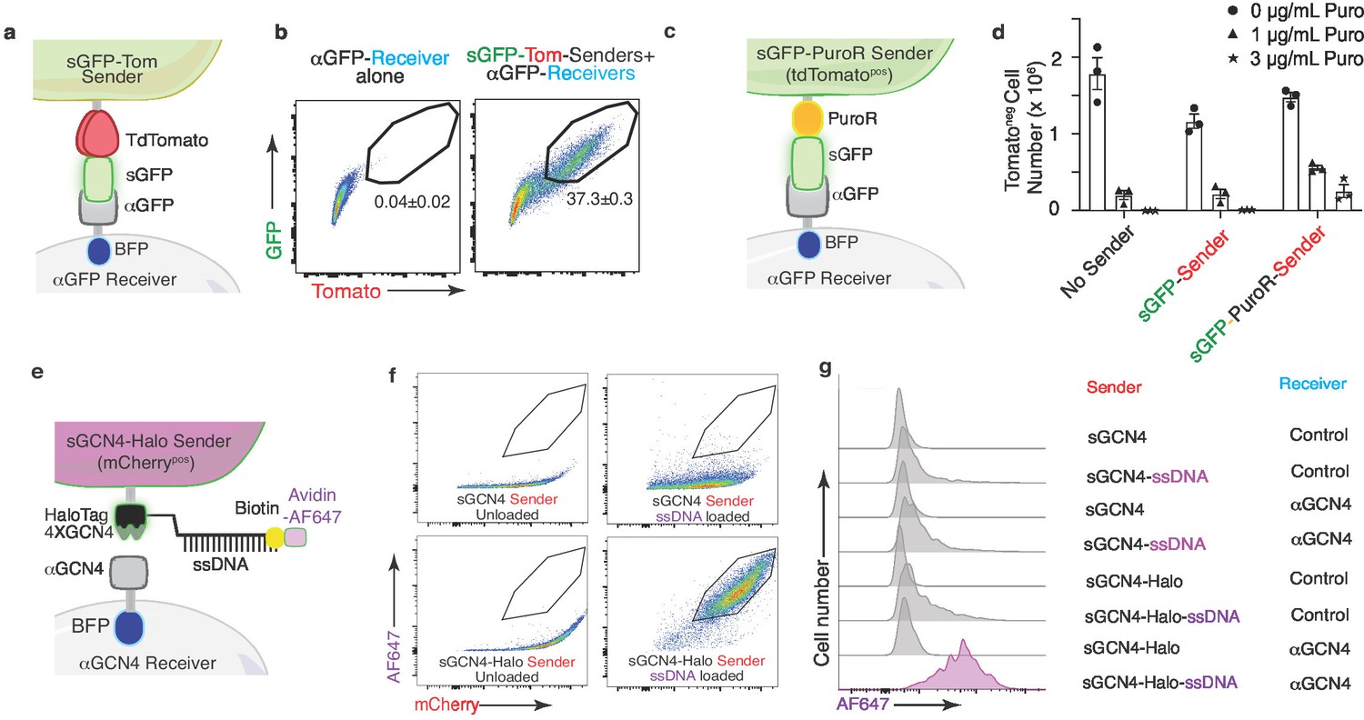
(a) Diagram of surface tdTomato-GFP (sGFP-Tom) co-transfer into touched receiver cells. Sender cells express sGFP-tdTomato and receiver cells express αGFP (tagged by intracellular BFP). (b) Representative FACS plots of αGFP 293 receiver cells cultured with the indicated sender cells at 1:1 ratio for 24 hr. Percent of GFP/Tomato dual-labeled receiver cells is indicated as mean +/- SD of BFPpos cells from triplicate cultures. (c) Diagram of GFP-PuroR co-transfer system. Sender cells express surface GFP associated with PuroR (marked by intracellular tdTomato), receiver cells express αGFP (tagged by intracellular BFP). PuroR: Gcn5-related N-acetyltransferase. (d) Co-transfer of GFP-PuroR from sGFP-PuroR sender cells to αGFP 293 receiver cells confers modest puromycin resistance to receiver cells. sGFP or sGFP-PuroR sender cells were co-cultured with αGFP 293 receiver cells for 24 hr at a 4:1 ratio before treatment with different doses of puromycin for 48 hr. tdTomatonegPInegBFPpos cell numbers were counted via FACS. (e) Diagram of using GCN4-HaloTag sender to transfer ssDNA into αGCN4 receiver cells. Sender cells (marked by intracellular 2A-mCherry) express surface 4XGCN4 associated with a HaloTag, loaded with 5’ HaloTag ligand, 3’ biotin dual conjugated ssDNA (21 nt), then stained with Avidin-AF647. Receiver cells express αGCN4 (tagged by intracellular BFP). (f) Loading of sender cells with ssDNA. Representative FACS plots of 293 sender cells expressing either sGCN4 or sGCN4-Halo were loaded with 5’ HaloTag-ligand, 3’ biotin dual-conjugated ssDNA (21nt), then stained with Avidin-AF647. AF647 specifically labeled loaded sGCN4-Halo sender cells but not sGCN4 sender cells. (g) Representative plot of AF647 intensity in αGCN4 293 receiver cells co-cultured with GCN4-HaloTag 293 sender cells at a 1:1 ratio for 6 hr. Receiver cells were defined as mCherrynegPInegBFPpos cells.
Videos
Video 1
Time-lapse movie of a sGFP sender cell transferring GFP into a αGFP receiver cell.
Additional files
-
Supplementary file 1
Plasmids used in each experiment in this study.
For each figure panel we list how the cells are referred to in the figure (Cell Name in Figure), the parental plasmid, the plasmid used to generate that cell line (N-terminal extracellular domain-transmembrane domain-cytoplastic domain; N-TM-C), whether they are the same plasmid used in a previous figure (Repeated Cell Line), whether the cells were selected with puromycin, and whether the nanobody had a Myc tag. The cells used in each experiment are listed in Supplementary file 2.
- https://cdn.elifesciences.org/articles/61080/elife-61080-supp1-v2.xlsx
-
Supplementary file 2
Cells used in each experiment in this study.
For each figure panel we list how the cells are referred to in the figure (Cell Name in Figure), the parent cell line or cell type, whether cDNA expression was stable (via lentiviral transduction) or transient (via transfection), the transgene expressed in those cells, whether they are the same cell line used in a previous figure (Repeated Cell Line), whether they are senders or receivers, whether the expression of GFP, BFP, mCherry or tdTomato is cell surface or intracellular, and whether the nanobody has a Myc tag. For each experiment, we list the Sender to Receiver cell ratio and the co-culture time. The vectors used for cDNA expression in each cell type in each experiment are listed in Supplementary file 1.
- https://cdn.elifesciences.org/articles/61080/elife-61080-supp2-v2.xlsx
-
Transparent reporting form
- https://cdn.elifesciences.org/articles/61080/elife-61080-transrepform-v2.docx
Download links
A two-part list of links to download the article, or parts of the article, in various formats.
Downloads (link to download the article as PDF)
Open citations (links to open the citations from this article in various online reference manager services)
Cite this article (links to download the citations from this article in formats compatible with various reference manager tools)
A versatile system to record cell-cell interactions
eLife 9:e61080.
https://doi.org/10.7554/eLife.61080


