ClpAP proteolysis does not require rotation of the ClpA unfoldase relative to ClpP
- Department of Biology, Massachusetts Institute of Technology, United States
- Department of Chemistry, Massachusetts Institute of Technology, United States
Figures
Figure 1
ClpAP structure and rotary translocation model.
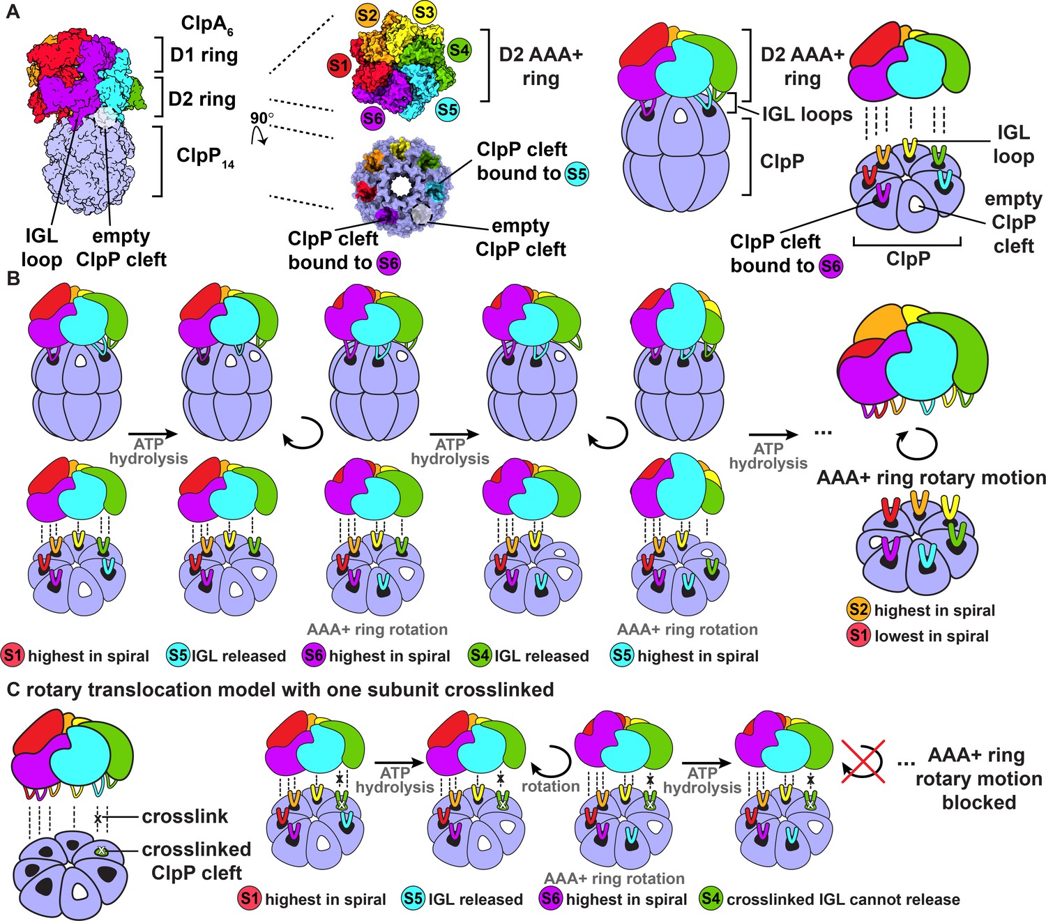
(A) Complex of ClpP with ClpA (PDB 6UQO). Subunits of ClpA, labeled S1 through S6, are ordered from the highest to the lowest position in the spiral relative to ClpP at the beginning of the mechanical cycle. The IGL loops of ClpA hexamers dock into a subset of the seven clefts in a heptameric ClpP ring. In this structure, there is an empty cleft between the second lowest and lowest subunits in the spiral (S5 and S6, respectively). The coloring of the ClpP clefts represents the docked position of the IGL loops from the corresponding AAA+ subunits; empty clefts are colored white. The rightmost panel is a generalized model of the ClpA D2 AAA+ ring docking into the ClpP interface. (B) Rotary translocation model with clockwise around-the-ring ATP hydrolysis and IGL loop release and rebinding (Ripstein et al., 2020; Lopez et al., 2020). When subunit S1 is highest in the spiral, ATP hydrolysis releases the IGL loop of subunit S5 and the AAA+ ring rotates clockwise with respect to ClpP. During rotation, subunit S6 moves to the top in of the spiral, and the IGL loop of subunit S5 takes a clockwise ‘step’ and rebinds to the adjacent empty ClpP cleft. Repetition of this sequence of ATP hydrolysis and IGL loop release and rebinding results in rotary motion of the AAA+ ring with respect to the ClpP ring. (C) Rotary translocation model with at least one crosslinked IGL loop. If one ClpA subunit is crosslinked to a ClpP cleft, the rotary motion of the AAA+ ring with respect to the ClpP ring is blocked. The crosslinked ClpA subunit cannot be released from the ClpP cleft and cannot sequentially move to each position in the ClpA spiral.
Figure 2 with 1 supplement
ClpA‒ClpP crosslinking and purification.
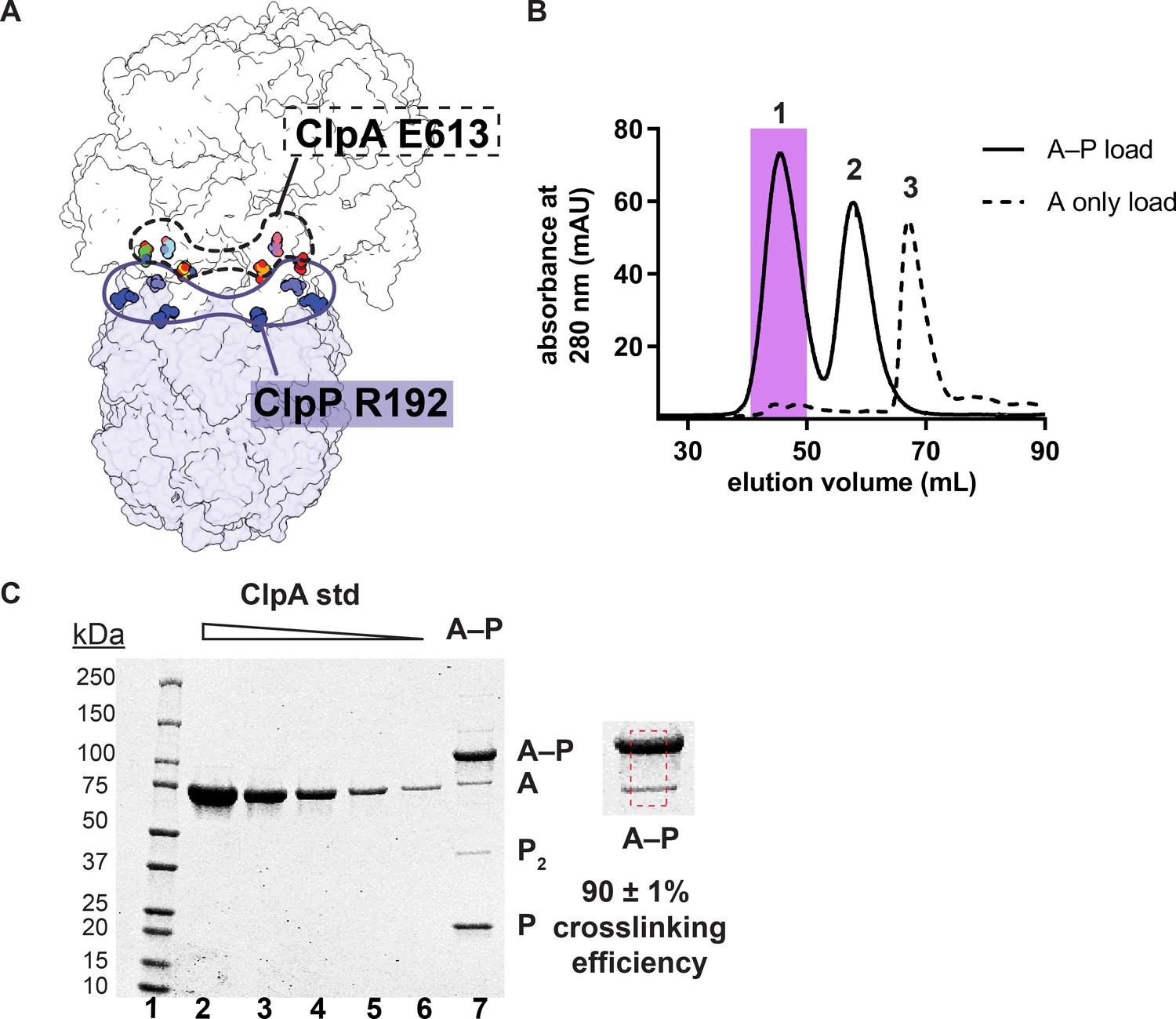
(A) Proximity of Glu613 residues in six subunits of the hexameric ClpA ring (shown in the dashed outline) to Arg192 residues in six of the seven subunits of a heptameric ClpP ring (shown in the solid outline). (B) Size-exclusion chromatograms of E613CClpA‡–ClpP+C following crosslinking (solid line; peaks 1 and 2) or uncrosslinked E613CClpA‡ (dashed line; peak 3), which is largely monomeric under the chromatography conditions. As shown in panel C, most ClpA in peak one is crosslinked to ClpP. The shaded area in peak 1 represents the crosslinked A–P that was pooled and used in all experiments in this study. Peak 2 corresponds to uncrosslinked ClpP+C remaining after the crosslinking reaction and chromatographs at the position expected for a tetradecamer. (C) Reducing SDS-PAGE of the peak-1 pool. Lanes 1–6 are MW standards or different concentrations of purified E613CClpA‡. Lane 7 is an aliquot of the peak-1 pool. The shift to higher molecular weight from uncrosslinked ClpA (A) to crosslinked ClpA–ClpP (A–P) is consistent with covalent linkage of a single ClpA monomer (~83 kDa) to a single ClpP monomer (~23 kDa). The dashed red box is a zoomed-in view of lane 7 used to calculate crosslinking efficiency of E613CClpA‡ to ClpP+C. Crosslinking efficiency was calculated as the mean ± 1 SD of four independent replicates. The quantification of SDS-PAGE bands used to calculate crosslinking efficiency is available in Figure 2—source data 1.
-
Figure 2—source data 1
Quantification of A–P crosslinking efficiency.
Values are relative fluorescent units (RFUs) calculated from the area under the curve of each species band on a Coomassie-stained SDS-PAGE gel. The volume of samples (3 or 5 µL) loaded in each lane is indicated and used to generate two standard curves for each sample load volume. No RFUs were recorded for the E613CClpA‡ samples containing only uncrosslinked ClpA that were used for the standard curves (represented by N/A). Concentrations of ClpA were calculated from the standard curve with matching sample load volumes.
- https://cdn.elifesciences.org/articles/61451/elife-61451-fig2-data1-v1.docx
Figure 2—figure supplement 1
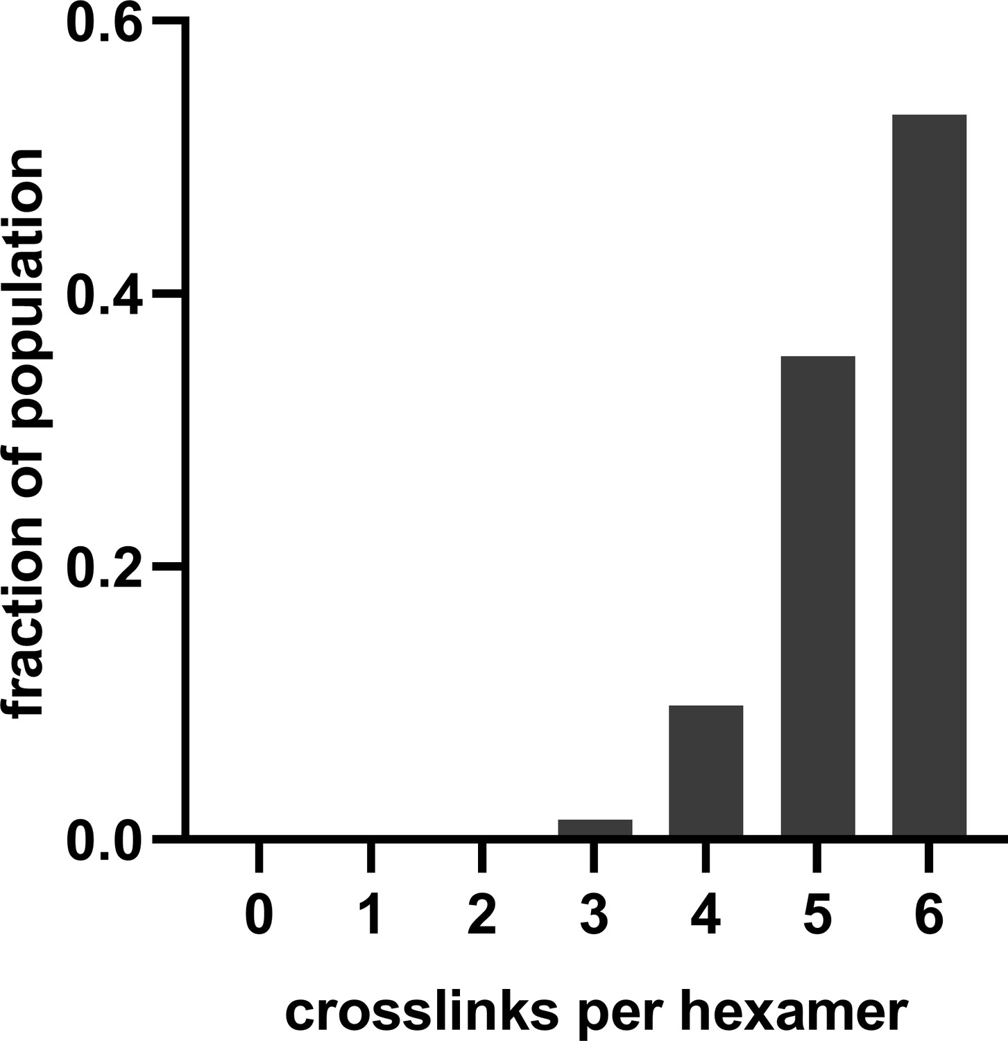
Histogram of the expected number of crosslinks between E613CClpA‡ and ClpP+C in the A–P pool assuming independent crosslinking of individual ClpA and ClpP subunits with 90 ± 1% efficiency.
Figure 3 with 1 supplement
Substrate degradation by crosslinked ClpAP (A–P) and uncrosslinked ClpAP (A•P).
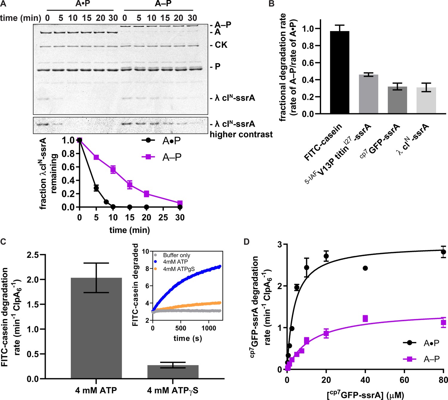
(A) Top, SDS-PAGE assay of the kinetics of λ cIN-ssrA degradation by A–P and the A•P control (CK is creatine kinase). Bottom, quantification of λ cIN-ssrA degradation. Values are means ± 1 SD (n = 3) and provided in Figure 3—source data 1. (B) Degradation of substrates of varying thermodynamic stability (18 µM) FITC-casein, 5 µM 5-IAFV13P titinI27-ssrA, 20 µM cp7GFP-ssrA, 15 µM λ clN-ssrA by A–P. Fractional degradation rates were calculated by dividing the degradation rates of A–P by the A•P rates. Values are means ± propagated error (n ≥ 3) and provided in Figure 3—source data 2. (C) Degradation of FITC-casein (18 µM) by A–P in the presence of ATP or ATPγS. FITC-casein degradation was quantified by normalizing the relative fluorescence units to the total FITC-casein degraded upon porcine elastase addition at the endpoint of the assay and subtracting the contributions of photobleaching from the buffer-only control. Values are means ± 1 SD (n = 3) and provided in Figure 3—source data 3. The inset shows representative degradation kinetics. (D) Michaelis-Menten analysis of cp7GFP-ssrA degradation kinetics by A–P and the A•P control. Values are means ±1 SD (n = 3) and provided in in Figure 3—source data 4. For A–P degradation, Vmax was 1.4 ± 0.07 min−1 ClpA6−1, KM was 13 ± 1.6 µM, and R2 was 0.96; for the A•P control, Vmax was 3.0 ± 0.10 min−1 ClpA6−1, KM was 3.7 ± 0.5 µM, and R2 was 0.96, where the errors are those of non-linear least-squares fitting to the Michaelis-Menten equation.
-
Figure 3—source data 1
Quantification of λ cIN-ssrA degradation kinetics.
Values are means of fraction remaining of λ cIN-ssrA from three technical replicates ± 1 SD. Values were not recorded (NR) for A–P at 8 min.
- https://cdn.elifesciences.org/articles/61451/elife-61451-fig3-data1-v1.docx
-
Figure 3—source data 2
Degradation of substrates of varying thermodynamic stability.
In columns 2–3, degradation rates are mean degradation rates from 3 to 4 technical replicates ±1 SD. In column 4, the fractional rates were calculated from dividing the mean A–P degradation rate by the mean A•P degradation rate, and the error is a propagated error calculated using the following formula.
- https://cdn.elifesciences.org/articles/61451/elife-61451-fig3-data2-v1.docx
-
Figure 3—source data 3
Degradation of FITC-casein (18 µM) by the purified A–P pool in the presence of ATP or ATPγS.
Values are mean FITC-casein degradation rates from three technical replicates ± 1 SD.
- https://cdn.elifesciences.org/articles/61451/elife-61451-fig3-data3-v1.docx
-
Figure 3—source data 4
Michaelis-Menten analysis of cp7GFP-ssrA degradation kinetics.
Values are mean degradation rates of cp7GFP-ssrA from three technical replicates ± 1 SD. Values were not recorded (NR) for A•P at 7.5 µM.
- https://cdn.elifesciences.org/articles/61451/elife-61451-fig3-data4-v1.docx
-
Figure 3—source data 5
ATPase and cp7GFP-ssrA degradation rates by ClpAP controls.
Values are mean ATP hydrolysis rates (5 mM ATP) or mean degradation rates (20 µM cp7GFP-ssrA) from three technical replicates ± 1 SD.
- https://cdn.elifesciences.org/articles/61451/elife-61451-fig3-data5-v1.docx
Figure 3—figure supplement 1
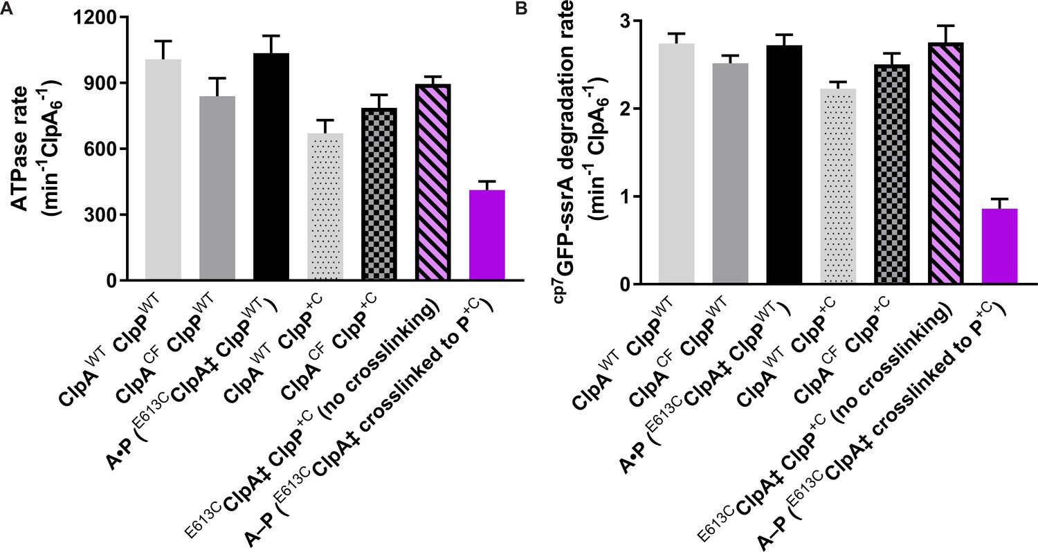
ATPase and degradation activities of uncrosslinked E613CClpA‡ClpP+C and E613CClpA‡ClpPWT (A•P) complexes are comparable to those of wild-type ClpAP.
(A) Hydrolysis of ATP (5 mM) by crosslinked ClpAP (A–P) and uncrosslinked controls consisting of wild-type ClpA (ClpAWT), cysteine-free ClpA (ClpACF), or E613CClpA‡ and either wild-type ClpP (ClpPWT) or ClpP+C. (B) Degradation of cp7GFP-ssrA (20 µM) by crosslinked (A–P) and uncrosslinked ClpAP (A•P) variants, as shown in panel (A). A•P and uncrosslinked E613CClpA‡ClpP+C degrade cp7GFP-ssrA (20 µM) at similar rates as wild-type ClpAP and other uncrosslinked controls, and faster than crosslinked A–P. Values are means ± 1 SD (n ≥ 3) and provided in Figure 3—source data 5.
Tables
Key resources table
| Reagent type (species) or resource | Designation | Source or reference | Identifiers | Additional information |
|---|---|---|---|---|
| Gene (Escherichia coli) | clpA | UniProtKB - P0ABH9 | ||
| Gene (Escherichia coli) | clpP | UniProtKB - P0A6G7 | ||
| Strain, strain background (Escherichia coli) | T7 Express | New England Biolabs | C2566I | Chemically competent cells |
| Recombinant DNA reagent | pT7 ClpP+C (plasmid) | This paper | For overexpression of C-terminally His6-tagged ClpP (C91V, C113A) with extra Cys residue for crosslinking. Progenitor: pT7 ClpP-TEV-cHis6(Stinson et al., 2013; Amor et al., 2016) | |
| Recombinant DNA reagent | pET23b His7SumoFLAG E613CClpA‡(plasmid) | This paper | For overexpression of ClpA with Cys substitution and C47S, C203S, C243S background for crosslinking. Progenitor: pET23b His7Sumo ClpAcfΔC9 (Zuromski et al., 2020) | |
| Recombinant DNA reagent | pET23b His7Sumo ClpAcfΔC9 (plasmid) | Zuromski et al., 2020 | For overexpression of cysteine-free ClpA (ClpACF) harbouring C47S, C203S, C243S mutations | |
| Recombinant DNA reagent | WT ClpA (plasmid) | Seol et al., 1994, Hou et al., 2008 | WT ClpA (M169T background) for overexpression | |
| Recombinant DNA reagent | ClpP-His6(plasmid) | Kim et al., 2001 | WT ClpP for overexpression | |
| Recombinant DNA reagent | cp7GFP-ssrA (plasmid) | Nager et al., 2011 | Circularly permutated variant of superfolder GFP-ssrA for overexpression | |
| Recombinant DNA reagent | V13P titinI27-ssrA (plasmid) | Kenniston et al., 2003 | ssrA-tagged I27 domain variant for overexpression | |
| Recombinant DNA reagent | His6SUMO λ cIN-ssrA (plasmid) | This paper | ssrA-tagged residues 1–93 of λ cI (UniProtKB - P03034) for overexpression | |
| Chemical compound, drug | Bismaleimidoethane | Thermo Fisher Scientific | Cat # 22323 | |
| Chemical compound, drug | Adenosine 5ʹ-O-(3-Thiotriphosphate), Tetralithium Salt | Millipore Sigma | Cat# 119120–25 MG | |
| Chemical compound, drug | 5-Iodoacetamidofluorescein | Thermo Fisher Scientific | Cat# I30451 | |
| Chemical compound, drug | Casein fluorescein isothiocyanate from bovine milk (FITC-casein) | Sigma-Aldrich | Cat# C0528-10MG |
Additional files
Download links
A two-part list of links to download the article, or parts of the article, in various formats.
Downloads (link to download the article as PDF)
Open citations (links to open the citations from this article in various online reference manager services)
Cite this article (links to download the citations from this article in formats compatible with various reference manager tools)
ClpAP proteolysis does not require rotation of the ClpA unfoldase relative to ClpP
eLife 9:e61451.
https://doi.org/10.7554/eLife.61451


