A robust method for particulate detection of a genetic tag for 3D electron microscopy
- The University of Queensland, Institute for Molecular Bioscience, Australia
- Mark Wainwright Analytical Centre, University of New South Wales, Australia
- School of Medical Sciences, University of New South Wales, Australia
- The University of Queensland, Centre for Microscopy and Microanalysis, Australia
- Division Microrobotics and Control Engineering, Department of Computing Science, University of Oldenburg, Germany
- Queensland University of Technology, Australia
- EMBL Australia Node for Single Molecule Sciences, University of New South Wales, Australia
- Monash Institute of Pharmaceutical Sciences, Monash University, Australia
Figures
Figure 1 with 5 supplements
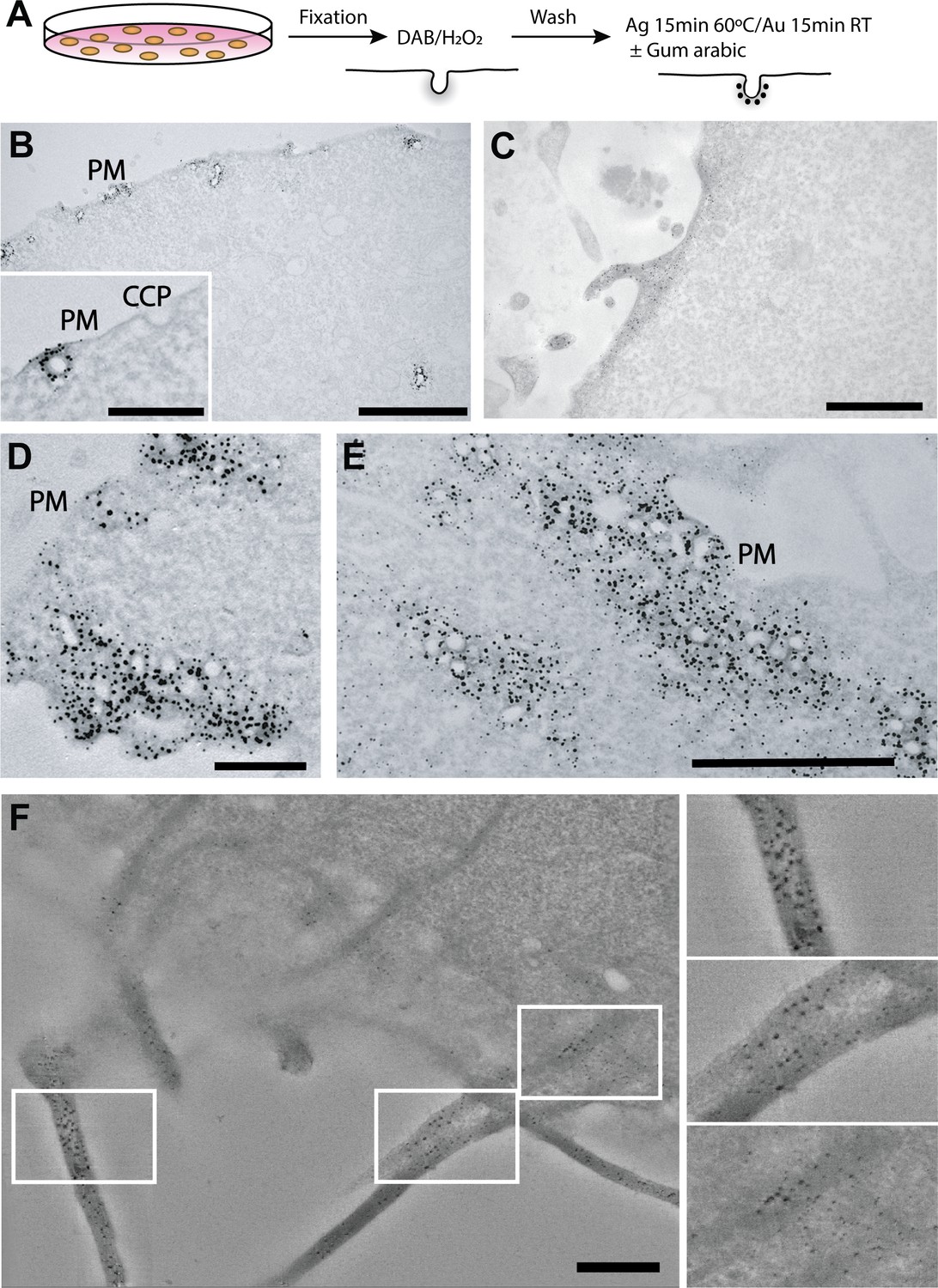
APEX-Gold particulate labelling of genetically-tagged proteins of interest.
(A) Schematic of the APEX-Gold method. Cells were transfected with Cavin4-APEX2 (B,D,E) (control light microscopy experiments are described in Figure 1—figure supplement 5) or LifeAct-APEX2 (C,F), fixed, treated with diaminobenzidine (DAB), and then incubated with Ag/Au reagents in the presence of gum arabic. (B,D,E) Low (B) and higher (D,E, inset in B) magnification views of caveolae labelling. (C) Labelled actin filaments. (F) Optical slice projection through tomogram of LifeAct-APEX2 expressing cells. APEX-Gold particulate reaction product can be observed tightly associated with and throughout the actin bundles in three dimensions. Note the uniform gold label, the lack of background, and high signal to noise. PM, plasma membrane; CCP, clathrin-coated pit. Bars, B, 2 µm (inset 500 nm); C, 1 µm; D, 500 nm; E, 1 µm; F, 500 nm.
Figure 1—figure supplement 1
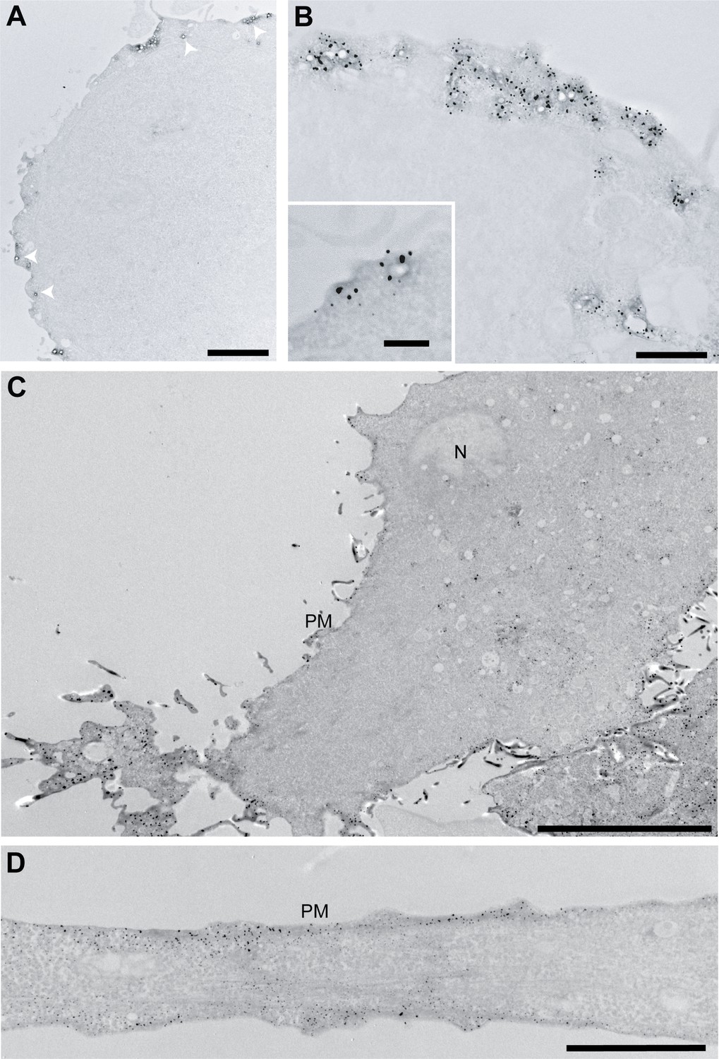
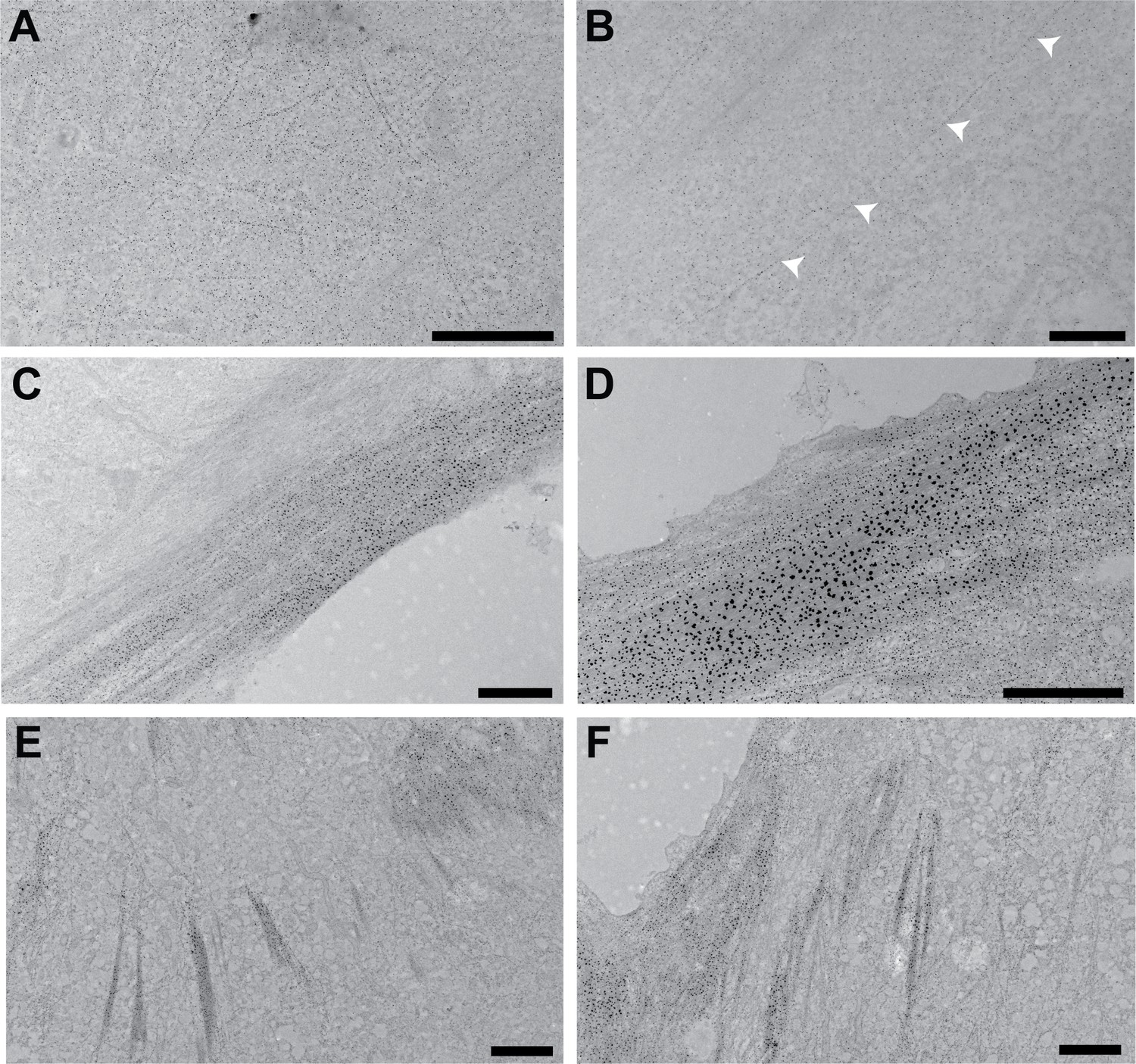
Cavin-4 and LifeAct APEX-Gold labelling.
(A,B) Low and higher magnification views respectively of Cavin4-APEX2 labelling using the APEX-Gold method without gum arabic. Arrows, caveolae labelled with gold. (C,D) Low and higher magnification examples respectively of labelling on actin filaments with LifeAct-APEX2 with APEX-Gold enhancement. N, nucleus; PM, plasma membrane. Bars, A, 1 µm; B, 1 µm (inset, 200 nm); C, 10 µm; D, 2 µm.
Figure 1—figure supplement 2
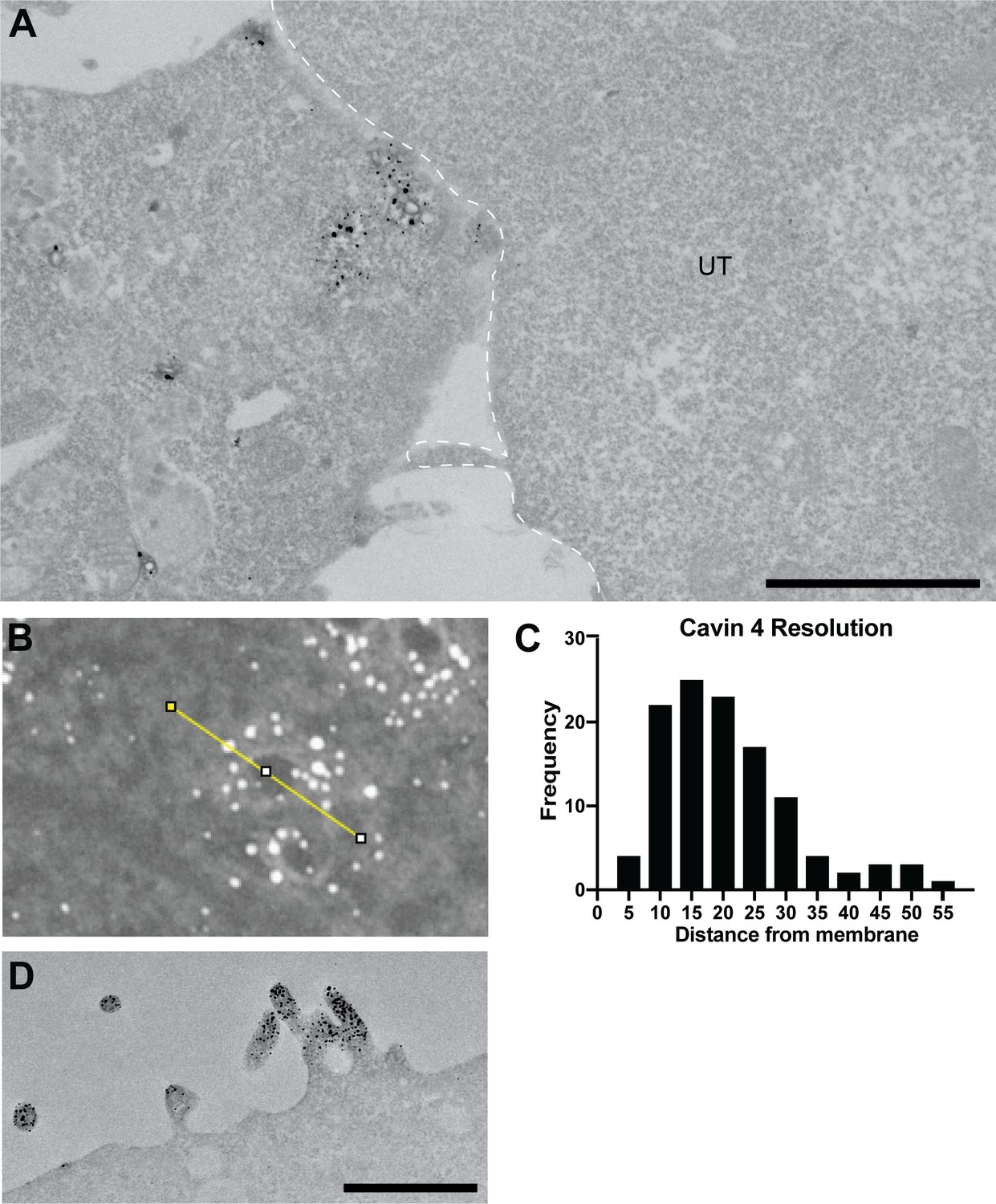
APEX-Gold produces an easy to identify signal with little background, high specificity and broad applicability.
(A) An example of a Cavin4-APEX2 transfected cell immediately adjacent to a untransfected cell (UT, highlighted by dashed line). Sections were poststained with uranyl acetate and Reynold’s lead citrate. Note the absence of particulate reaction product within the untransfected cell. (B) Micrograph of Cavin4-APEX2 labelling enhanced with APEX-Gold. Linescans (FIJI) across caveolae were used to measure the distance from centre of gold particles to the caveolar membrane. (C) The distribution of particle label resolution. (D) A1AR-APEX2 stably expressing CHO cells, processed with APEX-Gold enhancement without gum arabic showing labelling at specific areas of plasma membrane. UT, untransfected cell. Bars, A, 2 µm; D, 500 nm.
Figure 1—figure supplement 3
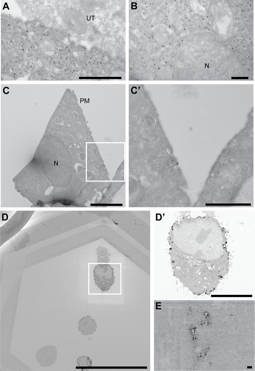
Cryo-sectioning and SEM array tomography of APEX-Gold-labelled cells.
(A,B) Cryosectioned cells showing LifeAct-APEX2 labelling. (C,C’) Cryosectioned cells showing Cavin4-APEX2 labelling. (D–E) Scanning electron microscopic (SEM) analysis of sections of BHK cells expressing Cavin4-APEX2 (Raith eLine Plus). UT, untransfected; N, nucleus; PM, plasma membrane. Bars, A, 1 µm; B, 500 nm; C, 2 µm; C’, 1 µm; D, 50 µm; D’, 10 µm; E, 100 nm.
Figure 1—figure supplement 4
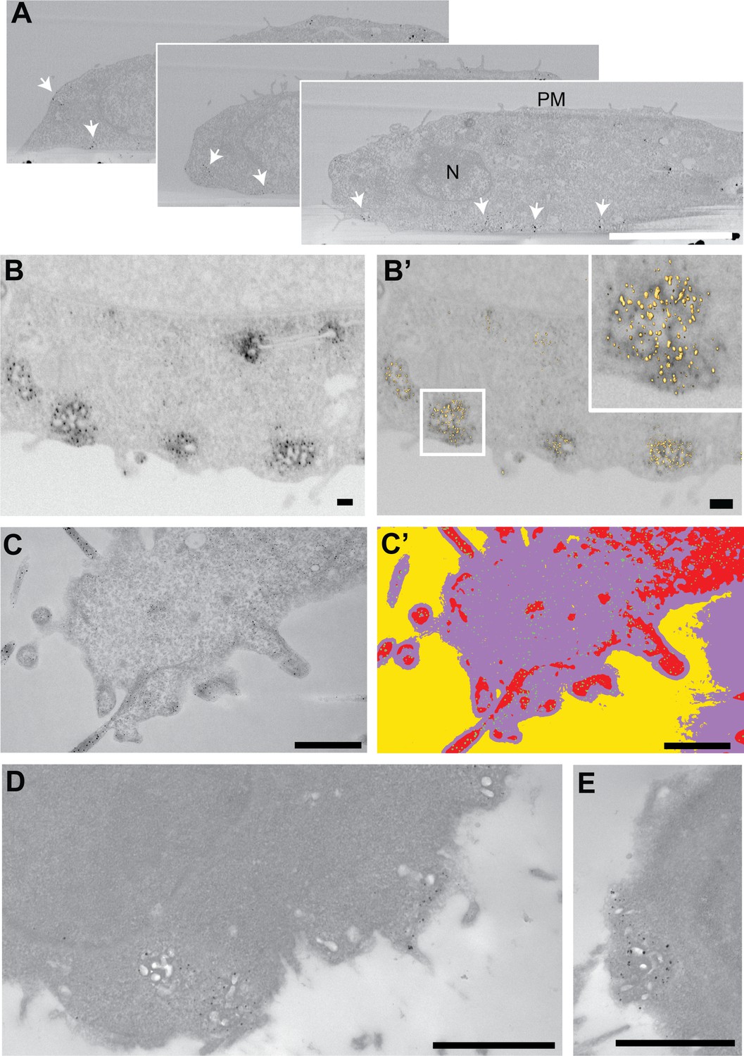
APEX-Gold is compatible with serial block-face SEM, FIB-SEM, image segmentation analysis and pre-embedding labelling techniques.
(A) Serial micrographs of Cavin4-APEX2 labelling obtained using serial block-face imaging scanning electron microscope (SEM). Arrows indicate areas of APEX-Gold labelling. (B–B’) Focussed ion beam (FIB) analysis of same experiment followed by supervised segmentation. (C) Actin filaments labelled with LifeAct-APEX2 followed by APEX-Gold enhancement. (C’) Segmentation of the diaminobenzidine (DAB) product and the gold labelling using FIJI/Weka automated segmentation of the image shown in C. (D,E) Fast-frozen and freeze-substituted cells embedded in lowicryl showing Cavin4-APEX2 labelling. Bars, A, 5 µm; B,B’, 200 nm; C,C’, 1 µm; D, 1 µm; E, 500 nm.
Figure 1—figure supplement 5
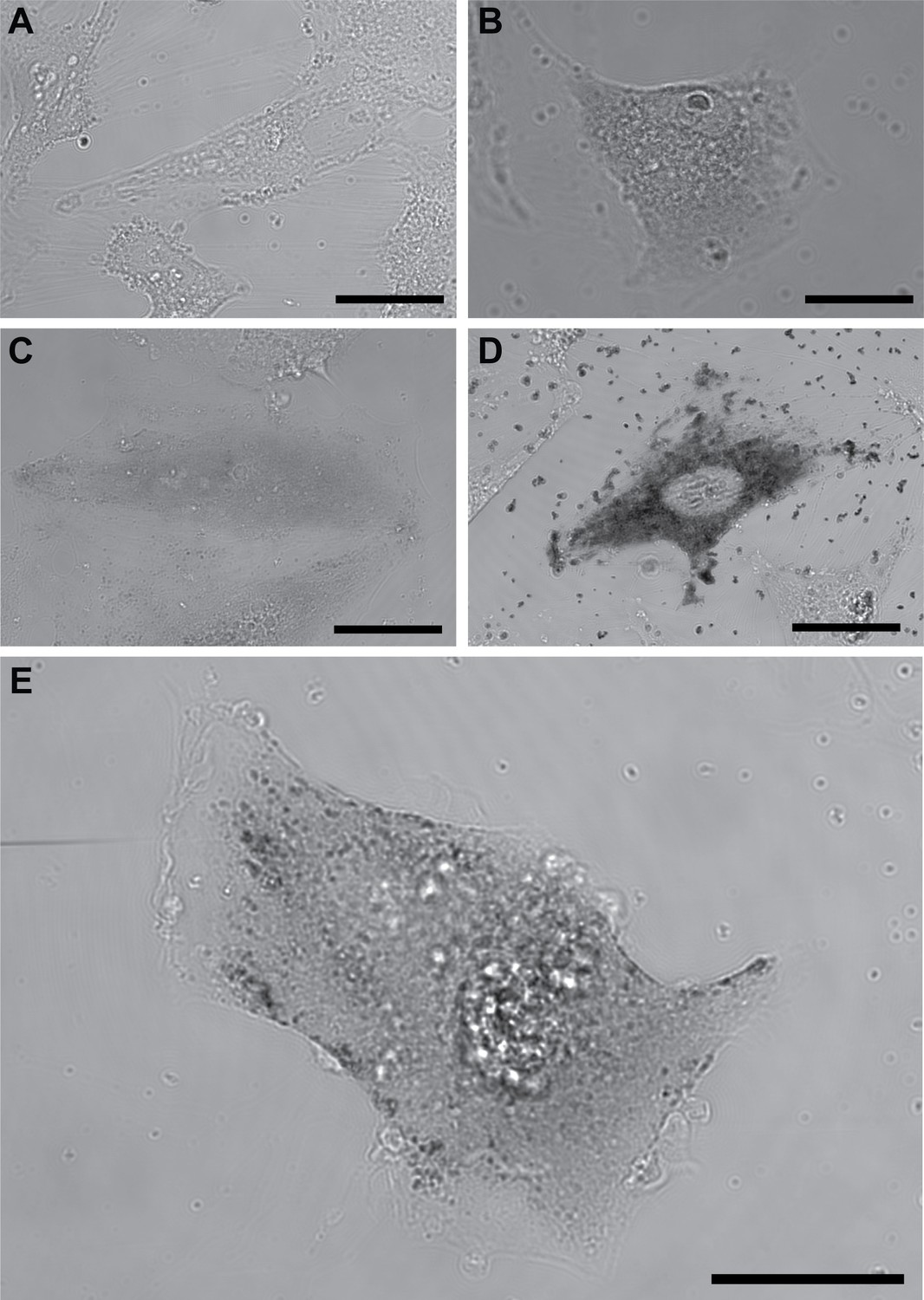
Brightfield images of BHK cells with various treatments.
(A) Untransfected BHK cell treated with diaminobenzidine (DAB), Ag/Au, and gum arabic. (B) BHK cell transfected with Cavin4-APEX2 treated with DAB. (C) BHK cell transfected with Cavin4-APEX2 treated with DAB, Ag, and gum arabic. (D) BHK cell transfected with Cavin4-APEX2 treated with DAB and Ag/Au. (E) BHK cell transfected with Cavin4-APEX2 treated with DAB, Ag/Au, and gum arabic. Bars, A-E, 50 µm.
Figure 2 with 3 supplements
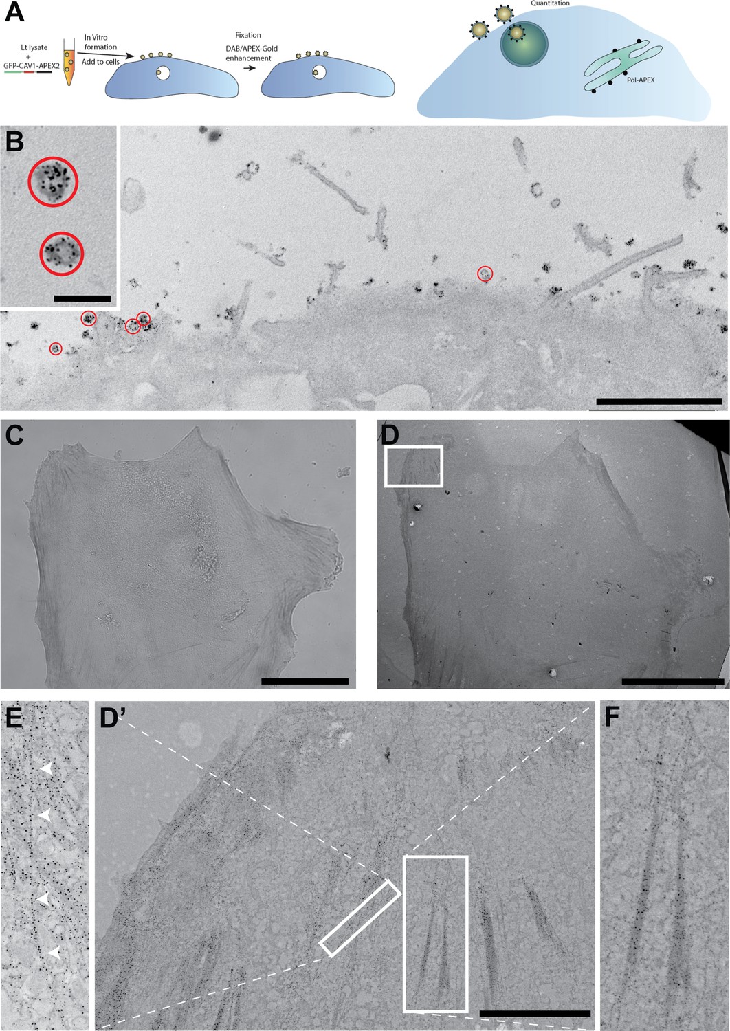
APEX-fusion protein density determination using an internal standard and low level protein detection.
(A) Schematic explaining the cell-free caveolae-APEX2-Gold system. (B) A431 cells were incubated with in vitro synthesized CAV1-APEX2 cell-free caveolae for 5 min at 37°C before fixation and processing for APEX-Gold detection. Note the Ag/Au labelling of the surface-associated cell-free caveolae circled in red and low background label within cell. (C) Light microscopic detection of tropomyosin 3.1 (Tpm3.1)-APEX2 after APEX-Gold DAB/Ag/Au detection. (D) Low magnification electron microscopy (EM) showing a basal section of the same cell. (D’,F) Higher magnification views of gold labelled stress fibres from boxed region. (D’,E) Gold labelling follows individual actin filaments from boxed region. Bars, B, 2 µm (inset 200 nm); C,D, 50 µm; D’, 5 µm.
Figure 2—figure supplement 1
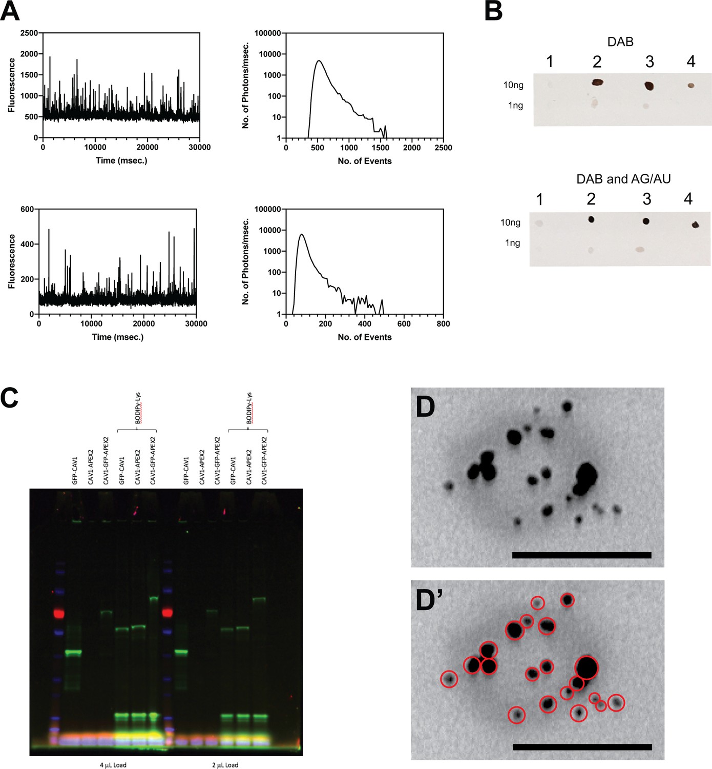
Cell free GFP-Cav1-Apex2 vesicle characterisation.
(A) Fluorescence correlation spectroscopic (FCS) analysis of cell-free synthesized GFP-CAV1-APEX2. (B) Dot blots comparing two concentrations of the indicated in vitro synthesized proteins (1; GFP-CAV1, 2; CAV1-APEX2, 3; GFP-CAV1-APEX2) or commercial horseradish peroxidase (4; HRP) treated with diaminobenzidine (DAB) or DAB and Ag/Au. (C) Expression of the indicated constructs followed by in-gel fluorescence detection using the GFP fluorescence or with fluorescently labelled lysine (BODIPY-lysine). (D) High magnification example micrograph of a single in vitro synthesized CAV1-APEX2 cell-free vesicle with APEX-Gold processing. (D’) Ag/Au particles circled in red. Bars, D,D’, 100 nm.
Figure 2—figure supplement 2
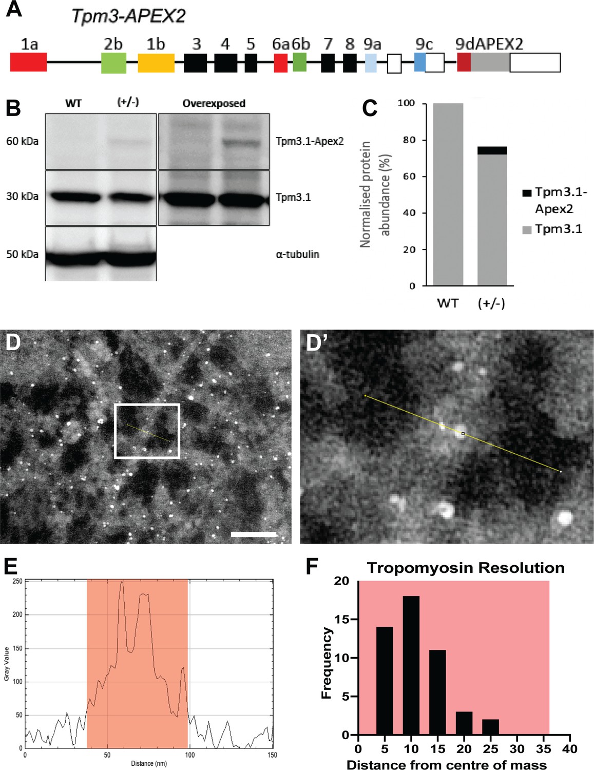
Low abundance Tpm3.1 labelling highlights APEX-Gold sensitivity and resolution.
(A) Genomic organization of the mouse line expressing tropomyosin 3.1 (Tpm3.1) C-terminally tagged with APEX2 (Meiring et al., 2019). (B) Western blot detection and (C) quantitation of endogenous and APEX2-tagged Tpm3.1. (D,D’) Tpm3.1-APEX2 labelling enhanced with APEX-Gold, also analysed with linescans to measure distance from particles centre to the actin bundle centre of mass. (E) Representative output from linescan showing edges of bundle highlighted in orange and the two peaks within indicative of APEX-Gold. (F) The distribution of Tpm3.1-APEX2 resolution, and half the average width of bundles highlighted in orange. Bars, D, 50 nm.
Figure 2—figure supplement 3
Tpm3.1 APEX-Gold labelling.
Arrows, particle labelling following individual actin filaments. Bars, A, 2 µm; B, 1 µm; C-I, 2 µm.
Tables
Key resources table
| Reagent type (species) or resource | Designation | Source or reference | Identifiers | Additional information |
|---|---|---|---|---|
| Strain, strain background (Leishmania tarentolae) | LEXSY host P10 | Jena Biosciences | LT-101 | |
| Genetic reagent (Mus musculus) | Tpm3.1-APEX2 ± heterozygous | PMID:31331962 | ||
| Cell line (Mesocricetus auratus) | BHK-21 | ATCC | CCL-10 | |
| Cell line (Homo sapiens) | A-431 | ATCC | CRL-1555 | |
| Cell line (Cricetulus griseus) | Flp-In-CHO | Invitrogen | R75807 | |
| Transfected construct (synthetic) | LifeAct-APEX2 | This study | RRID:170523 | |
| Transfected construct (Mus musculus) | Cavin4-APEX | This study | RRID:170524 | |
| Transfected construct (synthetic) | pCSDEST2 | PMID:17948311 | RRID:22424 | |
| Transfected construct (synthetic) | p3E-APEX2 | PMID:29621251 | RRID:108894 | |
| Transfected construct (synthetic) | p3E-APEX2-P2A-mKate2 | PMID:26585296 | RRID:61671 | |
| Transfected construct (Mus musculus) | pME-CAV1 | This study | RRID:170527 | |
| Transfected construct (synthetic) | pME-LifeAct | PMID:32709891 | RRID:109545 | |
| Transfected construct (Mus musculus) | pME-Cavin4 | This study | RRID:170528 | |
| Transfected construct (Homo sapiens) | A1AR-APEX2 | This study | RRID:170529 | |
| Antibody | Anti-tropomyosinthree mouse monoclonal | Sigma-Aldrich | MABT1335 | (1:1000) |
| Antibody | Anti-alpha tubulin rabbit monoclonal | Abcam | Ab52866 | (1:3000) |
| Recombinant DNA reagent | GFP-CAV1-APEX (cell-free) | This study | RRID:170525 | |
| Recombinant DNA reagent | CAV1-APEX (cell-free) | This study | RRID:170526 | |
| Recombinant DNA reagent | pCellFree_G03 | PMID:25529348 | ||
| Recombinant DNA reagent | pCellFree_G03 | PMID:25529348 | ||
| Sequence-based reagent | Antisplice leader oligonucleotide | PMID:19648909 | CAATAAAGTACAGAAACTGATACTTATATAGCGTT | |
| Commercial assay or kit | FluoroTect GreenLys in vitro Translation Labeling System | Promega | L5001 | |
| Commercial assay or kit | MycoAlert Mycoplasma Detection Kit | Lonza | LT07-418 | |
| Chemical compound, drug | 25% EM grade glutaraldehyde | Electron Microscopy Services | 16220 | |
| Chemical compound, drug | 16% EM grade paraformaldehyde | Electron Microscopy Services | 15710 | |
| Chemical compound, drug | Uranyl acetate | Electron Microscopy Services | 22400 | |
| Chemical compound, drug | Lead citrate | ProSciTech | C073 | |
| Chemical compound, drug | DAB | Sigma-Aldrich | D5905 | |
| Chemical compound, drug | Hydrogen peroxide solution | Sigma-Aldrich | H1009 | |
| Chemical compound, drug | Osmium tetroxide | ProSciTech | C010 | |
| Chemical compound, drug | LX 112 Embedding Kit | Ladd Research Industries | 21210 | |
| Chemical compound, drug | Horseradish peroxidase – 25 mg Type VI-A | Sigma-Aldrich | P6782 | |
| Chemical compound, drug | Silver nitrate | Sigma-Aldrich | 209139 | |
| Chemical compound, drug | Gum arabic | Electron Microscopy Services | 25574 | |
| Chemical compound, drug | Gold chloride | Electron Microscopy Services | 16583 | |
| Chemical compound, drug | Hexamethylenetetramine | Sigma-Aldrich | 398160 | |
| Chemical compound, drug | Sodium tetraborate decahydrate (borax) | Sigma-Aldrich | B9876 | |
| Chemical compound, drug | Sodium thiosulphate | Sigma-Aldrich | 72049 | |
| Chemical compound, drug | Bactotryptone | Beckton Dickinson | 211699 | |
| Chemical compound, drug | Hemin chloride | MP Biomedicals | 0219402505 | |
| Chemical compound, drug | ATP | Chem-Impex | 00015 | |
| Chemical compound, drug | GTP | Chem-Impex | 00348 | |
| Chemical compound, drug | Spermidine | Sigma-Aldrich | 85558–5G | |
| Chemical compound, drug | DTT | Sigma-Aldrich | D0632-10G | |
| Chemical compound, drug | Cr phosphate | Chem-Impex | 00072 | |
| Chemical compound, drug | PEG 3350 | Hampton Research | HR2-527 | |
| Chemical compound, drug | Prot Inhib C | Roche Diagnostics | 11 873 580 001 | |
| Chemical compound, drug | CTP | Chem-Impex | 00095 | |
| Chemical compound, drug | UTP | Chem-Impex | 00311 | |
| Chemical compound, drug | T7 polymerase | In-house purification | N/A | |
| Chemical compound, drug | Cr phosphokinase | Sigma-Aldrich | C3755-35KU | |
| Chemical compound, drug | Alanine | Sigma-Aldrich | A7627 | |
| Chemical compound, drug | Arginine | Sigma-Aldrich | A5006 | |
| Chemical compound, drug | Asparagine | Sigma-Aldrich | A0884 | |
| Chemical compound, drug | Aspartic acid | Sigma-Aldrich | A9256 | |
| Chemical compound, drug | Cysteine | Sigma-Aldrich | C7352 | |
| Chemical compound, drug | Glutamic acid | Sigma-Aldrich | 49449 | |
| Chemical compound, drug | Glutamine | Sigma-Aldrich | G3126 | |
| Chemical compound, drug | Glycine | Sigma-Aldrich | G7126 | |
| Chemical compound, drug | Histidine | Sigma-Aldrich | H8000 | |
| Chemical compound, drug | Isoleucine | Sigma-Aldrich | I2752 | |
| Chemical compound, drug | Leucine | Sigma-Aldrich | L8912 | |
| Chemical compound, drug | Lysine | Sigma-Aldrich | L5626 | |
| Chemical compound, drug | Methionine | Sigma-Aldrich | M9625 | |
| Chemical compound, drug | Phenylalanine | Sigma-Aldrich | P2126 | |
| Chemical compound, drug | Proline | Sigma-Aldrich | P0380 | |
| Chemical compound, drug | Serine | Sigma-Aldrich | S4500 | |
| Chemical compound, drug | Threonine | Sigma-Aldrich | T8625 | |
| Chemical compound, drug | Tryptophan | Sigma-Aldrich | T0524 | |
| Chemical compound, drug | Tyrosine | Sigma-Aldrich | T8566 | |
| Chemical compound, drug | Valine | Sigma-Aldrich | V0500 | |
| Chemical compound, drug | Penicillin-streptomycin | Life Technologies | 15070–063 | |
| Chemical compound, drug | Potassium acetate | Sigma-Aldrich | P1190 | |
| Chemical compound, drug | Magnesium acetate | Amresco | 0131–1 KG | |
| Chemical compound, drug | LR clonase II Plus | Invitrogen | 12538120 | |
| Chemical compound, drug | Hygromycin-B | Thermo Fisher/Invitrogen | 10687010 | |
| Chemical compound, drug | DMEM | Life Technologies | 11995065 | |
| Chemical compound, drug | L-glutamine | Life Technologies | 25030081 | |
| Chemical compound, drug | Fetal bovine serum | Sigma-Aldrich | F9423 | |
| Software, algorithm | ImageJ/FIJI | PMID:22743772 | https://imagej.nih.gov/ij/ | |
| Software, algorithm | ImageJ/FIJI Weka Plugin | ImageJ developers | https://imagej.net/Trainable_Segmentation | |
| Software, algorithm | iTEM | Olympus | https://www.emsis.eu/home/ | |
| Software, algorithm | AttoBright: LabView and GUI for data acquisition, Matlab code and GUI for data analysis | PMID:31827096 | https://gambinsiereckilab.github.io/AttoBright/ | |
| Software, algorithm | MAPs | Thermo Fisher | https://www.fei.com/software/maps/#gsc.tab=0 |
Additional files
Download links
A two-part list of links to download the article, or parts of the article, in various formats.
Downloads (link to download the article as PDF)
Open citations (links to open the citations from this article in various online reference manager services)
Cite this article (links to download the citations from this article in formats compatible with various reference manager tools)
A robust method for particulate detection of a genetic tag for 3D electron microscopy
eLife 10:e64630.
https://doi.org/10.7554/eLife.64630


