Studying evolution of the primary body axis in vivo and in vitro
- EMBL Barcelona, Spain
- EMBL Heidelberg, Developmental Biology, Germany
Figures
Figure 1
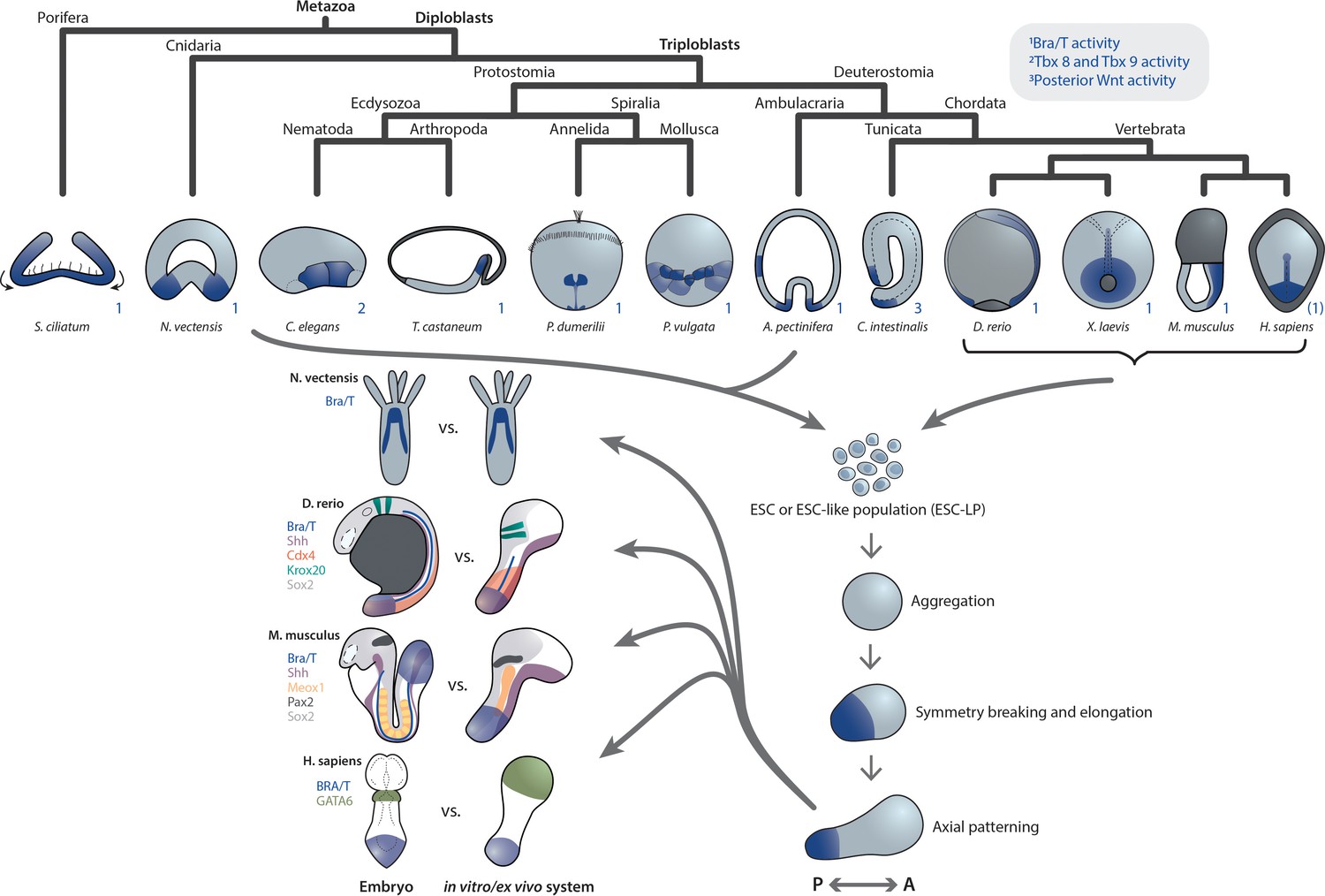
Primary axis formation during gastrulation in metazoan embryos and corresponding artificial systems.
Metazoan embryos around the gastrulation or an equivalent developmental phase exhibit distinct morphologies and associated overall tissue rearrangement dynamics. Yet, during this event they universally specify at least a primary body axis, demarcated by conserved expression of posterior (or oral in cnidarians) patterning determinants. Among them are T-box and Wnt genes, localized transcriptional activity of which is highlighted in blue. Note that marker gene expression patterns in the human embryo are speculative. Research efforts have shown that ESC or ESC-like populations from species across the animal kingdom can be (re-)aggregated in vitro and, although lacking the respective external environment and associated developmental cues, remain capable of recapitulating at least a basic transcriptional body plan with an anteroposterior (AP) and oral-aboral (OA) axis, respectively. Strikingly, comparison of examples of such in vivo or ex vivo systems highlights a remarkable overall similarity despite the varying geometry of the respective native embryo. This may point toward the existence of a conserved developmental mode that cells exhibit when released from their species-specific extraembryonic environment.
Figure 2

Diverse developmental trajectories accessible to ESC(-LP)s in the presence and absence of extraembryonic inputs.
Compared to the native embryo, (re-) aggregation of ESC-LPs reveals potentially alternative developmental modes to the same body plan. Evidence for this has been found in Nematostella reaggregates and mGastruloids (Kirillova et al., 2018; Hashmi et al., 2020; Vianello and Lutolf, 2020). (i)ETX embryos (Sozen et al., 2018; Zhang et al., 2019; Amadei et al., 2021) from mESCs, TSCs and XEN cells could point toward the existence of such an alternative mode, as they form structures resembling the actual gastrulating mouse embryo without closely mimicking blastocyst morphology (stages E3.5–4.5) earlier during their development.
Figure 3
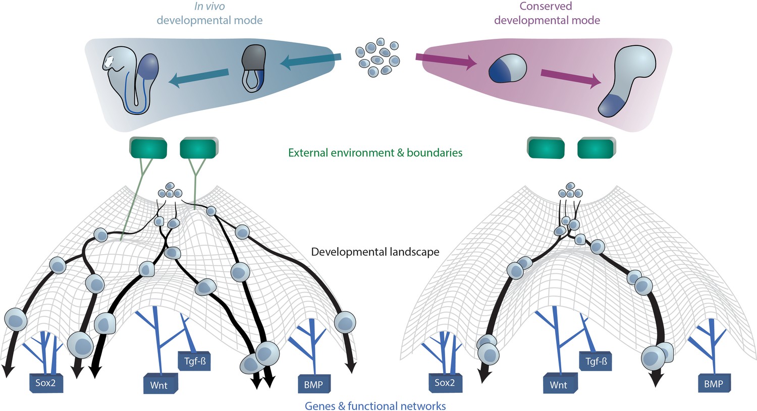
A conserved developmental mode emerges upon removal of species-specific extraembryonic environments.
The developmental trajectories which aggregates of ESC(-LP)s exhibit upon removal of external or extraembryonic and associated boundaries may constitute a conserved mode that is shared across species. On a cellular level, this can be visually approximated as cells undergoing differentiation within Waddington’s developmental landscape Waddington, 1957. The landscape is shaped by key gene networks which remain constant between species and in vitro (bottom) as well as the external (micro-)environment and embryo geometry (top), here represented as green tiles, which vary between species. In case the latter factors are not present as ESCs are removed from their native context and grown in vitro, cellular developmental trajectories revert to the aforementioned conserved mode since cells from different species now experience the same landscape.
Download links
A two-part list of links to download the article, or parts of the article, in various formats.
Downloads (link to download the article as PDF)
Open citations (links to open the citations from this article in various online reference manager services)
Cite this article (links to download the citations from this article in formats compatible with various reference manager tools)
Studying evolution of the primary body axis in vivo and in vitro
eLife 10:e69066.
https://doi.org/10.7554/eLife.69066


