A novel live-cell imaging assay reveals regulation of endosome maturation
- Biozentrum, University of Basel, Switzerland
Figures
Figure 1 with 6 supplements
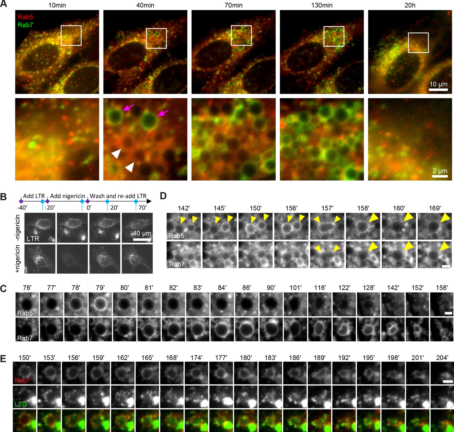
Rab conversion and completion of endolysosomal stages of endosome maturation can be observed in enlarged endosomes, induced with short nigericin treatment.
Nigericin was added to HeLa cells at 10 μM for 20 min and washed away, and cells were imaged by time-lapse microscopy. Recovery times are specified relative to removal of nigericin. (A,C,D) Cells stably expressing mApple-Rab5 and GFP-Rab7. (A) Images to show enlarged Rab5- (white arrows) and Rab7- (magenta arrows) positive compartments and return to normal morphology by 20 hr. (B) Lysotracker Red (LTR) was added to cells before, during and following nigericin treatment. Images show rapid re-accumulation of Lysotracker in treated cells (bottom row). Untreated cells were tracked in parallel (upper row). (C) The enlarged endosome was selected to show Rab5 recruitment, Rab conversion and endolysosomal maturation. (D) Example of homotypic fusion of two Rab5-positive endosome and subsequent Rab5 removal/weak Rab7 recruitment. (E) Cells stably expressing mApple-Rab7 were imaged in the presence of Lysotracker Green. An enlarged Rab7-positive endosome was selected to show accumulation of Lysotracker concomitant with the loss of spherical shape and a reduction in size of the maturing endolysome. (C–E) Scale bar = 2 μm. Time-lapse videos of the endosomes in (C–E) at 1 min interval are available in Figure 1—video 1, Figure 1—video 2, Figure 1—video 3, respectively.
Figure 1—figure supplement 1
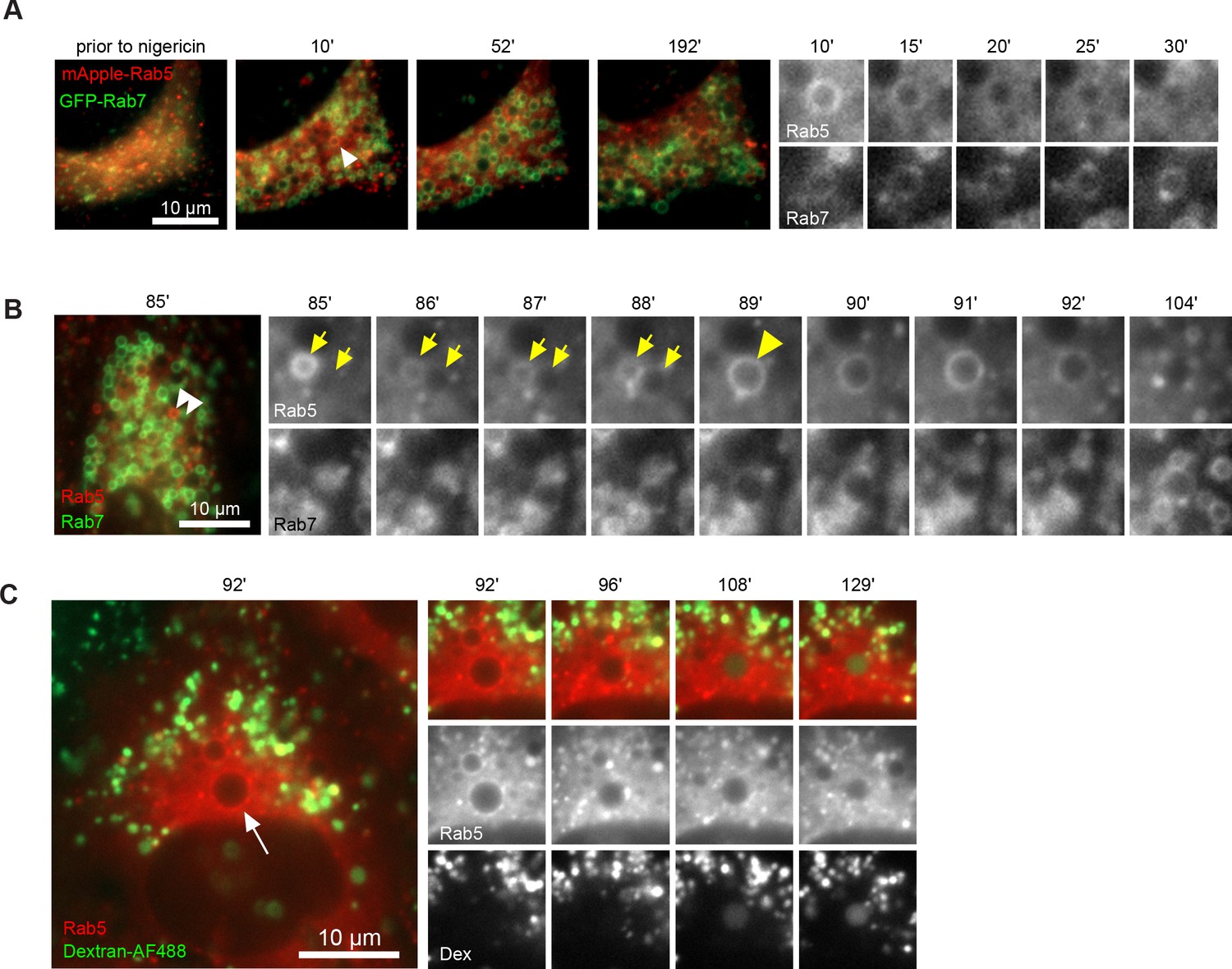
Enlarged compartments interact with early endosomes and acquire intraluminal content from the endolysosomal pathway.
Nigericin was added to HeLa cells, stably expressing mApple-Rab5 and GFP-Rab7 (A,B) or mApple-Rab5 alone (C), for 20 min and washed away, and cells were imaged by time-lapse microscopy. Recovery times are specified relative to removal of nigericin. (A) Example to show asynchronous nature of the transient Rab5-positive endosomes together with the closeup of a Rab5-positive endosome undergoing Rab conversion shortly after nigericin washout. (B) Example to show an enlarged compartment devoid of Rab5 fusing with Rab5-positive endosome to subsequently undergo Rab conversion. (C) Example to show an enlarged Rab5-positive endosome later acquiring Dextran-AF488, which had been pulse-chased into the endolysosomal pathway (4 hr pulse, 1.5 hr chase) prior to nigericin treatment. Recovery times are specified relative to removal of nigericin.
Figure 1—figure supplement 2
Enlarged endosomes that undergo Rab conversion can be induced by different treatments.
HeLa cells stably expressing mApple-Rab5 and GFP-Rab7 were treated for 4 hr with ammonium chloride (A) or for 20 min with monensin (B), washed and imaged at specified times after the wash. Representative images of endosomes to show Rab conversion. Homotypic fusion of two Rab5-positive endosomes is evident in (B).
Figure 1—figure supplement 3
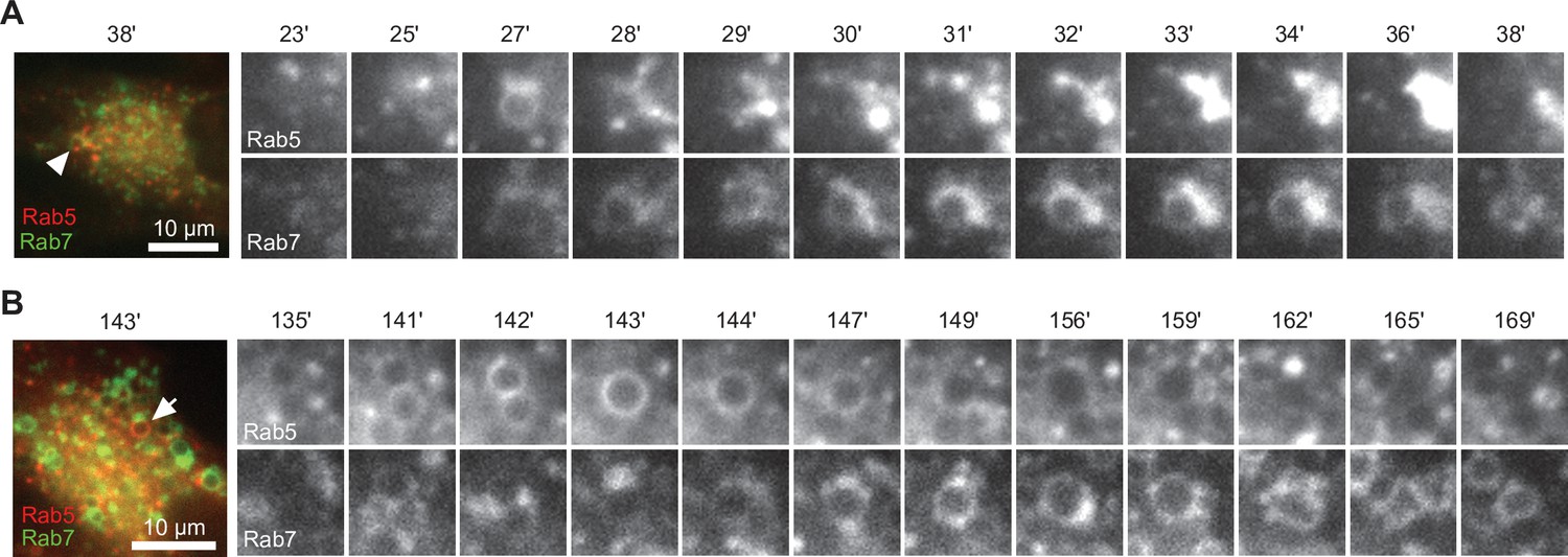
Enlarged endosomes that undergo Rab conversion can be induced in different cell types.
Cells were transiently transfected with mApple-Rab5 and GFP-Rab7, and treated 20 min with nigericin followed by recovery and time-lapse microscopy. (A) HEK293 cells, (B) Neuro2A cells, (C) Cos-1 cells. Images were selected to show Rab conversion in the enlarged endosomes.
Figure 1—video 1
Rab5 recruitment, Rab conversion and endolysosomal maturation.
Figure 1—video 2
Homotypic fusion of two Rab5-positive endosome and subsequent Rab5 removal/weak Rab7 recruitment.
Figure 1—video 3
An enlarged Rab7-positive endosome was selected to show accumulation of Lysotracker concomitant with the loss of spherical shape and a reduction in size of the maturing endolysosome.
Figure 2 with 1 supplement
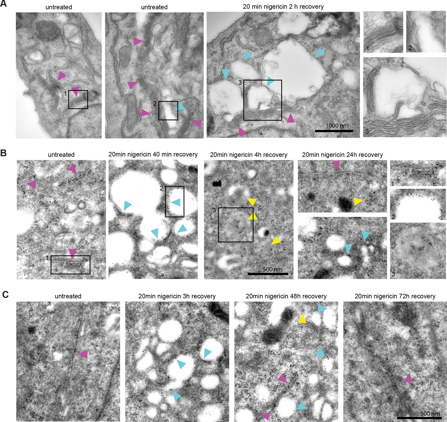
Nigericin-induced enlarged compartments originate at the Golgi and contain trans-Golgi marker GalT, later found in ILVs, with most enlarged compartments resolved by 48 hr.
Nigericin was added to HeLa cells for 20 min and washed away, and cells were processed for electron microscopy (A–C), imaged by time-lapse microscopy (D) or harvested for counting (E) at specified times after the wash. (A) Cells stained with osmium tetroxide and potassium hexacyanoferrate reveal large spherical compartments (cyan arrows) originating at the trans-face of the Golgi (magenta arrows) in nigericin-treated cells and, occasionally, in untreated cells. (B,C) Cells stably expressing GalT-GFP were stained with anti-GFP and 12 nM Gold-conjugated secondary antibody to reveal GalT-GFP at the Golgi (magenta arrows), the limiting membrane of the enlarged compartments (cyan arrows) as well as in ILVs of the enlarged MVBs at later time points (yellow arrows).
Figure 2—figure supplement 1
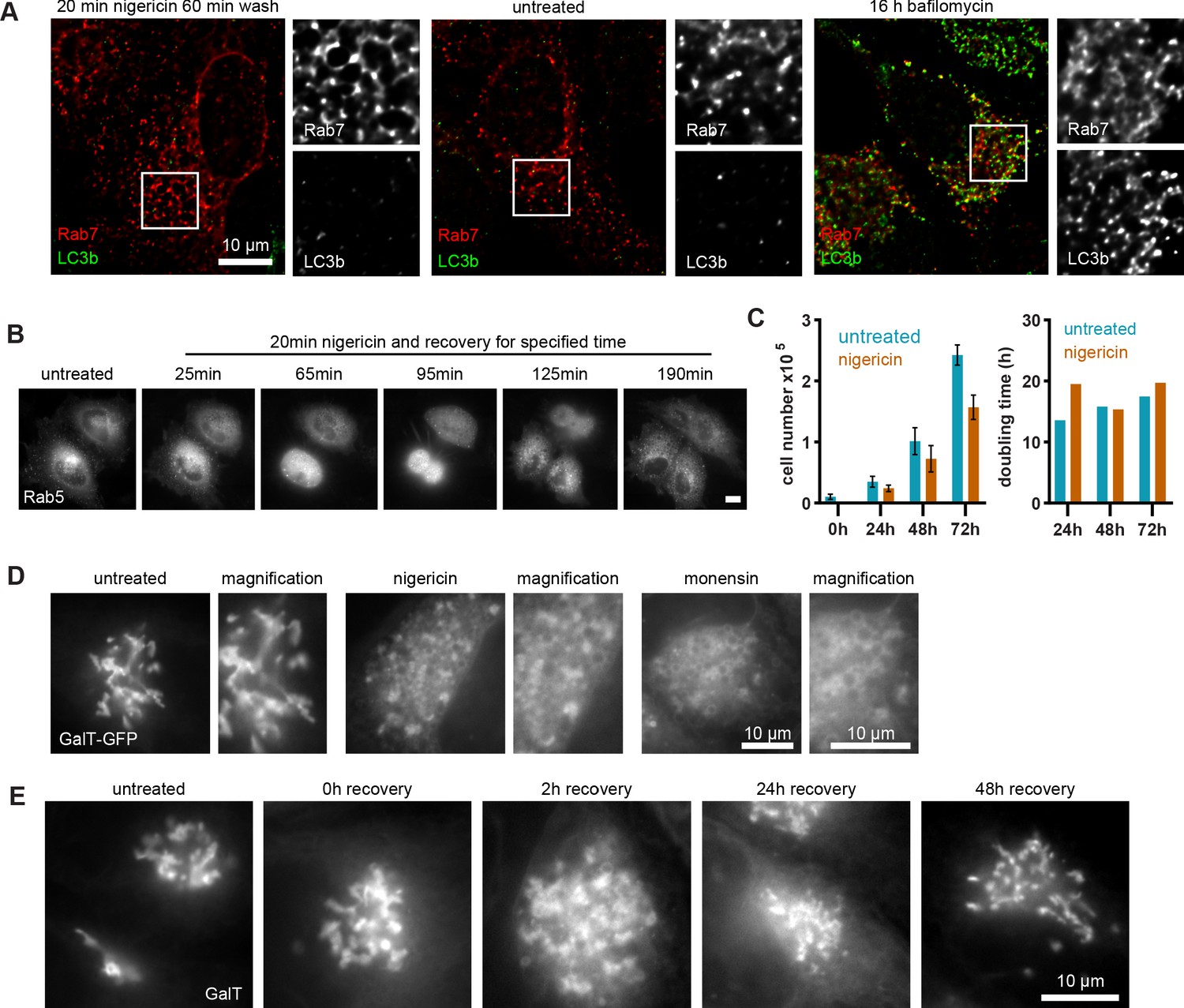
Cell growth is unaffected by nigericin treatment, which induces reversible Golgi vesiculation HeLa cells stably expressing mApple-Rab5 (A,B) or GalT-GFP (C–E) were treated for 20 min with 10 μM nigericin or 5 μM monensin, or left untreated.
(A) Images of cells stained with LC3b antibody to show absence of autophagy induction or presence of LC3B at the enlarged endosomes of nigericin-treated cells. Bafilomycin-treated cells are shown here as positive control. (B) Images to show cells dividing shortly after nigericin treatment, with recovery times specified. Scale bar = 10 μm. (C) Cells were plated in 12-well plates at 104 cells per well 24 hr prior to nigericin treatment, trypsinised and counted at specified recovery times. Triplicate wells were counted per time point per condition. Actual cells numbers and doubling times are presented. Representative graph of three independent experiments. Source data is available in Figure 2—figure supplement 1—source data 1. (D) Images to reveal extensive formation of GalT-positive enlarged compartments upon either treatment following 2 hr recovery. (E) Images to show Golgi vesiculation and return to ribbon morphology within 48 hr of recovery from nigericin treatment.
-
Figure 2—figure supplement 1—source data 1
Cell growth numbers and quantification post nigericin treatment.
- https://cdn.elifesciences.org/articles/70982/elife-70982-fig2-figsupp1-data1-v1.xlsx
Figure 3 with 3 supplements
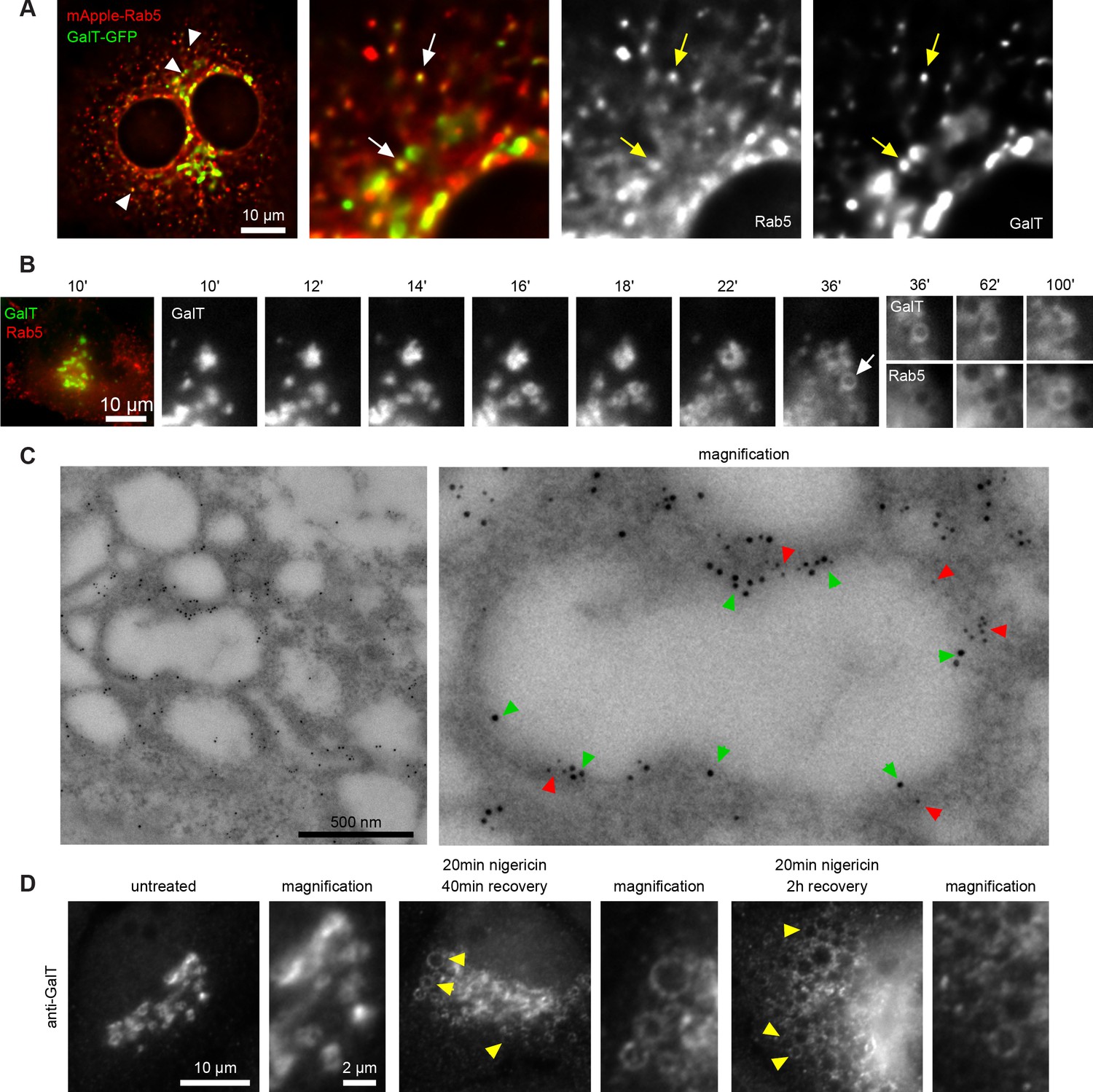
Short nigericin treatment leads to trans-Golgi vesiculation and subsequent acquisition of Rab5.
(A) Images of untreated HeLa cells stably expressing mApple-Rab5 and transiently expressing GalT-GFP. Arrows point to puncta positive for both markers. (B–D) Nigericin was added to HeLa cells for 20 min and washed away, and cells were imaged by time-lapse microscopy (B), processed for electron microscopy (C) or for immunofluorescence (D) at specified times after the wash. (B,C) HeLa cells stably expressing GalT-GFP and transiently transfected with mApple-Rab5. (B) Representative kinetic of Golgi vesiculation post nigericin treatment as visualised with GalT-GFP. The selected vesiculated compartment (arrow), initially negative for Rab5 subsequently becomes positive for both markers. A time-lapse video of the endosome at 2 min interval is available in Figure 3—video 1. (C) Immuno-EM image of a cell at 2 hr post recovery, with 12 nm Gold-labelled GFP (green arrows) and 5 nm Gold-labelled mApple (red arrows) present at the enlarged compartments. Scale bar = 500 nm. (D) Images of cells stained with anti-GalT to reveal endogenous GalT presence at the enlarged compartments.
Figure 3—figure supplement 1
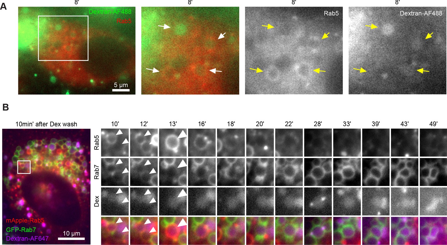
Endocytic cargo contributes to enlarged early endosome composition.
(A) HeLa cells stably expressing mApple-Rab5 were treated with nigericin, washed and 60 min later treated with 2.5 mg/mL Dextran-AF488 for 20 min. Cells were imaged at indicated times after washing away the Dextran. Images of a representative cell with one Rab5-negative and two of three Rab5-positive enlarged compartments containing endocytosed Dextran. (B) HeLa cells stably expressing mApple-Rab5 and GFP-Rab7 were treated with nigericin, washed and 75 min later treated with 1 mg/mL Dextran-AF647 for 20 min. Cells were imaged at indicated times after washing away the Dextran. Kinetic to show fusion of a Rab5-negative enlarged compartment devoid of Dextran with a large Rab5-positive endosome containing endocytosed Dextran, and the resulting enlarged early endosome acquiring further Dextran, undergoing Rab conversion and maturing into an endolysosome still containing the Dextran.
Figure 3—figure supplement 2
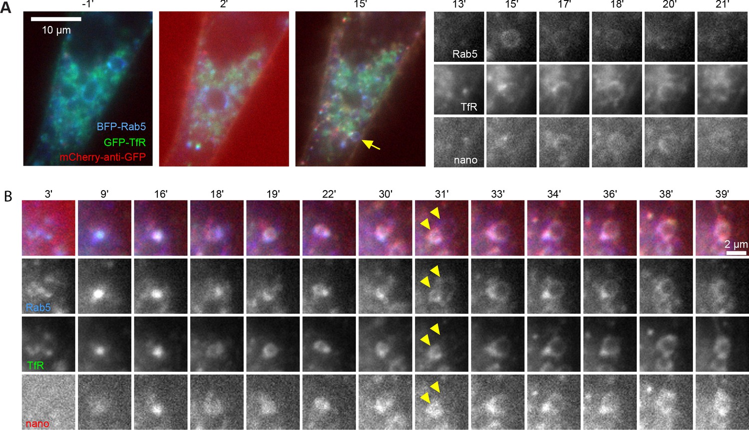
Endocytosed plasma membrane transferrin receptor contributes to enlarged early endosome composition.
HeLa cells transiently transfected with mTagBFP2-Rab5 and TfR-EGFP were treated with nigericin and washed, with mCherry- anti-GFP nanobody added 110 min later to label surface TfR. Times are relative to nanobody addition. (A) Kinetic to show uptake of nanobody, and concomitant acquisition of Rab5 and surface-derived TfR by the endosome. (B) Kinetic to show a Rab5- and surface-derived TfR-positive endosome fuse with the enlarged compartment (arrows) to form an enlarged endosome positive for both markers.
Figure 3—video 1
Kinetic to show fusion of a Rab5 negative enlarged compartment devoid of Dextran with a large Rab5 positive endosome containing endocytosed Dextran.
Figure 4 with 1 supplement
Enlarged endosomes recruit Rab5 and undergo Rab conversion with anticipated kinetics.
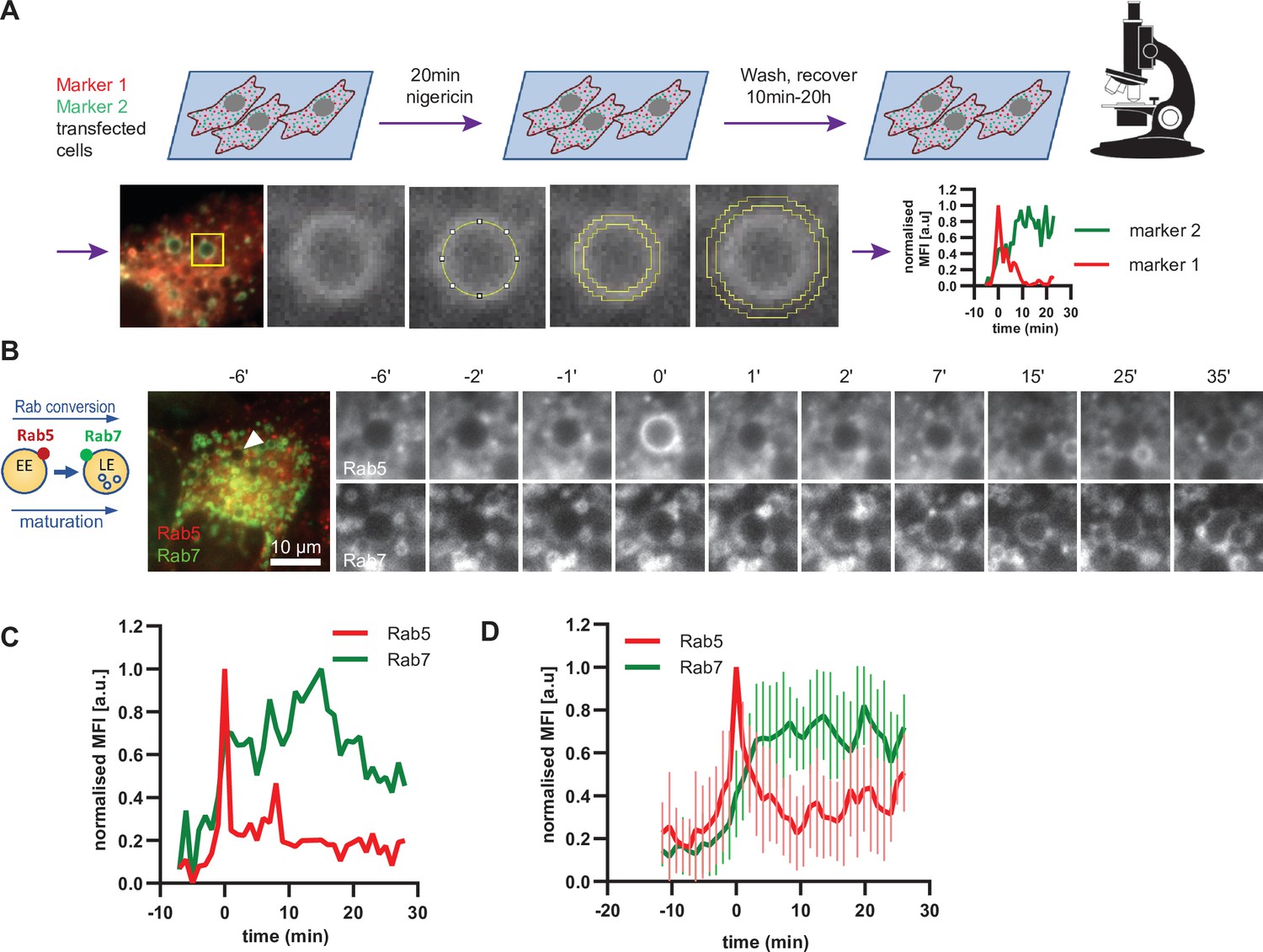
(A) Scheme to show experimental flow, starting with transfection of cells of choice with selected markers, followed by short nigericin treatment, and time-lapse microscopy during the recovery phase, with subsequent quantification of mean fluorescence intensity (MFI) of the chosen markers at the rim of the enlarged endosomes, and the resulting kinetic plots of background-subtracted MFI normalised for maximum and minimum values over the entire time course of the endosome. Since endosome maturation is asynchronous, relative time is calculated by using Rab5 peak as a reference for Rab conversion and set to t = 0. The plot shown in the scheme represents the kinetic of the images in Figure 1C (marker one as Rab5 and marker two as Rab7).(B,C,D) HeLa cells, stably expressing mApple-Rab5 and GFP-Rab7 were treated for 20 min with nigericin, washed and imaged over a 3 hr period.(B) Time-lapse images of a representative endosome to show transient Rab5 recruitment and its subsequent displacement by Rab7. (C) Corresponding graph of MFI of Rab5 and Rab7 at the rim of the endosome in (B) during and around the time of Rab conversion. Numerical data for all analyzed endosomes is available in Figure 4—source data 1. (D) Averaged Rab5 and Rab7 kinetics of 27 endosomes. Error bars represent standard deviation. Representative graph of three independent experiments.
-
Figure 4—source data 1
Quantification of Rab5 and Rab7 recruitment at endosomes.
- https://cdn.elifesciences.org/articles/70982/elife-70982-fig4-data1-v1.xlsx
Figure 4—figure supplement 1
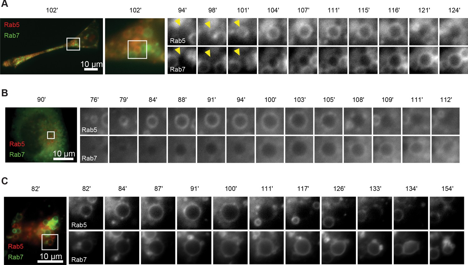
Enlarged endosomes occasionally display multiple Rab5 peaks and form Rab5 subdomains.
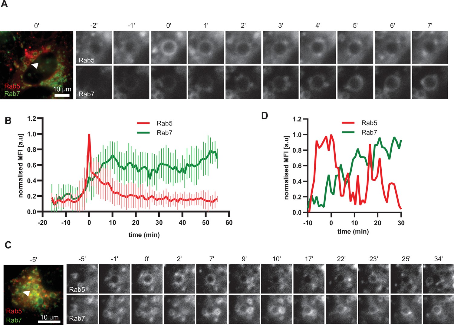
HeLa cells, stably expressing mApple-Rab5 and GFP-Rab7 were treated for 20 min with nigericin, washed and imaged over 3 hr, as described in Figure 4A. (A) Time-lapse images of an endosome displaying formation of Rab5 subdomains. (B) Averaged Rab5 and Rab7 kinetics of 21 endosomes. Error bars represent standard deviation. Representative graph of three independent experiments. (C–D) Time-lapse images (C) and a corresponding fluorescence quantification plot (D) of an endosome with multiple waves of Rab5 recruitment and a continuous increase in Rab7 levels.
Figure 5 with 2 supplements
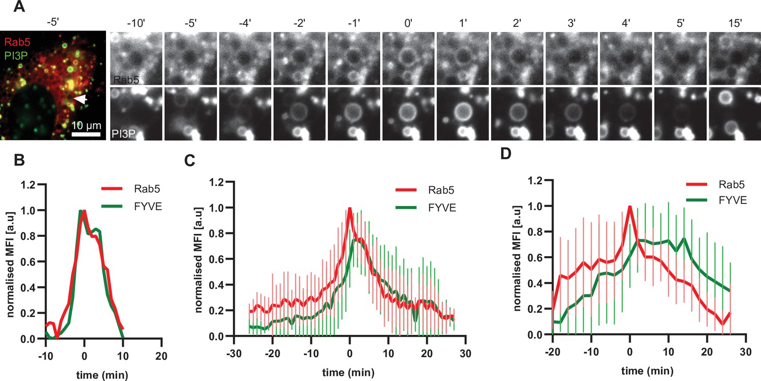
PI(3)P is recruited to endosomes concomitantly with Rab5.
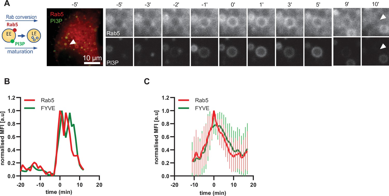
HeLa cells, stably expressing mApple-Rab5 and transiently transfected with the PI(3)P marker, GFP-FYVE, were treated for 20 min with nigericin, washed and imaged over 3 hr, as described in Figure 4A. (A) Time-lapse images of a representative endosome to show transient Rab5 recruitment accompanied by PI(3)P. A time-lapse video of the endosome at 1 min interval is available in Figure 5—video 1. (B) Corresponding graph of normalised mean fluorescence intensity of Rab5 and FYVE at the rim of the endosome in (A) over the time the endosome was detectable. (C) Averaged Rab5 and PI(3)P kinetics of 19 endosomes. Error bars represent standard deviation. Representative graph of three independent experiments. Numerical data for all analyzed endosomes is available in Figure 5—source data 1.
-
Figure 5—source data 1
Quantification of Rab5 and PI(3)P recruitment at endosomes.
- https://cdn.elifesciences.org/articles/70982/elife-70982-fig5-data1-v1.xlsx
Figure 5—figure supplement 1
PI(3)P is recruited to endosomes concomitantly with Rab5.
HeLa cells, stably expressing mApple-Rab5 and transiently transfected with the PI(3)P marker, GFP-FYVE, were treated for 20 min with nigericin, washed and imaged over 3 hr, as described in Figure 4A. (A) Time-lapse images of a representative endosome to show transient Rab5 recruitment accompanied by PI(3)P. (B) Corresponding graph of normalised mean fluorescence intensity of Rab5 and FYVE at the rim of the endosome in (A) over the time the endosome was detectable. (C) Averaged Rab5 and PI(3)P kinetics of 16 endosomes. Error bars represent standard deviation. (D) Averaged Rab5 and PI(3)P kinetics of 15 endosomes. Error bars represent standard deviation. (C) and (D) represent two independent experiments of a total of three.
Figure 5—video 1
Representative endosome to show transient Rab5 recruitment accompanied by PI(3)P.
Figure 6 with 2 supplements

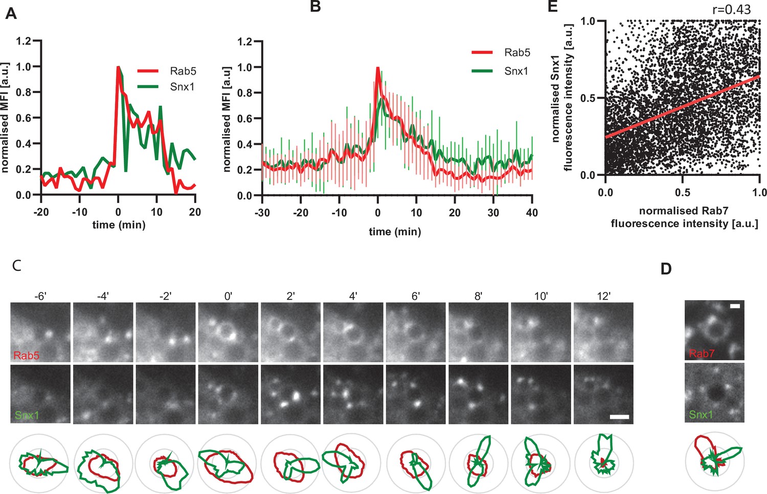
Snx1 subdomain formation at the endosomes initiates with Rab5 recruitment and peaks during Rab conversion stages.
HeLa cells, stably expressing mApple-Rab5 and transiently transfected with Snx1-GFP, were treated for 20 min with nigericin, washed and imaged over 3 hr, as described in Figure 4A. (A) Time-lapse images of a representative endosome to show Snx1 subdomain formation relative to Rab5 recruitment. A time-lapse video of the endosome at 1 min interval is available in Figure 6—video 1. (B) Corresponding graph of normalised mean fluorescence intensity (MFI) of Rab5 and Snx1 at the rim of the endosome in (A) over the time the endosome was detectable. (C) Averaged Rab5 and Snx1 kinetics of 12 endosomes. Error bars represent standard deviation. Representative graph of three independent experiments. Numerical data for all analyzed endosomes is available in Figure 6—source data 1. (D) Images of Rab5 and Snx1 at an enlarged endosome and a corresponding line profile of normalised fluorescence intensity along the rim to show co-existence as well as independence of subdomains of the two markers. Scale bar = 2 μm. (E) Correlation plot of normalised fluorescence intensity of Rab5 and Snx1 as measured in (D) for 14 endosomes for a total of 118 time points, and a corresponding regression line. Pearson’s correlation r = 0.43. Pooled data from two independent experiments. Numerical data for all analyzed endosomes is available in Figure 6—source data 2.
-
Figure 6—source data 1
Quantification of Rab5 and Snx1 recruitment at endosomes.
- https://cdn.elifesciences.org/articles/70982/elife-70982-fig6-data1-v1.xlsx
-
Figure 6—source data 2
Quantification of Rab5 and Snx1 subdomains at endosomes.
- https://cdn.elifesciences.org/articles/70982/elife-70982-fig6-data2-v1.xlsx
Figure 6—figure supplement 1
Dynamic Snx1 recruitment suggest active sorting at the enlarged endosome.
Nigericin was added to HeLa cells for 20 min and washed away, and cells were imaged by time-lapse microscopy, as described in Figure 4A. (A–E) Cells stably expressing mApple-Rab5 (A–C) or mApple-Rab7 (D–E) and transiently transfected with the Snx1-GFP. (A) Example graph of normalised mean fluorescence intensity (MFI) of Rab5 and Snx1 at the rim of the endosome over the time the endosome was detectable. (B) Averaged Rab5 and Snx1 kinetics of 14 endosomes. Error bars represent standard deviation. Representative graph of three independent experiments. Numerical data for all quantified endosomes is available in Figure 6—source data 1. (C) Images and corresponding line profiles of normalised fluorescence intensity of Rab5 and Snx1 along the rim of the maturing endosome at consecutive time points. Rab5 was adjusted for a single maximum and minimum values during the recorded kinetic to highlight its overall signal increase. Snx1 was adjusted for max and min values for each time point to highlight the dynamic nature of Snx1 subdomains. Scale bar = 2 μm. Numerical data for all quantified endosomes is available in Figure 6—figure supplement 1—source data 1. (D) Images of Rab7 and Snx1 at an enlarged endosome and a corresponding line profile of normalised fluorescence intensity along the rim to show co-existence as well as independence of subdomains of the two markers. Scale bar = 1 μm. (E) Correlation plot of normalised fluorescence intensity of Rab7 and Snx1 as measured in (E) for three endosomes for a total of 96 time points, and a corresponding regression line. Pearson’s correlation r = 0.43. Numerical data for all quantified endosomes is available in Figure 6—figure supplement 1—source data 2.
-
Figure 6—figure supplement 1—source data 1
Quantification of Rab5 and Snx1 subdomains at endosomes.
- https://cdn.elifesciences.org/articles/70982/elife-70982-fig6-figsupp1-data1-v1.xlsx
-
Figure 6—figure supplement 1—source data 2
Quantification of Rab7 and Snx1 subdomains at endosomes.
- https://cdn.elifesciences.org/articles/70982/elife-70982-fig6-figsupp1-data2-v1.xlsx
Figure 6—video 1
Snx1 subdomain formation relative to Rab5 recruitment.
Figure 7 with 1 supplement
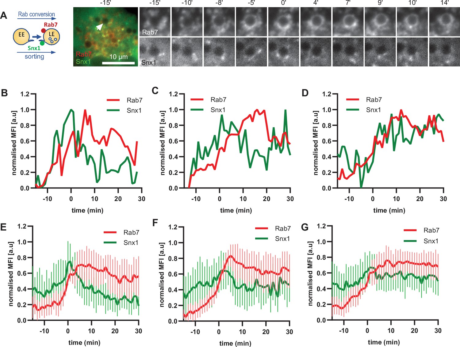
Snx1 subdomain formation at the endosomes initiates with Rab5 recruitment, peaks during Rab conversion stages and continues in late endosomes.
HeLa cells, stably expressing mApple-Rab7 and transiently transfected with Snx1-GFP, were treated for 20 min with nigericin, washed and imaged over 3 hr, as described in Figure 4A. (A) Time-lapse images of a representative endosome to show Snx1 subdomain formation relative to Rab7 recruitment. A time-lapse video of the endosome at 1 min interval is available in Figure 7—video 1. (B) Corresponding graph of MFI of Rab7 and Snx1 at the rim of the endosome in (A) over the time the endosome was detectable, to show Snx1 peaking during Rab conversion. (C,D) Additional graphs of MFI of Rab7 and Snx1 at the rim of endosomes to show the second Snx1 peak (C) or continuing Snx1 presence (D). (E,F,G) Averaged Rab7 and Snx1 kinetics binned into the three patterns of Snx1 recruitment as observed in (B,C,D), representing 19, 21, and 20 endosomes for the single peak, double peak and continuing presence of Snx1, respectively. Error bars represent standard deviation. Three independent experiments were performed, and data pooled. Numerical data for all analyzed endosomes is available in Figure 7—source data 1.
-
Figure 7—source data 1
Quantification of Rab7 and Snx1 recruitment at endosomes.
- https://cdn.elifesciences.org/articles/70982/elife-70982-fig7-data1-v1.xlsx
Figure 7—video 1
Snx1 subdomain formation relative to Rab7 recruitment.
Figure 8 with 5 supplements
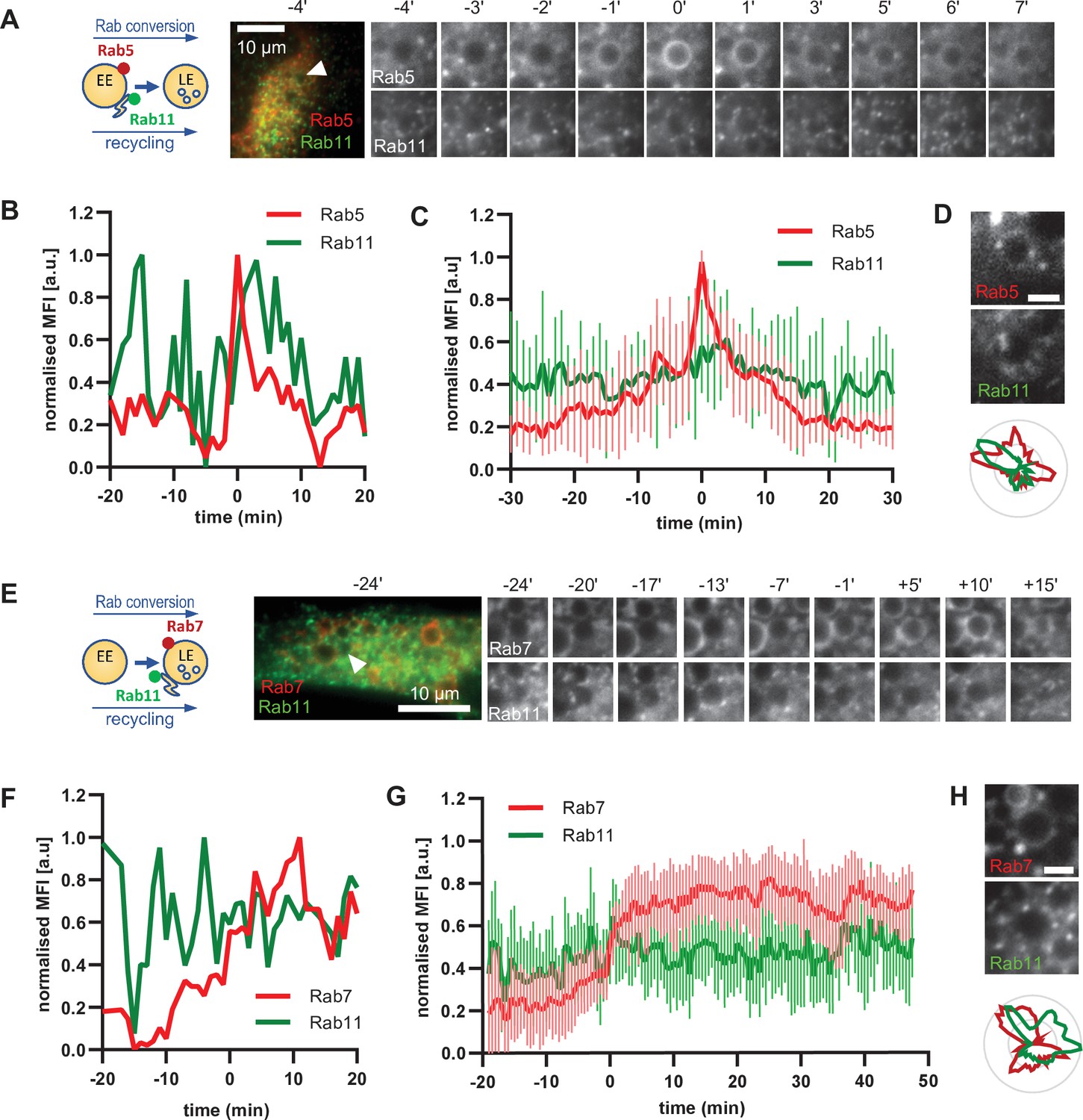
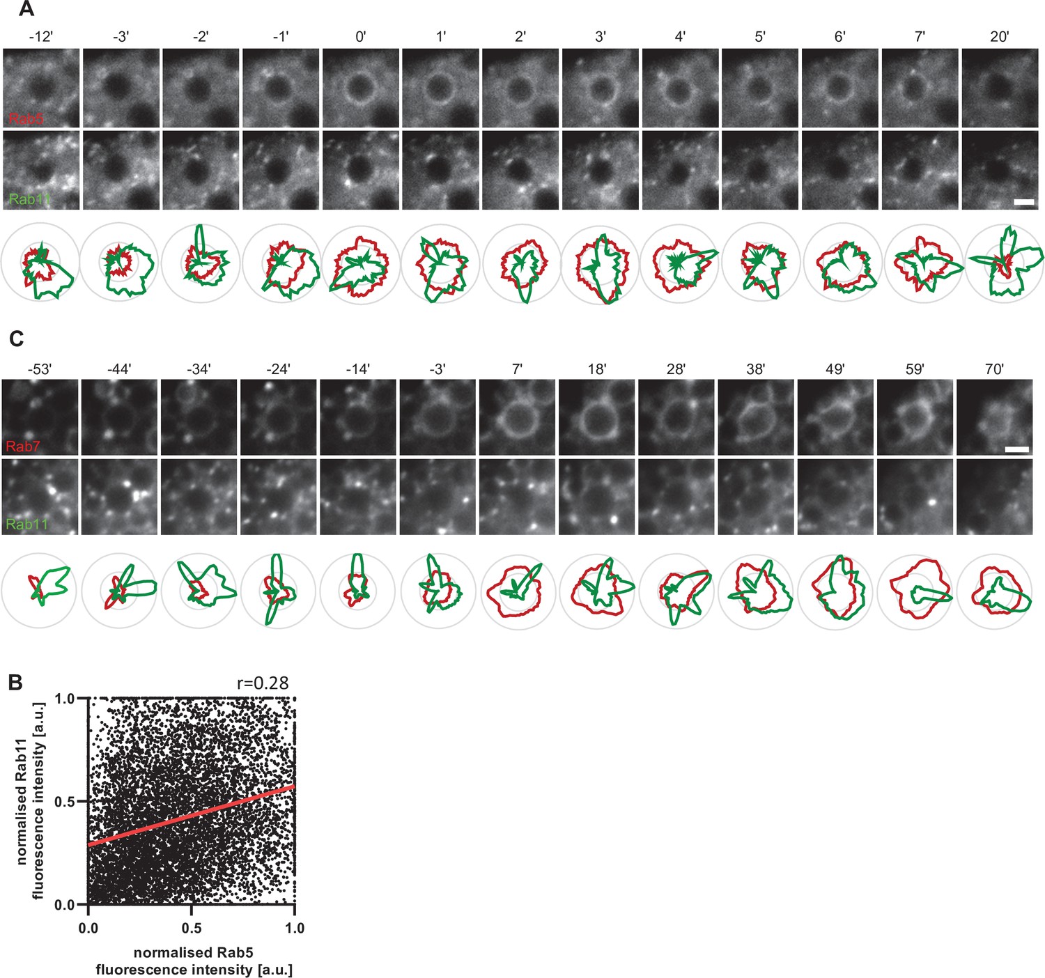
Rab11 interacts with the maturing endosome independently of Rab5 or Rab7.
HeLa cells, stably expressing mApple-Rab5 (A–D) or mApple-Rab7 (E–H) and transiently transfected with GFP-Rab11, were treated for 20 min with nigericin, washed and imaged over 3 h, as described in Figure 4A. (A,E) Time-lapse images of a representative endosome to show continuous Rab11 interaction with the maturing endosome relative to Rab5 (A) or Rab7 (E) recruitment. Time-lapse videos of the endosomes in (A,E) at 1 min interval are available in Figure 8—videos 1 and 4. Additional videos of endosomes at 2 s interval to show Rab11 circling around the enlarged Rab5 positive compartments are available in Figure 8—videos 2 and 3. (B,F) Corresponding graphs of normalised mean fluorescence intensity of Rab5 (B) or Rab7 (F) and Rab11 at the rim of the endosome in (A) or (E), respectively, over the time the endosome was detectable. (C,G) Averaged Rab5 (C) or Rab7 (G) and Snx1 kinetics of 16 and 15 endosomes, respectively. Error bars represent standard deviation. Representative graphs each of three independent experiments. Numerical data for all analyzed endosomes is available in Figure 8—source data 1 and Figure 8—source data 3. (D,H) Images of Rab5 (D) or Rab7 (H) and Rab11 at an enlarged endosome and corresponding line profiles of normalized fluorescence intensity along the rim to show co-existence as well as independence of subdomains of Rab11 and the two markers. Scale bar = 2 μm. Numerical data for analyzed endosomes is available in Figure 8—source data 2 and Figure 8—source data 4.
-
Figure 8—source data 1
Quantification of Rab5 and Rab11 recruitment at endosomes.
- https://cdn.elifesciences.org/articles/70982/elife-70982-fig8-data1-v1.xlsx
-
Figure 8—source data 2
Quantification of Rab5 and Rab11 subdomains at endosomes.
- https://cdn.elifesciences.org/articles/70982/elife-70982-fig8-data2-v1.xlsx
-
Figure 8—source data 3
Quantification of Rab7 and Rab11 recruitment at endosomes.
- https://cdn.elifesciences.org/articles/70982/elife-70982-fig8-data3-v1.xlsx
-
Figure 8—source data 4
Quantification of Rab7 and Rab11 subdomains at endosomes.
- https://cdn.elifesciences.org/articles/70982/elife-70982-fig8-data4-v1.xlsx
Figure 8—figure supplement 1
Rab11 interacts with the maturing endosome independently of Rab5 or Rab7.
HeLa cells, stably expressing mApple-Rab5 (A,B) or mApple-Rab7 (C) and transiently transfected with GFP-Rab11, were treated for 20 min with nigericin, washed and imaged over 3 hr, as described in Figure 4A. (A,C) Images and corresponding line profiles of normalised fluorescence intensity of Rab5 (A) or Rab7 (C) and Rab11 along the rim of the maturing endosome at consecutive time points. Rab5/Rab7 was adjusted for a single maximum and minimum values during the recorded kinetic to highlight its overall signal increase. Rab11 was adjusted for maximum and minimum values for each time point to highlight the dynamic nature of Rab11 interactions with the maturing endosome. Scale bar = 2 μm. (B) Correlation plot of normalised fluorescence intensity of Rab5 and Rab11 as measured in Figure 8D for 14 endosomes for a total of 193 time points, and a corresponding regression line. Pearson’s correlation r = 0.28. Numerical data for all analysed endosomes is available in Figure 8—figure supplement 1—source data 1 and 2.
-
Figure 8—figure supplement 1—source data 1
Quantification of Rab5 and Rab11 subdomains at endosomes in Figure 8—figure supplement 1A.
- https://cdn.elifesciences.org/articles/70982/elife-70982-fig8-figsupp1-data1-v1.xlsx
-
Figure 8—figure supplement 1—source data 2
Numerical data for all analysed endosomes plotted in Figure 8—figure supplement 1A.
- https://cdn.elifesciences.org/articles/70982/elife-70982-fig8-figsupp1-data2-v1.xlsx
Figure 8—video 1
Rab11 interaction with the maturing endosome relative to Rab5.
Figure 8—video 2
Rab11 circling around the enlarged Rab5 positive compartments.
Figure 8—video 3
Rab11 circling around the enlarged Rab5 positive compartments.
Figure 8—video 4
Rab11 interaction with the maturing endosome relative to Rab7.
Figure 9 with 4 supplements
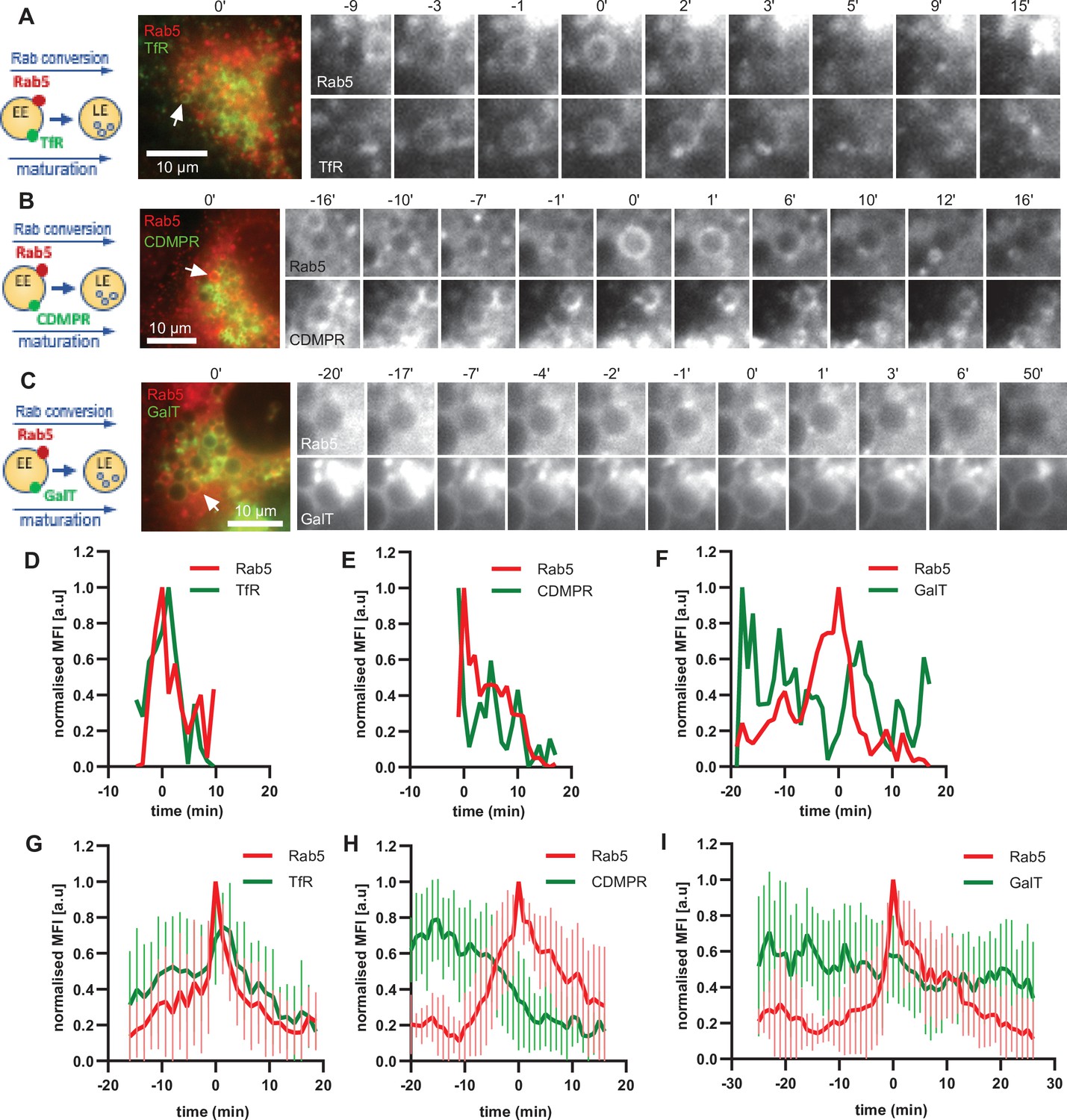
Selective cargo recycling takes place in enlarged Rab5-positive endosomes.
HeLa cells, stably expressing mApple-Rab5 and transiently transfected with the cargos TfR-GFP (A,D,G), GFP-CDMPR (B,E,H) or GalT-GFP (C,F,I), were treated for 20 min with nigericin, washed and imaged over 3 hr, as described in Figure 4A. (A–C) Time-lapse images of representative endosomes to show cargo acquisition and removal relative to Rab5 recruitment. Time-lapse videos of the endosomes in (A,D,G) at 1 min interval are available in Figure 9—video 1, Figure 9—video 2, and Figure 9—video 3, respectively. (D–F) Corresponding graphs of MFI of Rab5 and specified cargo at the endosome in (A,B,C), respectively, over the time the endosome was detectable, to show prompt removal of TfR and CDMPR, and GalT remaining unchanged. (G–I) Averaged Rab5 and specified cargo acquisition and removal, representing 24, 24, and 17 endosomes, respectively. Error bars represent standard deviation. Three independent experiments were performed. Numerical data for all analyzed endosomes is available in Figure 9—source data 1, Figure 9—source data 2, and Figure 9—source data 3.
-
Figure 9—source data 1
Quantification of acquisition and removal of TfR to and from endosomes in relation to Rab5.
- https://cdn.elifesciences.org/articles/70982/elife-70982-fig9-data1-v1.xlsx
-
Figure 9—source data 2
Quantification of acquisition and removal of CDMPR to and from endosomes in relation to Rab5.
- https://cdn.elifesciences.org/articles/70982/elife-70982-fig9-data2-v1.xlsx
-
Figure 9—source data 3
Quantification of acquisition and removal of GalT to and from endosomes in relation to Rab5.
- https://cdn.elifesciences.org/articles/70982/elife-70982-fig9-data3-v1.xlsx
Figure 9—figure supplement 1
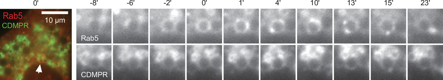
CDMPR can be acquired by Rab5-positive endosomes.
HeLa cells, stably expressing mApple-Rab5 and transiently transfected with CDMPR-GFP, were treated for 20 min with nigericin, washed and imaged over 3 hr, as described in Figure 4A. Time-lapse images of a representative endosome to show CDMPR acquisition at Rab5-positive enlarged endosome.
Figure 9—video 1
TfR acquisition and removal relative to Rab5 recruitment.
Figure 9—video 2
CDMPR acquisition and removal relative to Rab5 recruitment.
Figure 9—video 3
GalT acquisition and removal relative to Rab5 recruitment.
Figure 10 with 3 supplements

GalT-pHlemon sensor detects endosomal acidification.
HeLa cells were transiently transfected with the ratiometric pH sensor, GalT-pHlemon. (C,F) Cells were stably expressing mApple-Rab5. (B) Cells were incubated with Lysotracker Red (LTR) for 20 min prior to imaging. (A) Images of a representative cell to show Golgi-ribbon distribution of GalT-pHlemon in both YFP and CFP channels as well as cytosolic CFP-filled puncta in CFP channel only, representing highly acidified organelles. (B,C) Images to show CFP puncta mostly positive for LTR (B, arrows) and occasionally positive for Rab5 (C, arrows). (D) Graph to show robust response of GalT-pHlemon sensor to pH 4.5–7.0 range as displayed by YFP/CFP ratio measurements in cells incubated with calibration buffers of specified pH values. Numerical data for all analysed Golgi ROIs is available in Figure 10—source data 1. (E) YFP/CFP measurements of GalT-pHlemon in the Golgi ribbon, endo-/lysosomes (cytosolic CFP puncta), as well as in the enlarged endosomes post 20 min nigericin treatment and 100 min recovery. Numerical data for all analysed organelle ROIs is available in Figure 10—source data 2. (F) Cells were treated with nigericin, washed and imaged over 3 hr. Images show GalT-pHlemon sensor localising to the enlarged transiently Rab5-positive endosome and changing YFP and CFP intensity consistent with endosomal acidification. A time-lapse video of the endosome at 1 min interval is available in Figure 10—video 1.
-
Figure 10—source data 1
Quantification of GalT-pHlemon signal in cells in calibration buffers of known pH.
- https://cdn.elifesciences.org/articles/70982/elife-70982-fig10-data1-v1.xlsx
-
Figure 10—source data 2
Quantification of GalT-pHlemon signal in Golgi ribbon, endo-/lysosomal puncta, and enlarged endosomes.
- https://cdn.elifesciences.org/articles/70982/elife-70982-fig10-data2-v1.xlsx
Figure 10—figure supplement 1
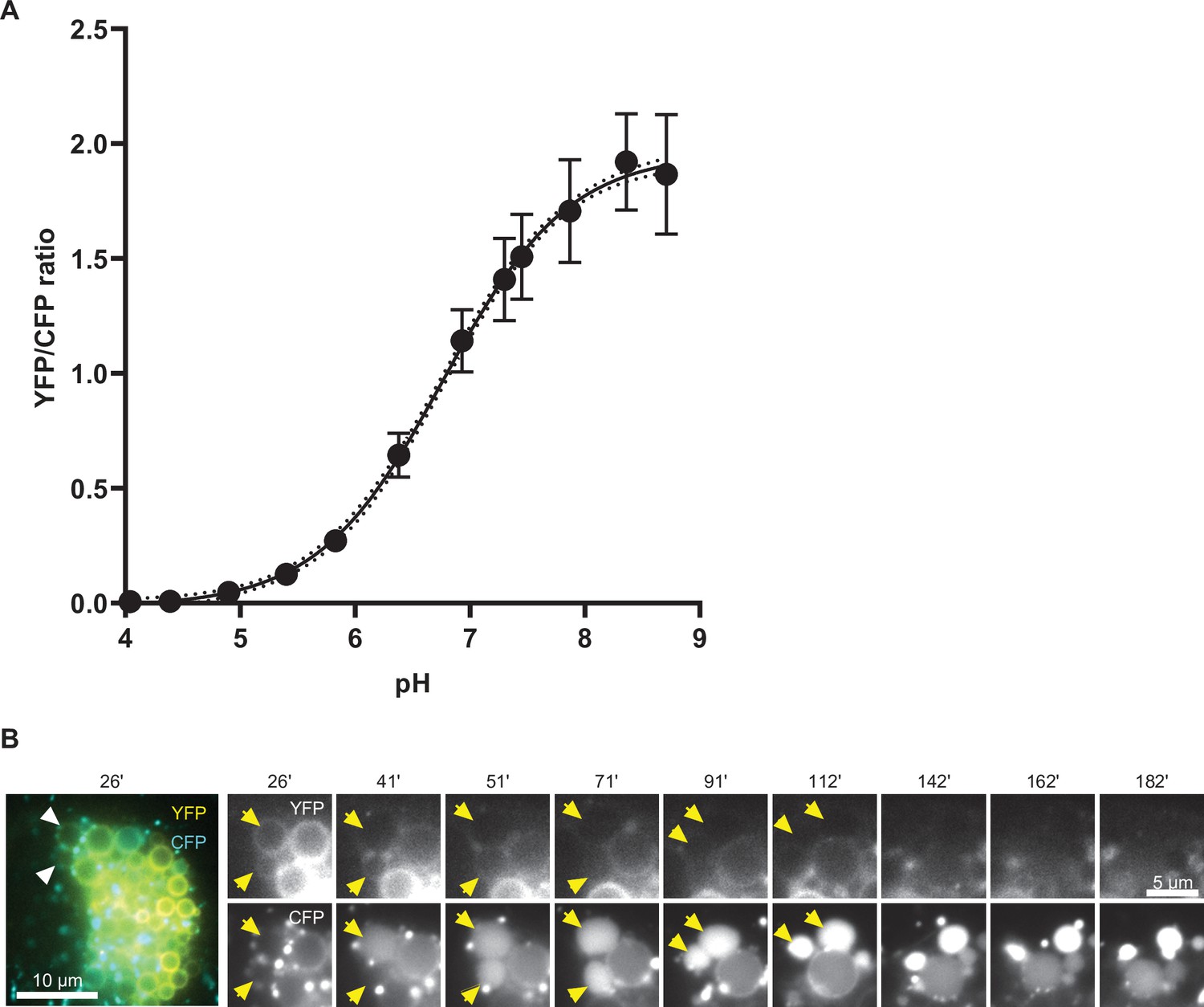
GalT-pHlemon sensor responds to pH changes in a sigmoidal dose-response manner, and its puncta exhibit lysosome-like behavior.
HeLa cells were transiently transfected with the ratiometric pH sensor, GalT-pHlemon. (A) Graph to show response of GalT-pHlemon sensor to pH 4.0–9.0 range as displayed by YFP/CFP ratio measurements in cells incubated with calibration buffers of specified pH values as well as the interpolation of sigmoidal dose-response curve. Numerical data for all quantified Golgi ROIs is available in Figure 10—figure supplement 1—source data 1. (B) Cells were treated with nigericin, washed and imaged over 3 hr. Time-lapse images show CFP-filled puncta fuse with the enlarged compartments, which shrink over time to re-obtain near-punctate morphology. A time-lapse video of the endosome at 1 min interval is available in Figure 10—video 1.
-
Figure 10—figure supplement 1—source data 1
Sigmoidal dose-response curve to define relationship between GalT-pHlemon signal and pH.
- https://cdn.elifesciences.org/articles/70982/elife-70982-fig10-figsupp1-data1-v1.xlsx
Figure 10—video 1
GalT-pHlemon sensor localising to the enlarged transiently Rab5-positive endosome and changing YFP and CFP intensity consistent with endosomal acidification.
Figure 10—video 2
CFP-filled puncta fuse with the enlarged compartments, which shrink over time to re-obtain near-punctate morphology.
Figure 11 with 2 supplements
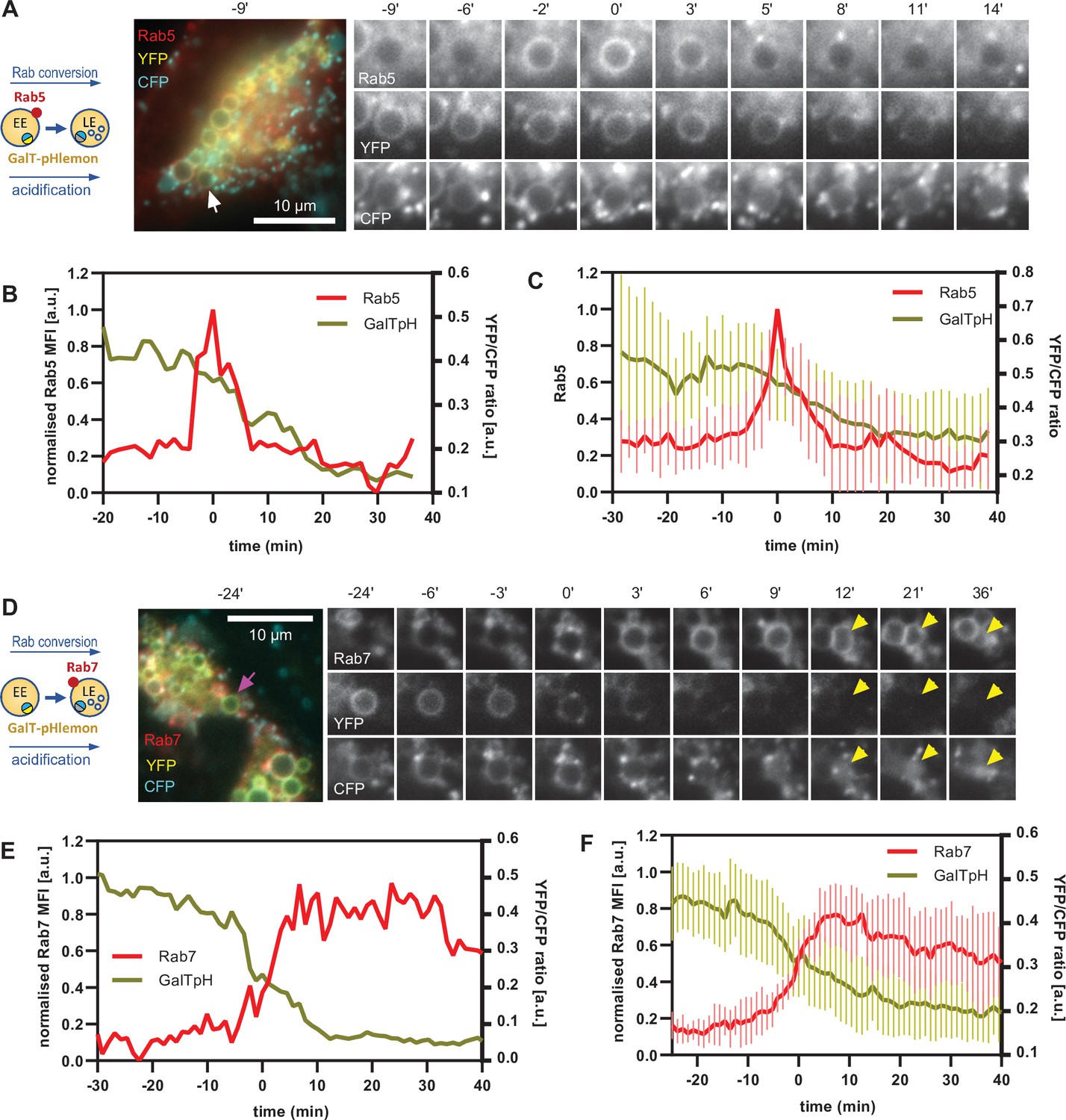
GalT-pHlemon sensor detects endosomal acidification, which correlates with Rab conversion.
HeLa cells, transiently expressing mApple-Rab5 (A–C) or stably expressing mApple-Rab7 (D–F) and transiently transfected with GalT-pHlemon, were treated for 20 min with nigericin, washed and imaged over 3 hr, as described in Figure 4A. (A,D) Time-lapse images of a representative endosome to show association of Rab conversion with acidification, as detected by the decrease in the YFP signal and relatively constant CFP at the rim. Time-lapse videos of the endosomes at 85 s interval are available in Figure 11—videos 1 and 2. (B,E) Corresponding graphs of normalised MFI of Rab5 (B) or Rab7 (E) at the rim and lumenal YFP/CFP ratio of GalT-pHlemon signal of the endosomes in (A) or (D), respectively, during and around the time of Rab conversion. (C,F) Averaged Rab5 (C) or Rab7 (F) and GalT-pH kinetics of 19 and 18 endosomes, respectively. Error bars represent standard deviation. Pooled data from two independent experiments. Numerical data for all analysed endosomes is available in Figure 11—source data 1 and Figure 11—source data 2.
-
Figure 11—source data 1
Quantification of Rab5 recruitment and GalT-pHlemon signal at endosomes.
- https://cdn.elifesciences.org/articles/70982/elife-70982-fig11-data1-v1.xlsx
-
Figure 11—source data 2
Quantification of Rab7 recruitment and GalT-pHlemon signal at endosomes.
- https://cdn.elifesciences.org/articles/70982/elife-70982-fig11-data2-v1.xlsx
Figure 11—video 1
Association of Rab conversion with acidification.
Figure 11—video 2
Association of Rab conversion with acidification.
Figure 12 with 5 supplements
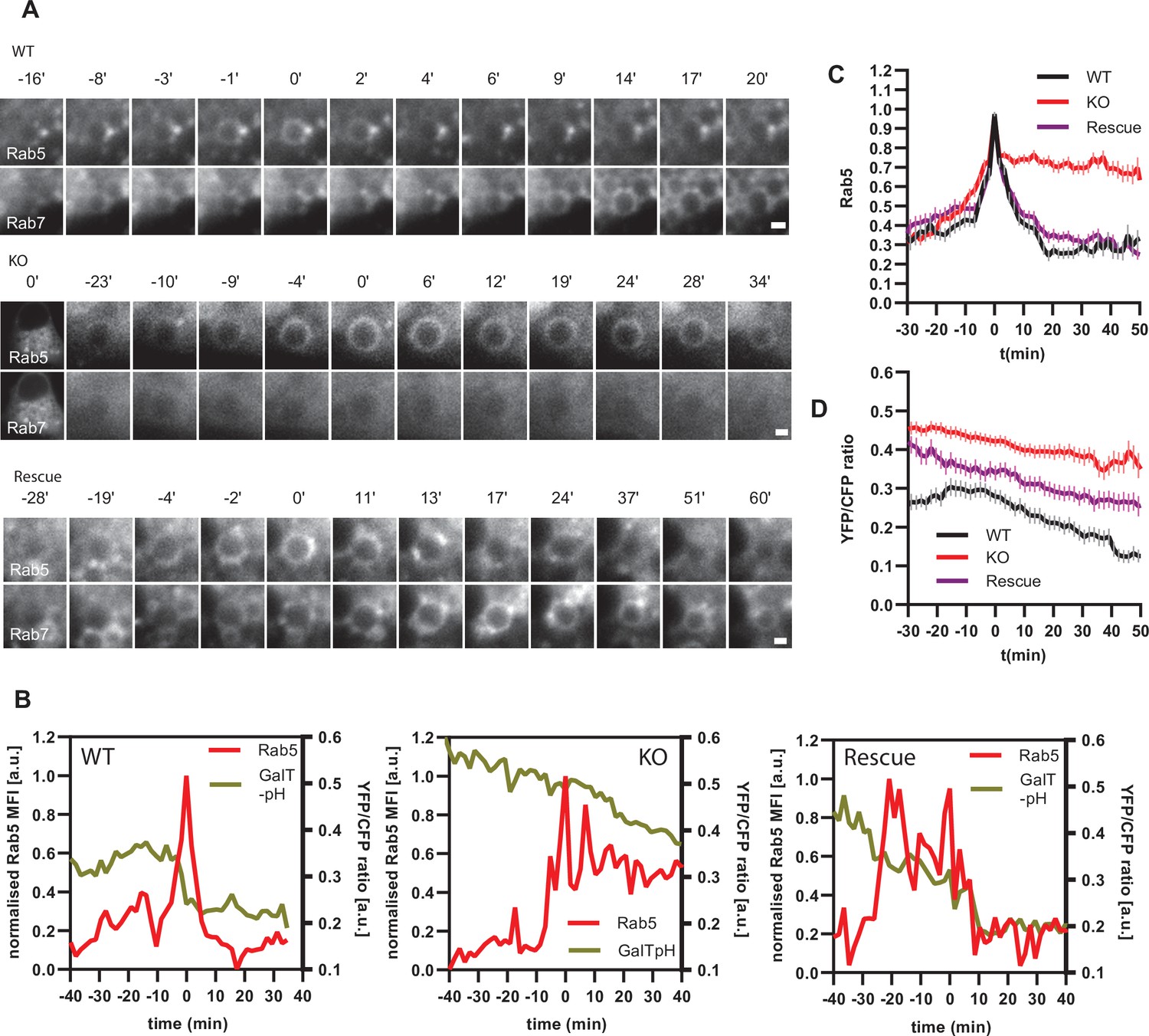
Ccz1 KO disrupts Rab conversion and delays endosomal acidification.
HeLa cell lines with wild-type (WT) Ccz1 and knocked-out Ccz1 (KO) were transiently transfected with mApple-Rab5 and either GFP-Rab7 (A) or GalT-pHlemon (B–D). Ccz1 expression plasmid was co-transfected for 72 hr for rescue experiments. Nigericin was added to cells for 20 min and washed away, and cells were imaged by time-lapse microscopy, as described in Figure 4A. (A) Time-lapse images of representative endosomes to show absence of Rab7 recruitment and lack of Rab5 displacement in KO cells, compared to the expected Rab conversion in WT and rescue cells. Scale bar = 1 μm. Time-lapse videos of the endosomes at 1 min interval are available in Figure 12—videos 1–3 for WT, KO and rescue, respectively. (B) Graphs of normalised mean fluorescence intensity of Rab5 and YFP/CFP ratio of the endosomal GalT-pHlemon signal in representative endosomes during and around the time of Rab conversion / Rab5 peak, in WT, KO and rescue cells. (C,D) Averaged kinetics of Rab5 recruitment (C) and corresponding GalT-pHlemon YFP/CFP ratios (D) for WT, KO and rescue cells, in 54, 54, and 56 endosomes, respectively. Error bars represent SEM. Pooled data from three independent experiments using different Ccz1 clones. Numerical data for all analysed endosomes is available in Figure 12—source data 1.
-
Figure 12—source data 1
Quantification of Rab5 recruitment and GalT-pHlemon signal at endosomes in Ccz1 WT, KO and rescue cells.
- https://cdn.elifesciences.org/articles/70982/elife-70982-fig12-data1-v1.xlsx
Figure 12—figure supplement 1
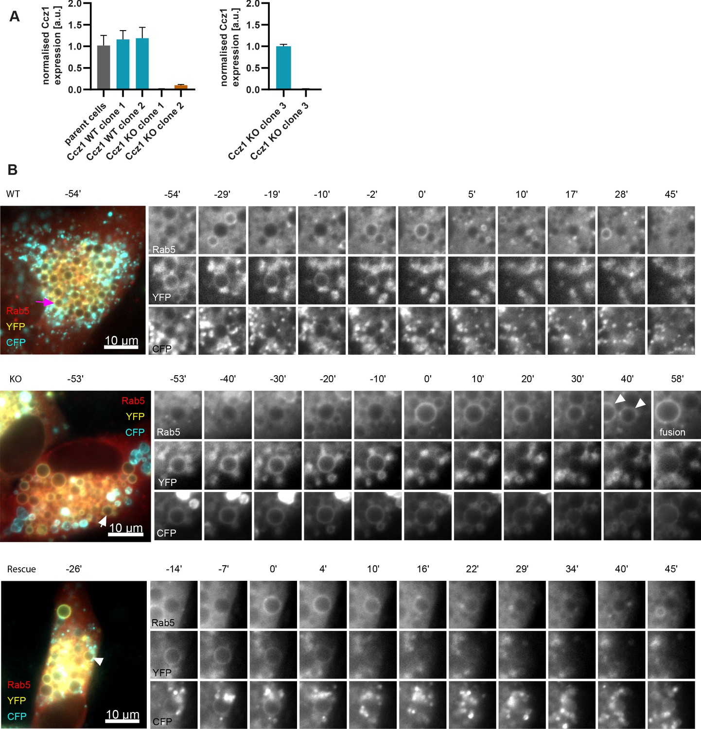
Validation and characterisation of Ccz1 knockout cell lines.
(A) Ccz1 mRNA expression levels in HeLa cell lines with wild-type (WT) Ccz1 and knocked-out Ccz1 (KO), three clones each, measured by qRT-PCR and normalised for actin. Raw RT-PCR data is available in Figure 12—figure supplement 1—source data 1. (B) HeLa cell lines with wild-type (WT) Ccz1 and knocked-out Ccz1 (KO) were transiently transfected with mApple-Rab5 and GalT-pHlemon. Ccz1 expression plasmid was co-transfected for 72 hr for rescue experiments. Time-lapse images of representative endosomes, corresponding to those quantified in Figure 12B, to show acidification in endosomes recruiting Rab5 in the three cell types.
-
Figure 12—figure supplement 1—source data 1
Raw RT-PCR data for Ccz1 expression levels in Ccz1 WT vs KO cells.
- https://cdn.elifesciences.org/articles/70982/elife-70982-fig12-figsupp1-data1-v1.xls
Figure 12—figure supplement 2
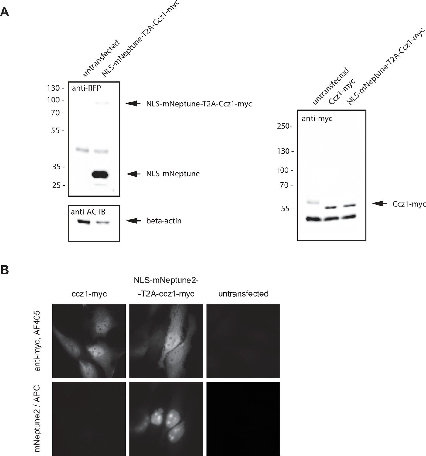
Validation and characterisation of Ccz1 the rescue construct.
(A) Western blot of the ccz1 rescue construct expressing NLS-mNeptune2-T2A-Ccz1-myc to show predominant production of two smaller products, NLS-mNeptune2 and ccz1-myc, as visualised by RFP and myc antibodies, respectively. Original uncropped and unformatted images are available in Figure 12—figure supplement 2—source data 1. (B) Immunofluorescence images of NLS-mNeptune2-T2A-Ccz1-myc transiently transfected into HeLa cells to reveal nuclear distribution of the mNeptune2 signal and cytosolic distribution of Ccz1-myc.
-
Figure 12—figure supplement 2—source data 1
Original western blot images for NLS-mNeptune-T2A-myc expression.
- https://cdn.elifesciences.org/articles/70982/elife-70982-fig12-figsupp2-data1-v1.pdf
Figure 12—video 1
Rab conversion in Ccz1 WT cells.
Figure 12—video 2
Rab conversion in Ccz1 KO cells.
Figure 12—video 3
Rab conversion in Ccz1 KO cells expressing the Ccz1 rescue construct.
Figure 13 with 1 supplement
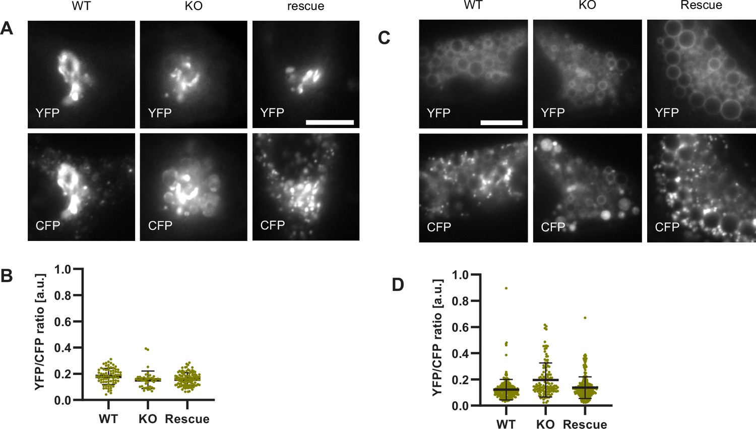
Lysosomes of Ccz1 knockout cells can acidify to the same extent as wild-type cells, with some delay.
HeLa cell lines with wild-type (WT) Ccz1 and knocked-out Ccz1 (KO) were transiently transfected with GalT-pHlemon. Ccz1 expression plasmid was co-transfected for 72 hr for rescue experiments. WT, KO and rescue cells were left untreated. (A,B) or treated with 20 min nigericin followed by 50 min recovery (C,D). (A) Images of cells showing Golgi and highly acidified organelles as visualised with GalT-pHlemon. Acidified organelles appear as puncta in WT and rescue cells and as larger round CFP-filled compartments in KO cells. Scale bar = 10 μm. (C) Images of cells pre-treated with nigericin have dispersed trans-Golgi and re-acidified organelles as in (A). Scale bar = 10 μm. (B,D) Corresponding measurements of YFP/CFP ratio of the GalT-pHlemon sensor in the acidified organelles. Numerical data for all analysed endosomes is available in Figure 13—source data 1 and Figure 13—source data 2.
-
Figure 13—source data 1
Quantification of GalT-pHlemon signal in endo-/lysosomal puncta in Ccz1 WT, KO and rescue cells.
- https://cdn.elifesciences.org/articles/70982/elife-70982-fig13-data1-v1.xlsx
-
Figure 13—source data 2
Quantification of GalT-pHlemon signal in endo-/lysosomal puncta in Ccz1 WT, KO and rescue cells post nigericin treatment.
- https://cdn.elifesciences.org/articles/70982/elife-70982-fig13-data2-v1.xlsx
Figure 13—figure supplement 1
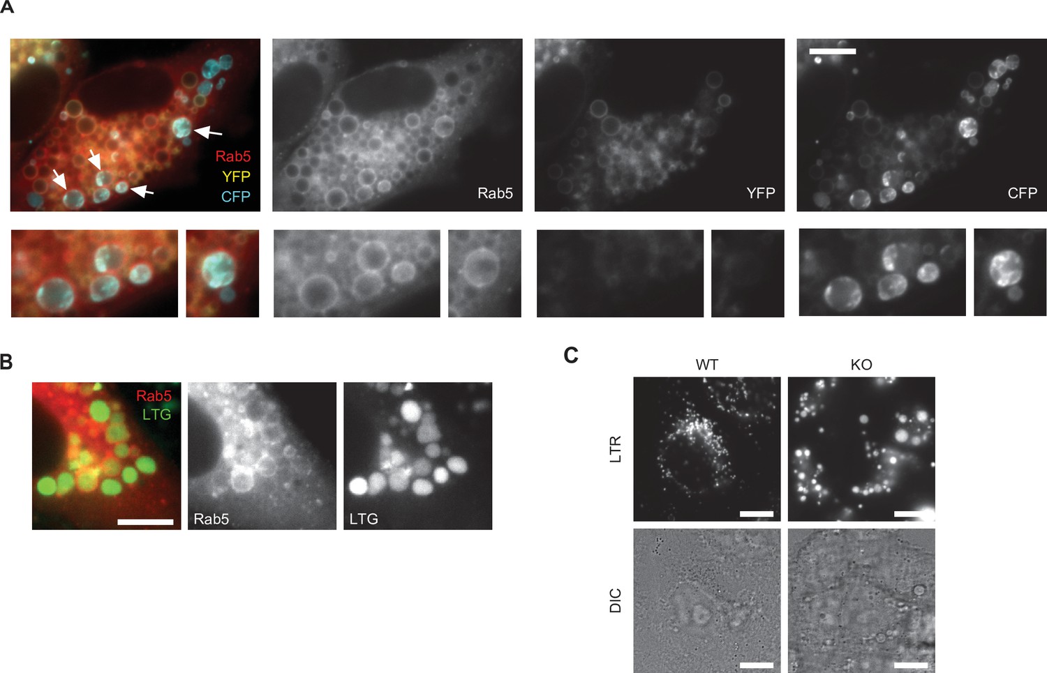
Characterisation of Ccz1 knockout cell lines.
(A) Images of Ccz1 KO cells pre-treated with nigericin, recorded at 150 min recovery, showing Rab5-positive acidified hybrid compartments (arrows). Cells were transiently transfected with mApple-Rab5 and GalT-pHlemon. Scale bar = 10 μm. (B) Images of Ccz1 KO cells pre-treated with nigericin, recorded at 24 hr recovery, showing Rab5-positive acidified hybrid compartments, as visualised with Lysotracker Green (LTG). Cells were transiently transfected with mApple-Rab5. Scale bar = 10 μm. (C) Images of untreated Ccz1 KO cells stained with Lysotracker Red (LTR) to reveal large, acidified compartments. Scale bar = 10 μm.
Tables
Key resources table
| Reagent type (species) or resource | Designation | Source or reference | Identifiers | Additional information |
|---|---|---|---|---|
| cell line (Homo sapiens) | HeLa CCL2 | ATCC | RRID:CVCL_0030 | |
| cell line (Homo sapiens) | HeLa Kyoto | ATCC | RRID:CVCL_1922 | |
| cell line (Homo sapiens) | Neuro-2a | ATCC | RRID:CVCL_0470 | |
| cell line (Homo sapiens) | HEK293 | ATCC | RRID:CVCL_6910 | |
| cell line (monkey) | Cos-1 | ATCC | RRID:CVCL_023 | |
| antibody | anti-GFP (Rabbit polyclonal) | Abcam | RRID:AB_305564 | IEM(1:100) |
| antibody | anti-LC3b (Rabbit polyclonal) | Cell Signalling Technology | RRID:AB_2137707 | IF(1:400) |
| antibody | anti-GalT (Rabbit polyclonal) | Sigma | RRID:AB_1078254 | IF(1:50) |
| antibody | anti-mCherry (Goat polyclonal) | St John’s laboratory | STJ140001 | IEM(1:100) |
| antibody | anti-myc (Mouse monoclonal, 9E10) | Sigma | RRID:AB_2533008 | WB(1:2000) |
| antibody | anti-rabbit coupled to 10 nm Gold (Goat, IgG) | BB International | RRID:AB_2715527 | IEM (1:100) |
| antibody | anti-mouse coupled to 5 nm Gold (Goat, IgG) | BB International | RRID:AB_1769168 | IEM (1:100) |
| antibody | anti-rabbit coupled to 5 nm Gold (Donkey, IgG) | Jackson Immuno Research | RRID:AB_2340610 | IEM (1:100) |
| antibody | anti-goat (Mouse, IgG) | Jackson Immuno Research | RRID:AB_2339054 | IEM (1:100) |
| recombinant DNA reagent | mApple-Rab5a (Plasmid) | RRID:Addgene_54944 | ||
| recombinant DNA reagent | mApple-Rab7a (Plasmid) | RRID:Addgene_54945 | ||
| recombinant DNA reagent | GalT-mCherry (Plasmid) | RRID:Addgene_55052 | ||
| recombinant DNA reagent | mNeptune2-C1 (Plasmid) | RRID:Addgene_54836 | ||
| recombinant DNA reagent | mApple-Lamp1-pHluorin (Plasmid) | RRID:Addgene_54918 | ||
| recombinant DNA reagent | GFP-Rab11a (Plasmid) | RRID:Addgene_12674 | ||
| recombinant DNA reagent | EGFP-2xFYVE (Plasmid) | RRID:Addgene_140047 | ||
| recombinant DNA reagent | Lamp1-GFP (Plasmid) | RRID:Addgene_34831 | ||
| recombinant DNA reagent | pSpCas9(BB)–2A-GFP (pX458) (Plasmid) | RRID:Addgene_48138 | ||
| recombinant DNA reagent | pSpCas9(BB)–2A-puro (pX459) (Plasmid) | RRID:Addgene_48139 | ||
| recombinant DNA reagent | Snx1-turboGFP (Plasmid) | Origene | Origene # RG201844 | |
| recombinant DNA reagent | Ccz1-myc (Plasmid) | Origene | Origene # RC222195 | |
| recombinant DNA reagent | mTagBFP2-Rab5 (Plasmid) | RRID:Addgene_55322 | ||
| recombinant DNA reagent | mCherry-tagged anti-GFP (VHH) (Plasmid) | RRID:Addgene_109421 |
Additional files
-
Transparent reporting form
- https://cdn.elifesciences.org/articles/70982/elife-70982-transrepform1-v1.docx
-
Supplementary file 1
Oligonucleotide sequences used to generate specified constructs.
Table to specify oligonucleotide sequences and their description and purpose in generating constructs as outlined in Materials and methods.
- https://cdn.elifesciences.org/articles/70982/elife-70982-supp1-v1.docx
Download links
A two-part list of links to download the article, or parts of the article, in various formats.
Downloads (link to download the article as PDF)
Open citations (links to open the citations from this article in various online reference manager services)
Cite this article (links to download the citations from this article in formats compatible with various reference manager tools)
A novel live-cell imaging assay reveals regulation of endosome maturation
eLife 10:e70982.
https://doi.org/10.7554/eLife.70982


