The DNA sensors AIM2 and IFI16 are SLE autoantigens that bind neutrophil extracellular traps
- Johns Hopkins University School of Medicine, Division of Rheumatology, United States
- Nephrology Unit, Parma University Hospital, Department of Medicine and Surgery, Italy
- Johns Hopkins University School of Medicine, Division of Pathology, United States
- Johns Hopkins University School of Medicine, Department of Biophysics and Biophysical Chemistry, United States
Figures
Figure 1 with 1 supplement
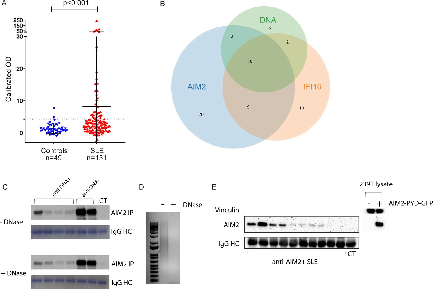
Anti-AIM2 antibodies are associated with anti-IFI16 and anti-DNA antibodies in SLE.
AIM2 antibodies were detected using immunoprecipitation (IP) of 35S-methionine labeled, in vitro transcribed and translated protein. Data are presented as OD units calibrated to a known positive reference serum. Dotted line indicates positive threshold value determined as the mean + 2 standard deviations of control serum samples. AIM2 autoantibodies were identified in 2/49 controls and 41/131 SLE patients. Statistical significance was determined using the Mann-Whitney test for nonparametric values (A). Relationship between anti-AIM2, -IFI16, and –DNA antibodies in the SLE cohort (B). Anti-AIM2 +SLE and control (CT) sera and AIM2 protein were each treated with or without DNase prior to being combined in the IP reaction. Coomassie stain of IgG heavy chain (HC) is shown below each IP result (C). 1 µg of Poly(dA:dT) was treated with DNase as in (C) and analyzed by SYBR Green staining in agarose gel (D). 293T cells were transfected with AIM2-PYD-GFP expression plasmid, and lysate was used in IP reaction with anti-AIM2 +SLE and CT sera (E). IP products and 293T lysates were blotted for AIM2 using anti-N terminal antibody (Cell Signaling D5X7K).
Figure 1—figure supplement 1
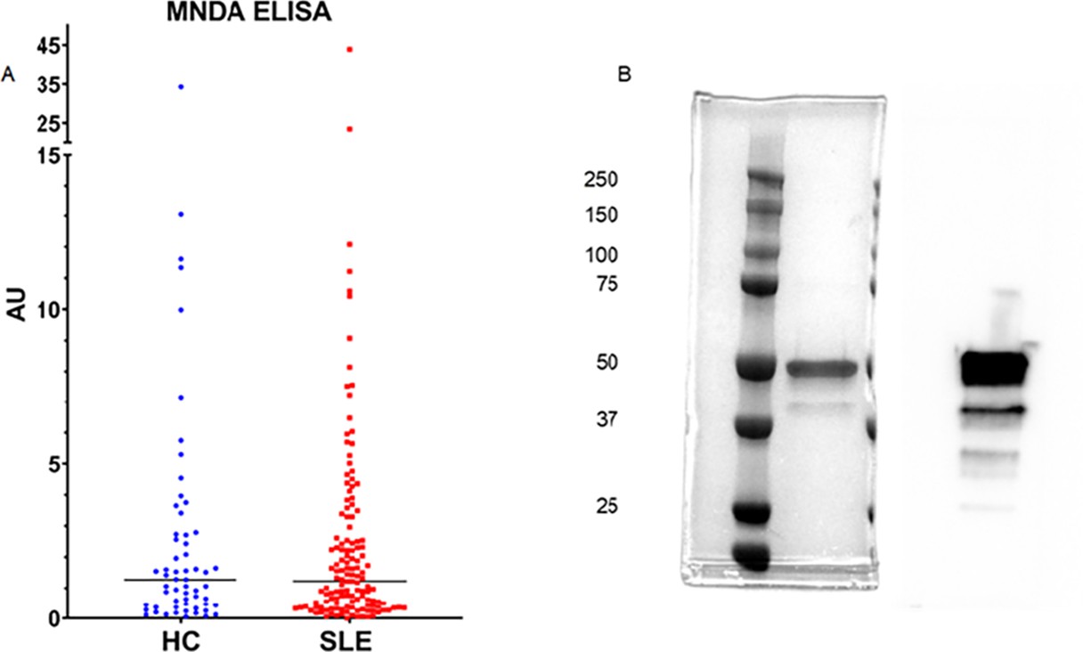
MNDA autoantibodies are not enriched in SLE.
Anti-MNDA antibodies were analyzed by ELISA using 59 healthy control (HC) and 137 SLE sera. (A). MNDA protein utilized in ELISA analyzed by Coommasie staining (left) and Western botting (right) (B). AU = arbitrary units.
Figure 2

IFI16 and AIM2 bind NETs and prevent NET degradation by DNase I.
NETs were induced in neutrophils using PMA 100 nM for 3 hr, then left untreated (A) or incubated with fluorescently labeled AIM2 (pink) and IFI16 (red) at 200 nM at RT for 1 hr (B). Following ALR incubation, samples were stained with anti-MPO-FITC antibody (green) and DAPI (blue), then imaged by confocal microscopy. NETs were treated with DNase I at 20 U/mL at RT for 1 hr (C). NETs incubated with ALRs as in (B) were then treated with 20 U/mL DNase I for 1 hr (D). Scale bars = 20 µm. NETs in 96 well plates were incubated with ALRs at 200 nM (or buffer only) for 1 hr at RT, then treated with DNase I at 0, 20, and 100 U/mL for 30 min at RT. NETs were then stained with Sytox-Green 5 µM, and samples analyzed by fluorimetry (E). RFU = fluorescence units. Mean and standard deviation of 4 replicate wells are indicated. Mann-Whitney test was used to compare groups. P > 0.05 = not significant (ns). P < 0.05 = significant (*). IFI16 and the catalytic domain of cGAS were combined with FAM-labeled 72 bp VACV dsDNA for 30 min, then DNase I added at concentration of 20 U/mL at time = 0 and the fraction of bound dsDNA was monitored via the fluorescence anisotropy of dsDNA•protein complex (F).
Figure 3 with 1 supplement
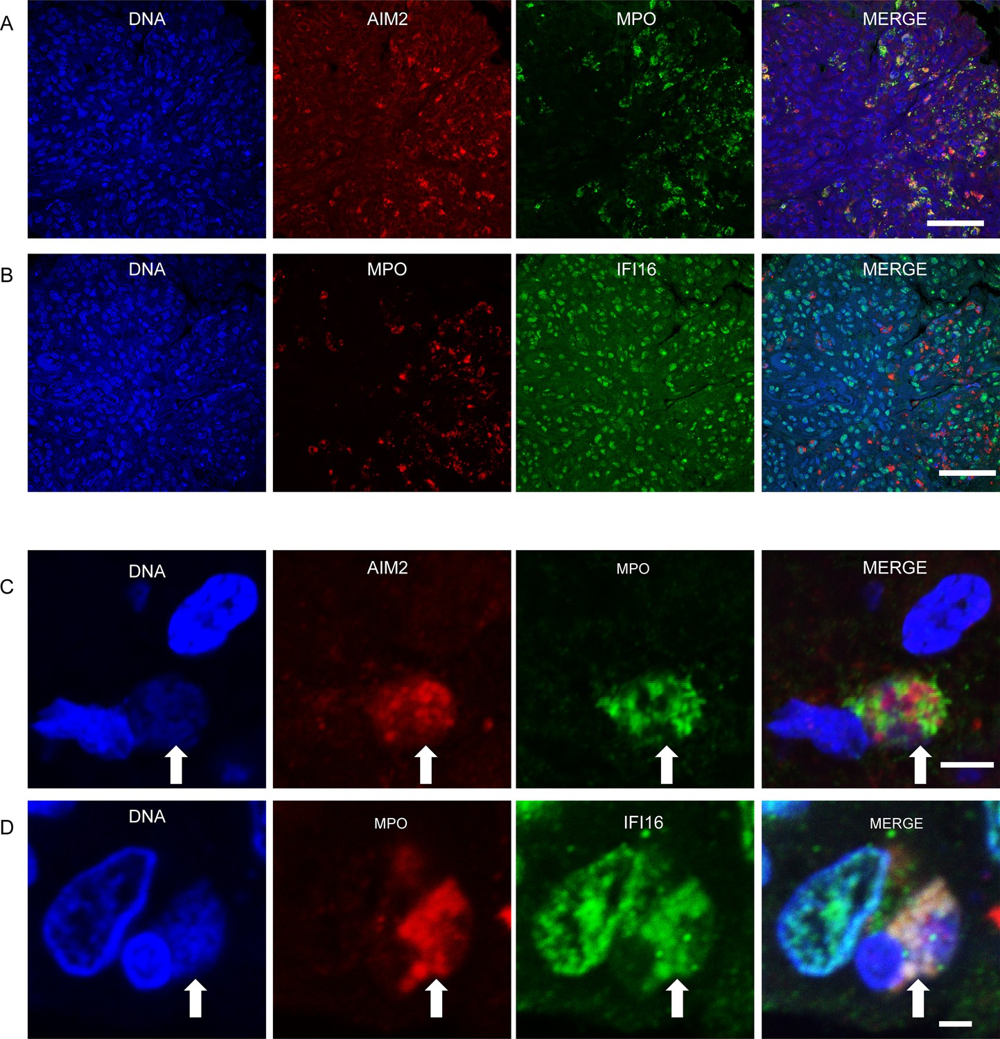
IFI16 and AIM2 bind NETs in diffuse proliferative lupus nephritis.
Representative images of ALR expression and ALR-NETs identified in patients with class IV lupus nephritis. AIM2 (A) expression was largely detected in MPO expressing neutrophils, while IFI16 (B) was more broadly distributed. NETs (arrows) demonstrating co-localizing staining for DNA, MPO, and AIM2 (C) or IFI16 (D) visualized by confocal microscopy. Scale bars: 50 µm (A, B) 5 µm (C), 2 µm (D).
Figure 3—figure supplement 1
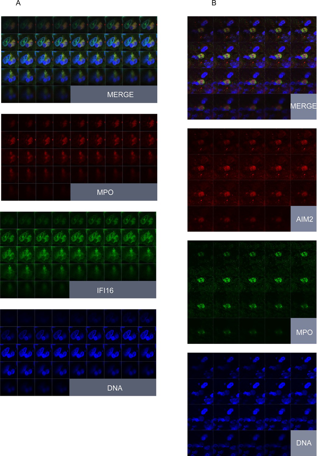
Z-stack imaging of AIM2/IFI16-NETs in lupus nephritis.
Renal biopsy paraffin section stained for DNA, MPO, and IFI16/AIM2 and imaged using z-stacking to identify extracellular DNA-containing structures containing MPO and IFI16 or AIM2. Individual squares within each panel represent adjacent focal planes, proceeding sequentially from top left to bottom right in each area imaged.
Tables
Table 1
Day of visit phenotypic characteristics of SLE patients related to AIM2 autoantibody status.
| Autoantibody | Anti-AIM2+n = 41 | Anti-AIM2-n = 90 | P value |
|---|---|---|---|
| IFI16 Positive | 19/41 (46%) | 12/90 (13%) | < 0.0001 |
| DNA Positive | 12/41 (29%) | 7/89 (8%) | 0.0026 |
| Disease Activity Feature | |||
| Physician Global Disease Activity | 0.63 ± 0.55 | 0.43 ± 0.51 | 0.0333 |
| SLEDAI | 2.29 ± 2.3 | 1.05 ± 1.61 | 0.0026 |
| C3 | 114.7 ± 36.9 | 121.4 ± 29.0 | 0.1352 |
| C4 | 19.5 ± 8.2 | 25 ± 9.3 | 0.0005 |
| Urine Protein/Creatinine ratio | 0.134 ± 0.15 | 0.107 ± 0.11 | 0.4372 |
-
Numerators correspond to number of patients with indicated feature positive and denominators to total number of patients with indicated feature recorded in the cohort, followed by percent (%) positive.
Additional files
-
Supplementary file 1
AIM2 autoantibody correlations and renal biopsy findings in SLE cohort.
(a) Phenotypic Characteristics of SLE Patients Related to AIM2 Autoantibody Level. Numerators correspond to number of patients with indicated feature positive and denominators to total number of patients with indicated feature recorded in the cohort, followed by percent (%) positive. (b) Immunologic phenotype of SLE patients related to AIM2 autoantibody status. Numerators correspond to number of patients with indicated feature positive and denominators to total number of patients with indicated feature, followed by percent (%) positive.
- https://cdn.elifesciences.org/articles/72103/elife-72103-supp1-v1.docx
-
Transparent reporting form
- https://cdn.elifesciences.org/articles/72103/elife-72103-transrepform1-v1.pdf
Download links
A two-part list of links to download the article, or parts of the article, in various formats.
Downloads (link to download the article as PDF)
Open citations (links to open the citations from this article in various online reference manager services)
Cite this article (links to download the citations from this article in formats compatible with various reference manager tools)
The DNA sensors AIM2 and IFI16 are SLE autoantigens that bind neutrophil extracellular traps
eLife 11:e72103.
https://doi.org/10.7554/eLife.72103


