Muscle systems and motility of early animals highlighted by cnidarians from the basal Cambrian
- Qingdao Institute of Marine Geology, China Geological Survey, China
- Function Laboratory of Marine Mineral Resources, Qingdao National Laboratory for Marine Science & Technology, China
- Université de Lyon, Université Claude Bernard Lyon 1, CNRS UMR 5276, Laboratoire de géologie de Lyon: Terre, Planètes, Environnement, Bâtiment GEODE, France
- State Key Laboratory of Continental Dynamics, Shaanxi Key laboratory of Early Life & Environments, Department of Geology, Northwest University, China
- Sorbonne Université, CNRS, Laboratoire de Biologie du Développement de Villefranche-sur-Mer (LBDV), France
- Early Life Evolution Laboratory, School of Earth Sciences & Resources, China University of Geosciences, China
- State Key Laboratory of Marine Environmental Science, College of Ocean and Earth Sciences, Xiamen University, China
- Department of Earth Science & Astronomy, Graduate School of Arts & Sciences, The University of Tokyo, Japan
Figures
Figure 1 with 4 supplements
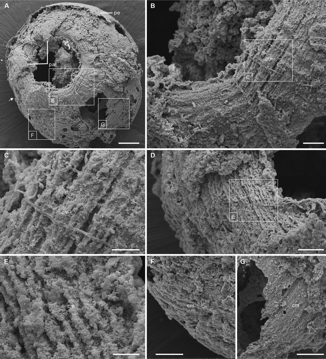
Post-embryonic stage of Olivooides sp. from the early Cambrian Kuanchuanpu Formation (ca. 535 Ma; South China), showing exposed muscle fibers. ELISN150-278. Scanning electron micrographs.
(A) General view of oral side. (B) Details of fiber bundles around aperture (location indicated in A). (C) Close-up showing individual fibers within each bundle. (D) Dense network of fibers (location indicated in A). (E) Close-up of individual fibers coated with fine grains of calcium phosphate. (F), (G) Circular fibers approximately half way between the oral and aboral poles. Abbreviations: cm, circular muscle; pal, perradial apertural lobe; pe, periderm; *, perradii; →, interradii. Scale bars: 100 μm in (A); 20 μm in (B); 10 μm in (C), (E); 20 μm in (D), (F) and (G).
Figure 1—figure supplement 1

Three scanning electron micrographs of the Olivooides specimens.
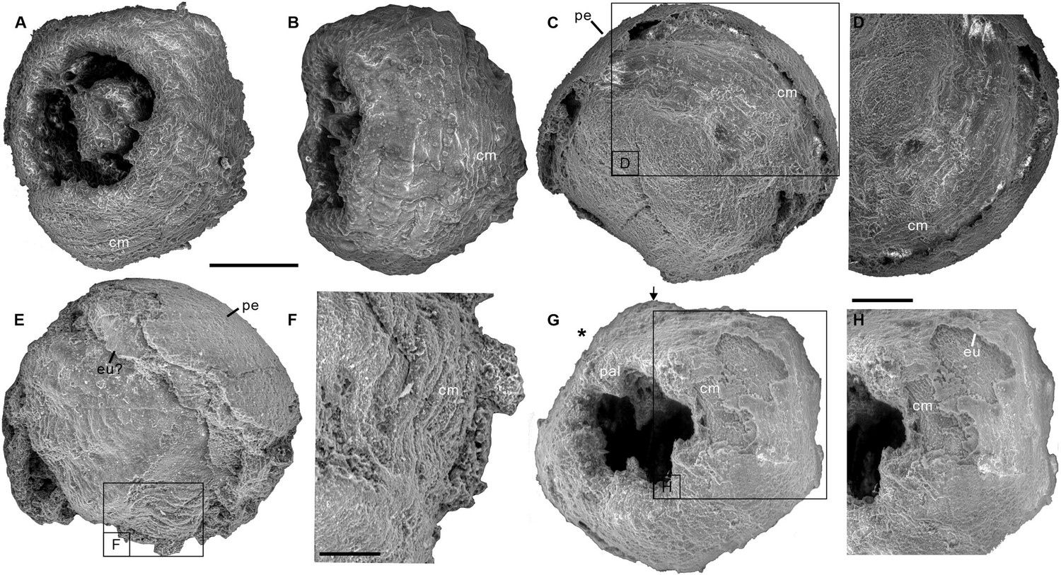
ELISN012-16, ELISN045-143, and ELISN111-54 from the Kuanchuanpu Formation, south China. (A) Oral view showing the external morphology of ELISN045-143 without periderm. (A), (D) ELISN012-16. General view of oral side (A), details of muscles fibers in perradial apertural lobes and general view of aboral side (D). (B) Oral view showing the external morphology of ELISN045-143 without periderm. (C) Close-up view of a showing the circular muscles. (E) Oral view showing the external morphology of ELISN111-54. (F) The enlarged view of c showing that the circular muscles extending into the triangular perradial apertural lobes marked by the giant white arrows. Abbreviations: cm, circular muscle; pal, perradial apertural lobe; *, perradii; →, interradii. Scale bars represent: 200 μm in (A), (B), (D) and (E); 50 μm in (C); 25 μm in (F).
Figure 1—figure supplement 2
Four scanning electron micrographs of the Olivooides specimens.
ELISN087-64 (A, B), ELISN088-48 (C, D), ELISN087-33 (E, F) and ELISN98-19 (G, H) from the Kuanchuanpu Formation, south China. (A) General view of oral side. (B) Lateral view. (C) General view of aboral side. (D) The enlarged view of (C) showing the circular muscles. (E) Lateral view. (F) The enlarged view of (E) showing that the circular muscles. (G) General view of oral side. (H) The enlarged view of (G) showing the circular muscles. Abbreviations: cm, circular muscle; eu, exumbrella; pal, perradial apertural lobe; *, perradii; →, interradii. Scale bars represent: 200 μm in (A–C), (E) and (G); 50 μm in (F), (H); 25 μm in (D).
Figure 1—figure supplement 3
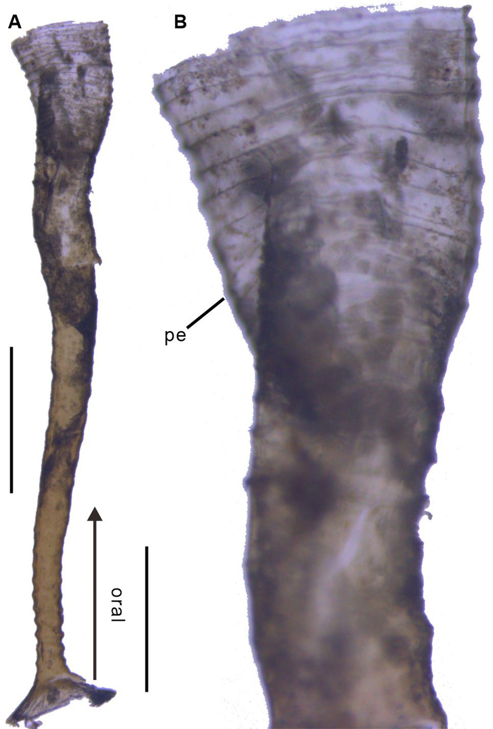
Polyp of Coronatae sp. from South China Sea showing the periderm.
(A), (B) Lateral view and details showing corrugations. Abbreviations are as follows: pe, periderm. Scale bars: 400 μm in (A); 100 μm in (B).
Figure 1—figure supplement 4

Origin of the fossil material.
Location map (Ningqiang area, Southern Shaanxi Province, China), photograph of outcrop (Shizhonggou Section; see red star) and stratigraphic position of fossil-bearing rock samples (A), (B) within the Kuanchuanpu Formation. Modified from Steiner et al., 2014.
Figure 2 with 2 supplements
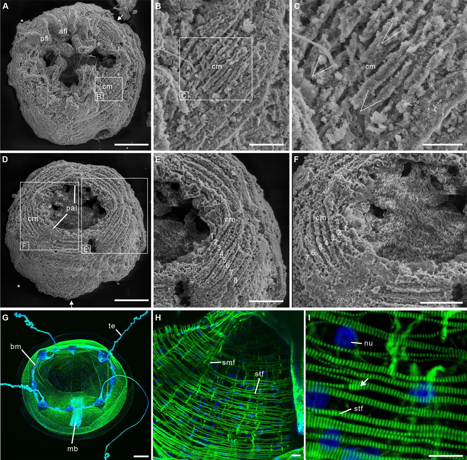
Post-embryonic stage of Olivooides sp. from the early Cambrian Kuanchuanpu Formation (ca. 535 Ma; South China), showing exposed muscle fibers by SEM.
(A–F) and myoepithelial muscle network in extant hydrozoan jellyfish by fluorescence microscopy (G–I). (A–C) ELISN052-33. General view of oral side and details of apertural circular muscle fibers and the V-shaped interconnection between the fibers in (C). (D–F) ELISN061-19. General view of oral side and details of apertural, circular muscles fibers. (G–I) Eirene sp. (Hydrozoa) young medusa, general oral view, circular muscles along subumbrella and details of striated fibers; white arrow (I) indicates bifurcating fibers. Green and blue in (G–I) correspond to actin (phalloidin) and DNA (Hoechst) staining. Abbreviations: afl, adradial fold lappet; bm, bell margin; cm, circular muscle; mb, manubrium; nu, nucleus; pal, perradial apertural lobe; pfl, perradial fold lappet; smf, smooth (radial) muscle fiber; stf, striated (circular) muscle fiber; te, tentacle *, perradii; →, interradii. Scale bars: 200 μm in (A), (D); 100 μm in (G); 50 μm in (E), (F); 20 μm in (B); 10 μm in (C), (H) and (I).
Figure 2—figure supplement 1
Two scanning electron micrographs of the Olivooides specimens.
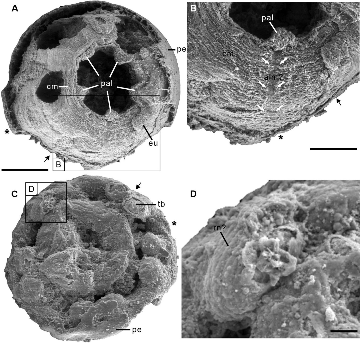
ELISN115-39 (Sinaster petalon; Wang et al., 2017) and ELISN107-470 (Hanagyroia orientalis; Wang et al., 2020) from the Kuanchuanpu Formation, south China. (A) Oral view showing the external morphology of ELISN115-39. (B) Close-up view of (A) showing the possible longitudinal muscle bands marked by the two rows of giant white arrows. (C) Oral view showing the external morphology of ELISN107-470. (D) The enlarged view of (C) showing the possible rings of nematocysts on the tentacles. Abbreviations: alm?, possible longitudinal muscle bands at adradii; cm, circular muscle; eu, exumbrella; pal, perradial apertural lobe; pe, periderm; *, perradii; →, interradii. Scale bars represent: 200 μm in (A), (C); 50 μm in (B); 25 μm in (D).
Figure 2—figure supplement 2
Myoepithelial muscle network in extant medusozoan cnidarians.
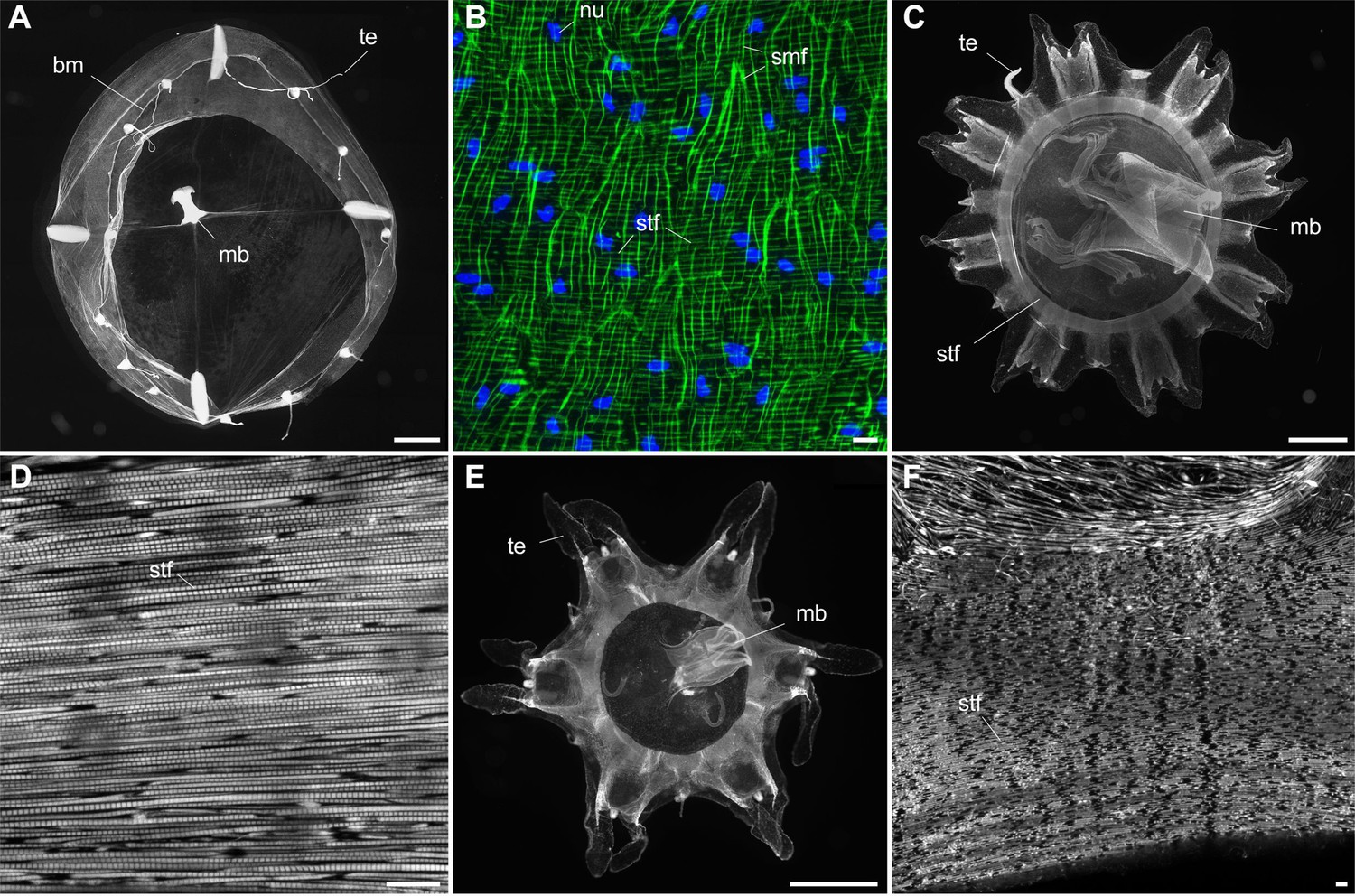
(A), (B) Clytia hemisphaerica (Hydrozoa) mature medusa, general oral view and details of fiber network (subumbrella). (C), (D) Pelagia noctiluca (Scyphozoa) metaephyra, general oral view and details of closely-packed circular striated muscle fibers (subumbrella). (E), (F) Chrysaora colorata (Scyphozoa) metaephyra, general oral view and details of circular striated muscle fibers. Green and blue colors (B) correspond to actin (phalloidin) and DNA (Hoechst) staining. Specimens represented by black and white images (A) and (C–F) were stained for actin (phalloidin). Abbreviations are as follows: bm, bell margin; mb, manubrium; nu, nucleus; smf, smooth (radial) muscle fiber; stf, striated (circular) muscle fiber; te, tentacle. Scale bars: 1000 µm in (A), (C) and (E); 10 µm in (B), (D) and (F).
Figure 3
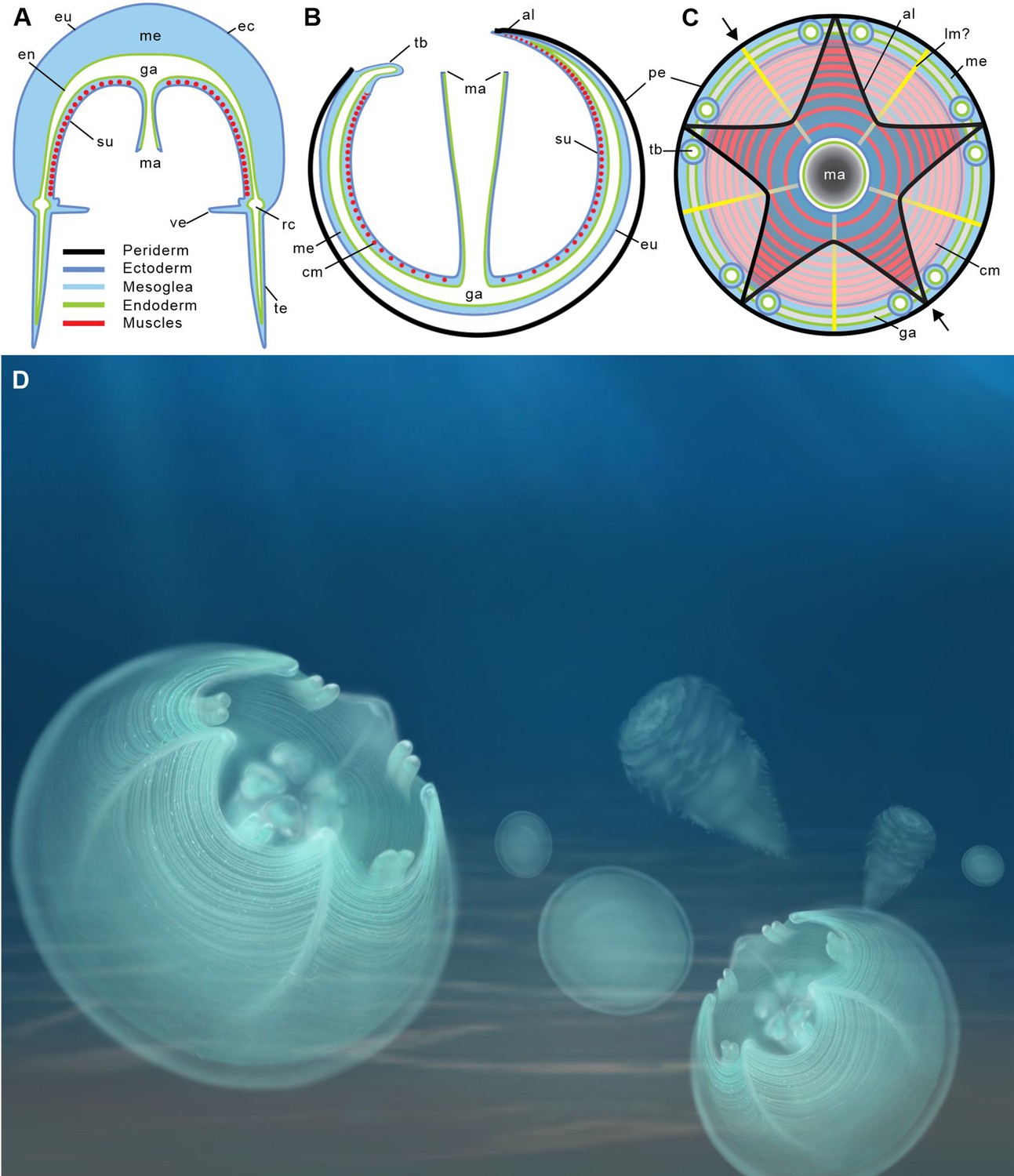
Location of epithelial muscles in extant hydromedusae (A) and early Cambrian Olivooidae medusozoans (B, C).
(A), (B) Simplified radial sections through body. (C) In oral view. (D) Artistic reconstruction of 535-million-year-old olivooid cnidarians showing eggs (the prehatched, background), post-embryonic (foreground) and polyp (background) stages. The circular muscle system is visible through the translucent periderm. The location of section (B) is indicated in (C) by small black arrows. Abbreviations: al, apertural lobe; cm, circular (coronal) muscle; en, endoderm; eu, exumbrella; ga, gastrovascular cavity; lm?, possible longitudinal muscle; ma, manubrium; me, mesoglea; pe, periderm; rc, radial canal; su, subumbrella; tb, tentacular bud; te, tentacle; ve, velum. Not to scale.
Figure 4 with 2 supplements
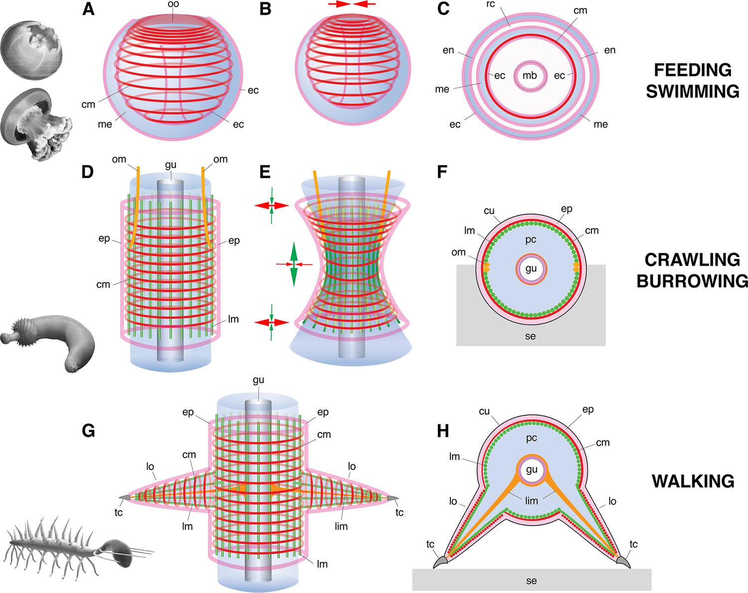
Basic muscle systems in early Cambrian animals and their main functions.
(A–C) Contractile epithelial muscles (MEC, myoepithelial, predominantly circular) and antagonistic mesoglea exemplified by olivooiids and free-swimming jellyfish; idealized relaxed (A) and contracted (B) states and simplified transverse section (C). (D–F) Grid-like network of circular and longitudinal subepidermal muscle fibers (HMS) around a cylindrical body filled with antagonistic internal fluid (hydroskeleton) exemplified by scalidophoran worms: idealized relaxed state (D); peristaltic contractions along body (E) and transverse section (F). (G–H) Longitudinal, circular muscles and extrinsic retractor muscles in lobopodians: idealized relaxed state (G); transverse section (H). Images (from top to bottom) represent an olivooid cnidarian, an extant jellyfish, the scalidophoran worm Ottoia (see Vannier, 2012) and the lobopodian Hallucigenia (see Chen and Zhou, 1997). Drawings and images not to scale. Abbreviations: cm, circular muscle; cu, cuticle; ec, ectoderm; en, endoderm; ep, epidermal layer; gu, gut; lc, lobopod claw; lim, limb muscle; lm, longitudinal muscle; lo, lobopod (soft leg); mb, manubrium; me, mesoglea; om, oblique muscle; oo, oral opening (bell margin); pc, primary cavity filled with fluid; rc, radial canal; se, sediment; tc, terminal claw.
Figure 4—figure supplement 1
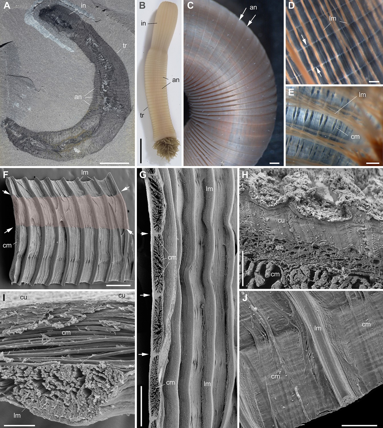
Muscle system in priapulid worms (Ecdysozoa).
(A) Ottoia prolifica from the mid-Cambrian Burgess Shale, general view showing annulated body. (B–J) Priapulus caudatus from Sweden. (B) General view of live specimen in sea water. (C) Annulated trunk. (D) Muscles (trunk). (E) Muscles (introvert). (F) Fragment of trunk showing the internal body wall with strong longitudinal muscles (pink area corresponds to one annulation). (G) Transverse section through circular muscles (one bundle per annulation). (H) Transverse section through body wall across cuticle, epithelial cells, and circular muscles. (I) Transverse section through body wall showing circular muscles overlying longitudinal ones. (J) Inner wall of body showing both circular and longitudinal muscles. Small white arrows indicate annulation boundaries. (A–E) are light photographs. (F–J) are SEM images. Abbreviations are as follows: an, annulation; cm, circular muscle; cu, cuticle; ec, epithelial cells; introvert; lm, longitudinal muscle; tr, trunk. Scale bars: 1 cm in (A), (B); 1 mm in (C); 500 µm in (D–G); 200 µm in (J); 50 µm in (I) and 10 µm in (H).
Figure 4—figure supplement 2
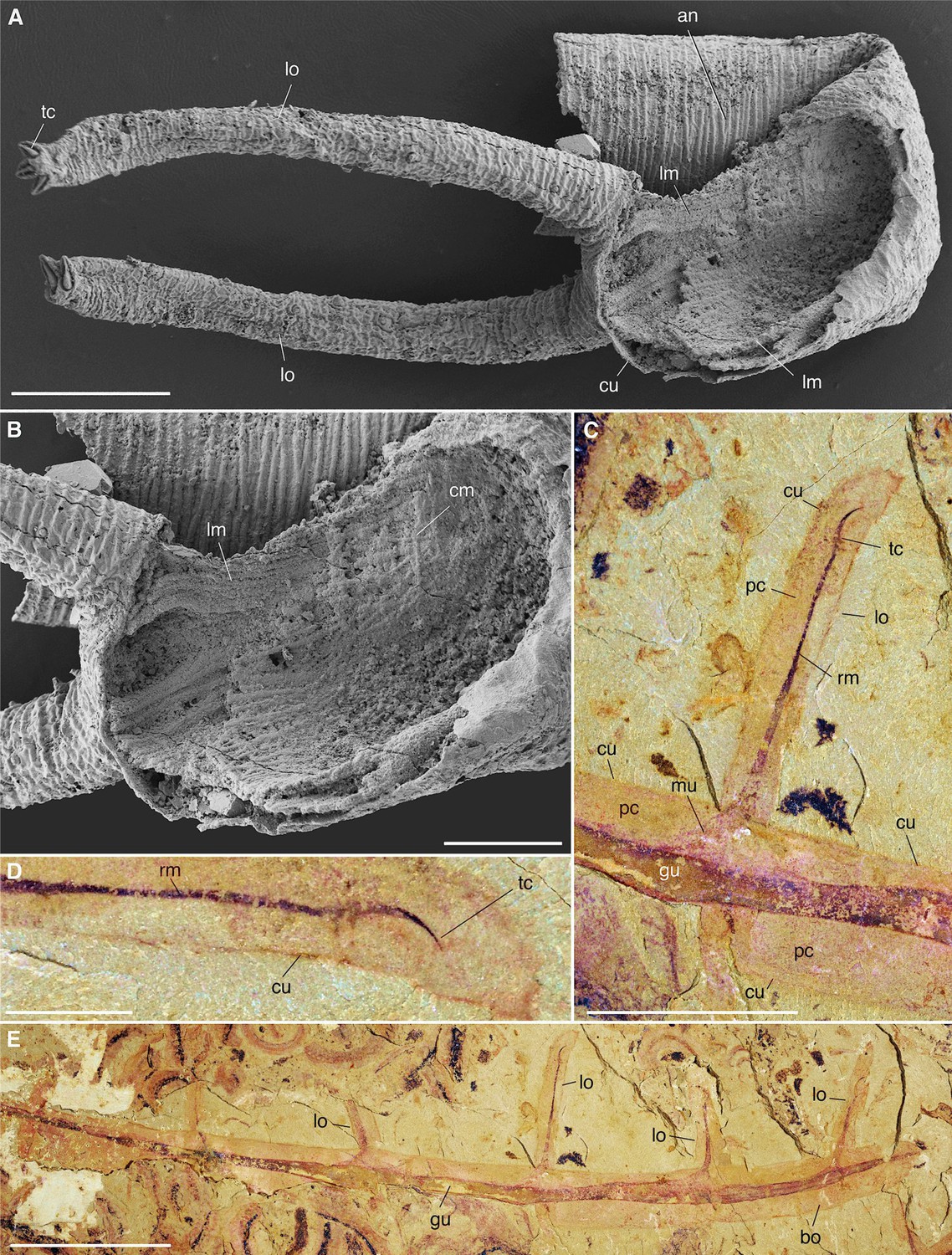
Muscle system in Cambrian lobopodians (Ecdysozoa, Panarthropoda).
(A), (B) Tritonychus phanerosarkus from the Yu’anshan Formation (Xiaotan section, Cambrian Series 2, Stage 3), Yongshan, Yunnan Province, China; general view and details of a secondarily phosphatized specimen showing a pair of walking appendages (lobopods) and the inner body wall lined with muscle fibers (see Zhang et al., 2016). (C–E) Paucipodia haikouensis from the Chengjiang Lagerstätte, ELI-JS0001a (see Vannier and Martin, 2017); close-up of lobopod and terminal claw, and general view; note muscle bridging claw to gut area. (A), (B) are SEM images (Courtesy Prof. Xiguang Zhang; see Zhang et al., 2016). Abbreviations: an, annuli; cm, circular muscle; cu, cuticle; gu, gut; lm, longitudinal muscle; lo, lobopod; mu, muscle; pc, primary cavity; rm, retractor muscle; tc, terminal claw. Scale bars: 10 mm in (E), 5 mm in (C), 1 mm in (D), 200 µm in (A) and 100 µm in (B).
Additional files
Download links
A two-part list of links to download the article, or parts of the article, in various formats.
Downloads (link to download the article as PDF)
Open citations (links to open the citations from this article in various online reference manager services)
Cite this article (links to download the citations from this article in formats compatible with various reference manager tools)
Muscle systems and motility of early animals highlighted by cnidarians from the basal Cambrian
eLife 11:e74716.
https://doi.org/10.7554/eLife.74716


