The VINE complex is an endosomal VPS9-domain GEF and SNX-BAR coat
- Department of Medical Genetics, University of British Columbia, Canada
- Centre for Molecular Medicine and Therapeutics, British Columbia Children’s Hospital Research Institute, University of British Columbia, Canada
Figures
Figure 1 with 2 supplements
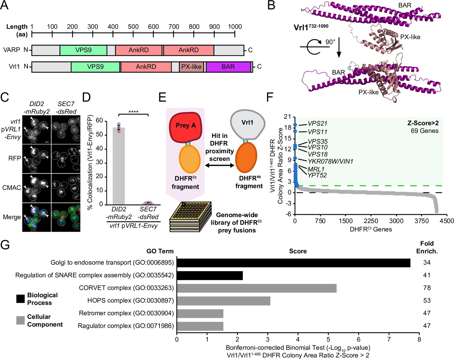
Vrl1 is a predicted PX-BAR protein that interacts with conserved machinery at the endosome.
(A) Schematic of Vrl1 and VARP domain architecture. (B) ColabFold predicts the Vrl1 C-terminus has a SNX-BAR-like PX and BAR domain fold. (C) Vrl1-Envy colocalizes with Did2-mRuby2-labeled endosomes, but not with the Sec7-dsRed Golgi marker. (D) Quantification of colocalization as the percentage of Vrl1 puncta overlapping RFP puncta in C. Two-tailed equal variance t test; n=3, cells/strain/replicate ≥1395; ****=p < 0.0001. (E) Schematic of DHFR proximity screen methodology. (F) Z-score distribution of the ratio of colony areas from genome-wide DHFR screens of full-length and truncated Vrl1 baits that localize to the endosome and cytosol, respectively. (G) Gene Ontology (GO) functional enrichment analysis of Vrl1 DHFR interactors (Z-score >2; http://geneontology.org). GO terms of the most specific hierarchical subclass with a fold enrichment value >25 are presented as the negative base 10 log of the associated p-value from a Bonferroni-corrected binomial test of significance. Scale bars, 2 µm. Error bars report standard error of the mean (SEM). Enrich., enrichment. aa, amino acids.
-
Figure 1—source data 1
Data associated with Figure 1D.
- https://cdn.elifesciences.org/articles/77035/elife-77035-fig1-data1-v2.xlsx
-
Figure 1—source data 2
Data associated with Figure 1G.
- https://cdn.elifesciences.org/articles/77035/elife-77035-fig1-data2-v2.xlsx
Figure 1—figure supplement 1
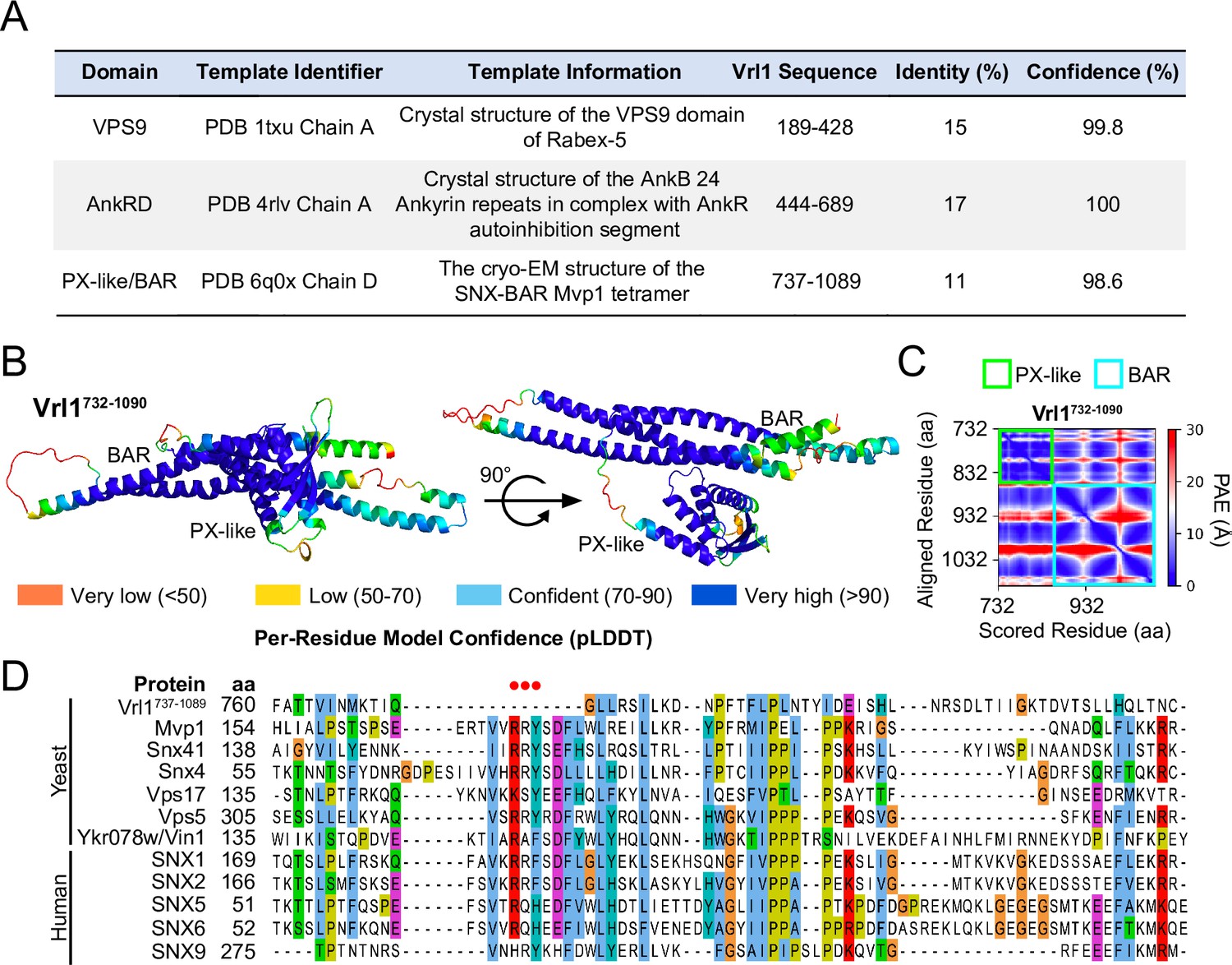
The Vrl1 PX-like domain is missing key PI3P-binding residues.
(A) Results from Phyre2 analysis of Vrl1 sequences (Intensive mode, http://www.sbg.bio.ic.ac.uk/phyre2). (B) ColabFold-predicted Vrl1 C-terminus with the predicted local-difference distance test (pLDDT; Jumper et al., 2021; Mirdita et al., 2022) scores mapped to each residue. (C) ColabFold-generated predicted alignment error (PAE; Jumper et al., 2021; Mirdita et al., 2022) plot for the Vrl1 C-terminus demonstrates a high confidence folding prediction. (D) Sequence alignment of Vrl1 C-terminus (aa 737–1089) with yeast and human PX domain-containing proteins. The canonical PI3P-binding ‘RRY’ motif is absent in Vrl1 (highlighted with red dots above alignment). aa, amino acids.
Figure 1—figure supplement 2
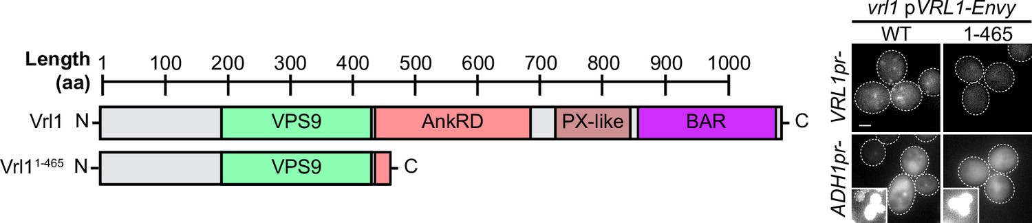
The Vrl1 N-terminus and VPS9 domain are not sufficient to localize to puncta.
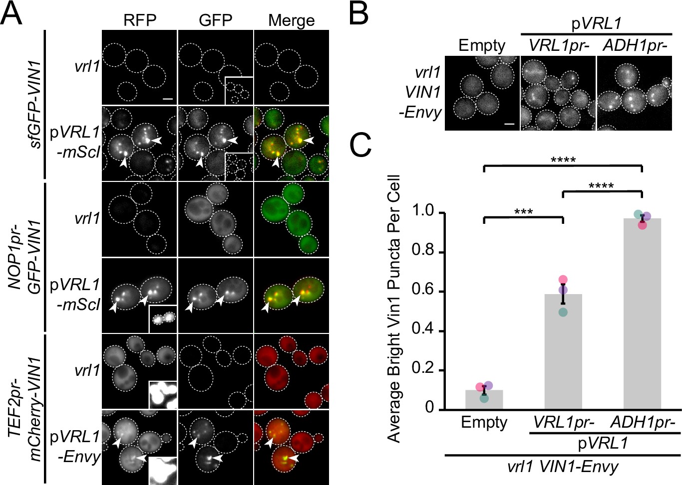
The Vrl1 N-terminus (aa 1–465) does not localize to puncta when expressed from the endogenous VRL1 promoter (VRL1pr) or the strong ADH1pr and instead accumulates in the cytosol. Insets are scaled to match other images in the same channel (see materials and methods for details). Scale bars, 2 µm. WT, wild type.
Figure 2 with 1 supplement
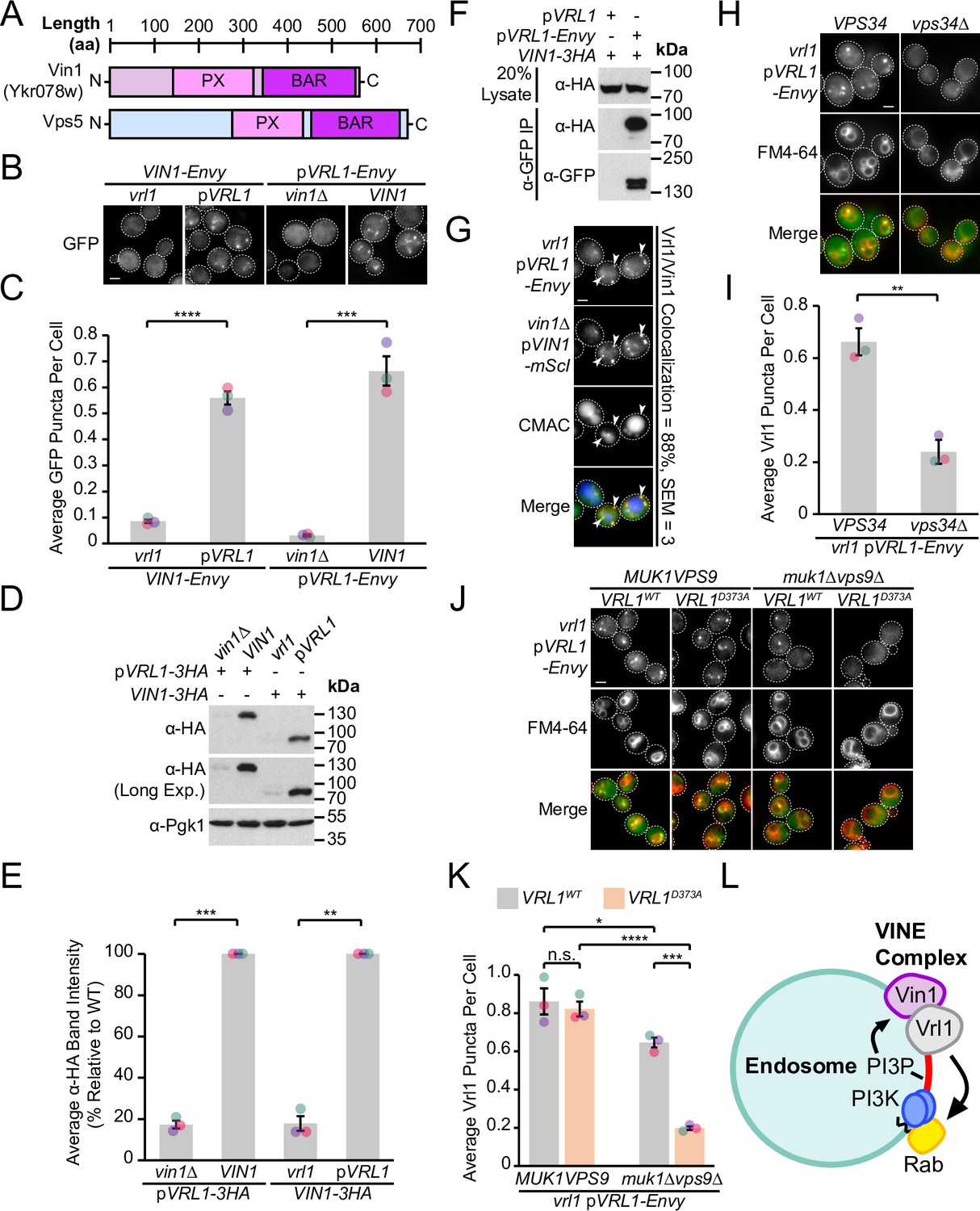
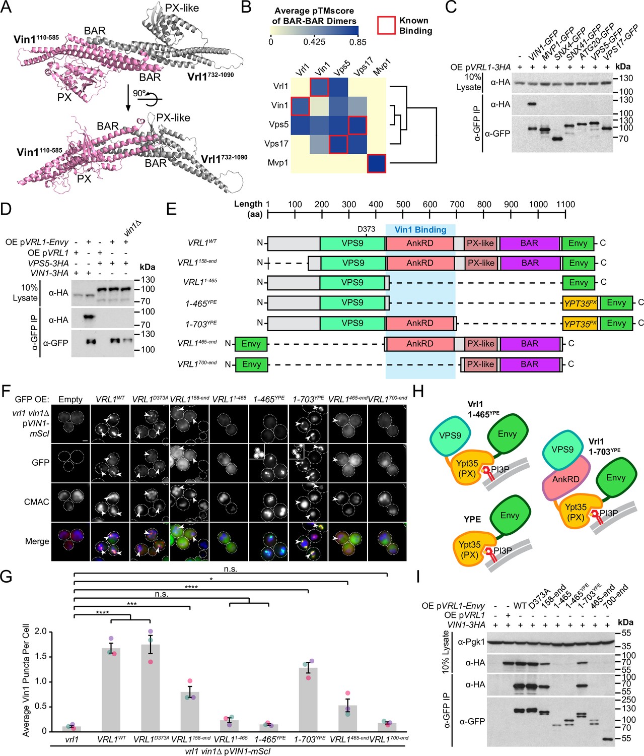
Vrl1 and the Vps5 paralog Vin1 form the VINE complex.
(A) Schematic of Ykr078w (Vin1) and its paralog Vps5. (B) Vin1-Envy and Vrl1-Envy require Vrl1 and Vin1, respectively, for localization to puncta. (C) Quantification of Vin1-Envy and Vrl1-Envy puncta per cell in B. Two tailed equal variance t tests; n=3, cells/strain/replicate ≥1,879; ***=p < 0.001, ****=p < 0.0001. (D) Vrl1-3HA and Vin1-3HA require Vin1 and Vrl1, respectively, for protein stability by western blot. Pgk1 serves as a loading control. (E) Quantification of Vrl1-3HA and Vin1-3HA levels in D by densitometry. Two tailed Welch’s t tests; n=3, **=p < 0.01, ***=p < 0.001. (F) Co-immunoprecipitation (CoIP) of Vin1-3HA with Vrl1-Envy suggests stable complex formation. (G) Vrl1-Envy colocalizes with Vin1-mScI at perivacuolar puncta. (H) Vrl1-Envy requires the PI3K catalytic subunit Vps34 for punctate localization. (I) Quantification of Vrl1-Envy puncta per cell in H. Two-tailed equal variance t test; n=3, cells/strain/replicate ≥897; **=p < 0.01. (J) Vrl1-Envy localization in the absence of VPS9-domain GEFs is dependent on the Vrl1 catalytic residue D373. (K) Quantification of Vrl1-Envy puncta per cell in J. One-way ANOVA with Tukey’s multiple comparison test; n=3, cells/strain/replicate ≥1705; not significant, n.s.=p > 0.05, *=p < 0.05, ***=p < 0.001, ****=p < 0.0001. (L) Model of the Vin1 and Vrl1-containing VINE complex at endosomes. Scale bars, 2 µm. Error bars report SEM. Exp., Exposure. WT, wild type.
-
Figure 2—source data 1
Data associated with Figure 2C.
- https://cdn.elifesciences.org/articles/77035/elife-77035-fig2-data1-v2.xlsx
-
Figure 2—source data 2
Data associated with Figure 2E.
- https://cdn.elifesciences.org/articles/77035/elife-77035-fig2-data2-v2.xlsx
-
Figure 2—source data 3
Data associated with Figure 2G.
- https://cdn.elifesciences.org/articles/77035/elife-77035-fig2-data3-v2.xlsx
-
Figure 2—source data 4
Data associated with Figure 2I.
- https://cdn.elifesciences.org/articles/77035/elife-77035-fig2-data4-v2.xlsx
-
Figure 2—source data 5
Data associated with Figure 2K.
- https://cdn.elifesciences.org/articles/77035/elife-77035-fig2-data5-v2.xlsx
-
Figure 2—source data 6
Uncropped blot data associated with Figure 2D.
- https://cdn.elifesciences.org/articles/77035/elife-77035-fig2-data6-v2.zip
-
Figure 2—source data 7
Uncropped blot data associated with Figure 2F.
- https://cdn.elifesciences.org/articles/77035/elife-77035-fig2-data7-v2.zip
Figure 2—figure supplement 1
Vrl1 is indispensable for Vin1 puncta localization.
(A) N-terminally tagged Vin1 requires Vrl1 for localization to puncta at three different expression levels. By relative fluorescence, the NOP1 promoter (NOP1pr) is stronger than the VIN1pr and the TEF2pr is stronger than the NOP1pr. Insets are scaled to match other images in the same channel (see Materials and methods for details). (B) Over-expression of Vrl1 from the ADH1pr increases the average number of bright Vin1-Envy puncta per cell. (C) Quantification of Vrl1-Envy puncta per cell in B. One-way ANOVA with Tukey’s multiple comparison test; n=3, cells/strain/replicate ≥1495; ***=p < 0.001, ****=p < 0.0001. Scale bars, 2 µm. Error bars report SEM.
-
Figure 2—figure supplement 1—source data 1
Data associated with Figure 2—figure supplement 1C.
- https://cdn.elifesciences.org/articles/77035/elife-77035-fig2-figsupp1-data1-v2.xlsx
Figure 3 with 3 supplements
Vrl1 interacts with Vin1 primarily through the AnkRD.
(A) ColabFold-predicted physical interaction of Vrl1 and Vin1 BAR domains along the canonical BAR-BAR dimerization interface. pTMscore = 0.75. (B) Matrix of ColabFold-predicted BAR-BAR dimers for select yeast SNX-BARs. Hierarchical clustering was performed using an uncentered Pearson correlation with average linkage. (C) Vin1 is the only yeast SNX-BAR that interacts with over-expressed Vrl1-3HA by CoIP. (D) Vps5-3HA does not bind to Vrl1-Envy in a strain lacking Vin1. (E) Schematic of Envy-tagged Vrl1 truncations and chimeras in F. (F) The Vrl1 AnkRD is necessary to recruit Vin1-mScI to puncta. Images with very bright signals use custom settings to show protein localization; insets are scaled identically to other images in the same channel (see materials and methods for details). (G) Quantification of Vin1-mScI puncta per cell in F. One-way ANOVA with Dunnett’s multiple comparison test; n=3, cells/strain/replicate ≥764; not significant, n.s.=p > 0.05, *=p < 0.05, **=p < 0.01, ***=p < 0.001, ****=p < 0.0001. (H) Diagram of chimeric Vrl1 fusion proteins that are artificially recruited to the endosomal system by the PX domain of sorting nexin Ypt35. (I) The Vrl1 AnkRD is necessary for physical interaction with Vin1-3HA by CoIP. Pgk1 serves as a loading control. Scale bars, 2 µm. Error bars report SEM. OE, over-expressed. YPE, Ypt35(PX)-Envy. WT, wild type.
-
Figure 3—source data 1
Data associated with Figure 3B.
- https://cdn.elifesciences.org/articles/77035/elife-77035-fig3-data1-v2.xlsx
-
Figure 3—source data 2
Data associated with Figure 3G.
- https://cdn.elifesciences.org/articles/77035/elife-77035-fig3-data2-v2.xlsx
-
Figure 3—source data 3
Uncropped blot data associated with Figure 3C.
- https://cdn.elifesciences.org/articles/77035/elife-77035-fig3-data3-v2.zip
-
Figure 3—source data 4
Uncropped blot data associated with Figure 3D.
- https://cdn.elifesciences.org/articles/77035/elife-77035-fig3-data4-v2.zip
-
Figure 3—source data 5
Uncropped blot data associated with Figure 3I.
- https://cdn.elifesciences.org/articles/77035/elife-77035-fig3-data5-v2.zip
Figure 3—figure supplement 1
Confidence measures of yeast SNX-BAR dimer predictions.
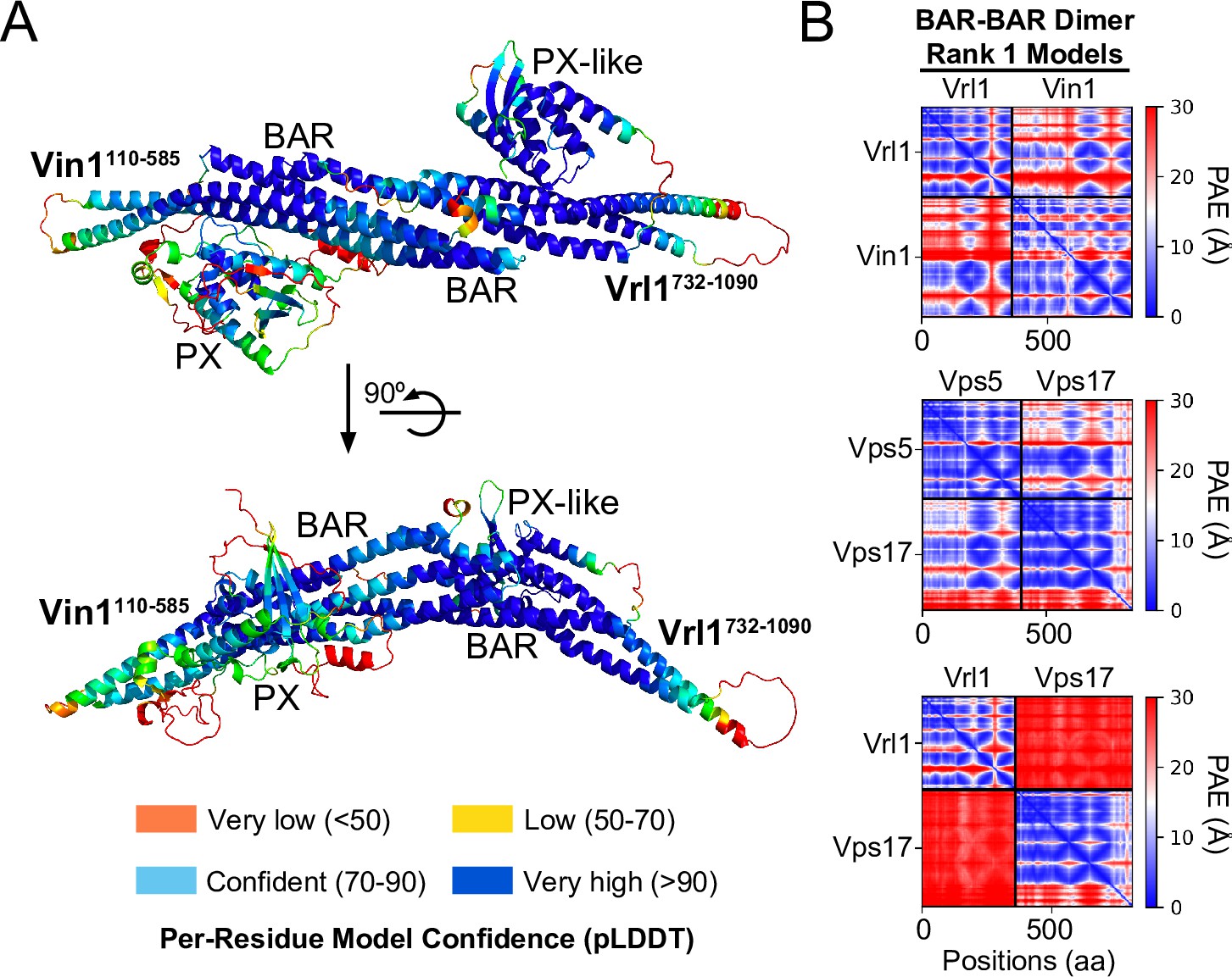
(A) ColabFold-predicted physical interaction of Vrl1 and Vin1 BAR domains with the predicted local-difference distance test (pLDDT; Jumper et al., 2021; Mirdita et al., 2022) scores mapped to each residue. (B) ColabFold-generated predicted alignment error (PAE; Jumper et al., 2021; Mirdita et al., 2022) plots for yeast SNX-BAR pairs demonstrate confidence of inter-chain contacts, indicating the relative likelihood of functional pairings. On-diagonal boxes with low PAE values reflect confidently predicted intramolecular interactions while off-diagonal boxes with low PAE values reflect confidently predicted intermolecular interactions between two separate polypeptide sequences. aa, amino acids.
Figure 3—figure supplement 2
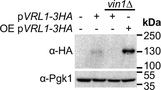
Over-expression of VRL1 restores protein levels in a vin1Δ mutant.
Vrl1 protein levels are restored in a vin1Δ mutant by western blot when VRL1 is over-expressed on the ADH1pr. Pgk1 serves as a loading control.
-
Figure 3—figure supplement 2—source data 1
Uncropped blot data associated with Figure 3—figure supplement 2.
- https://cdn.elifesciences.org/articles/77035/elife-77035-fig3-figsupp2-data1-v2.zip
Figure 3—figure supplement 3
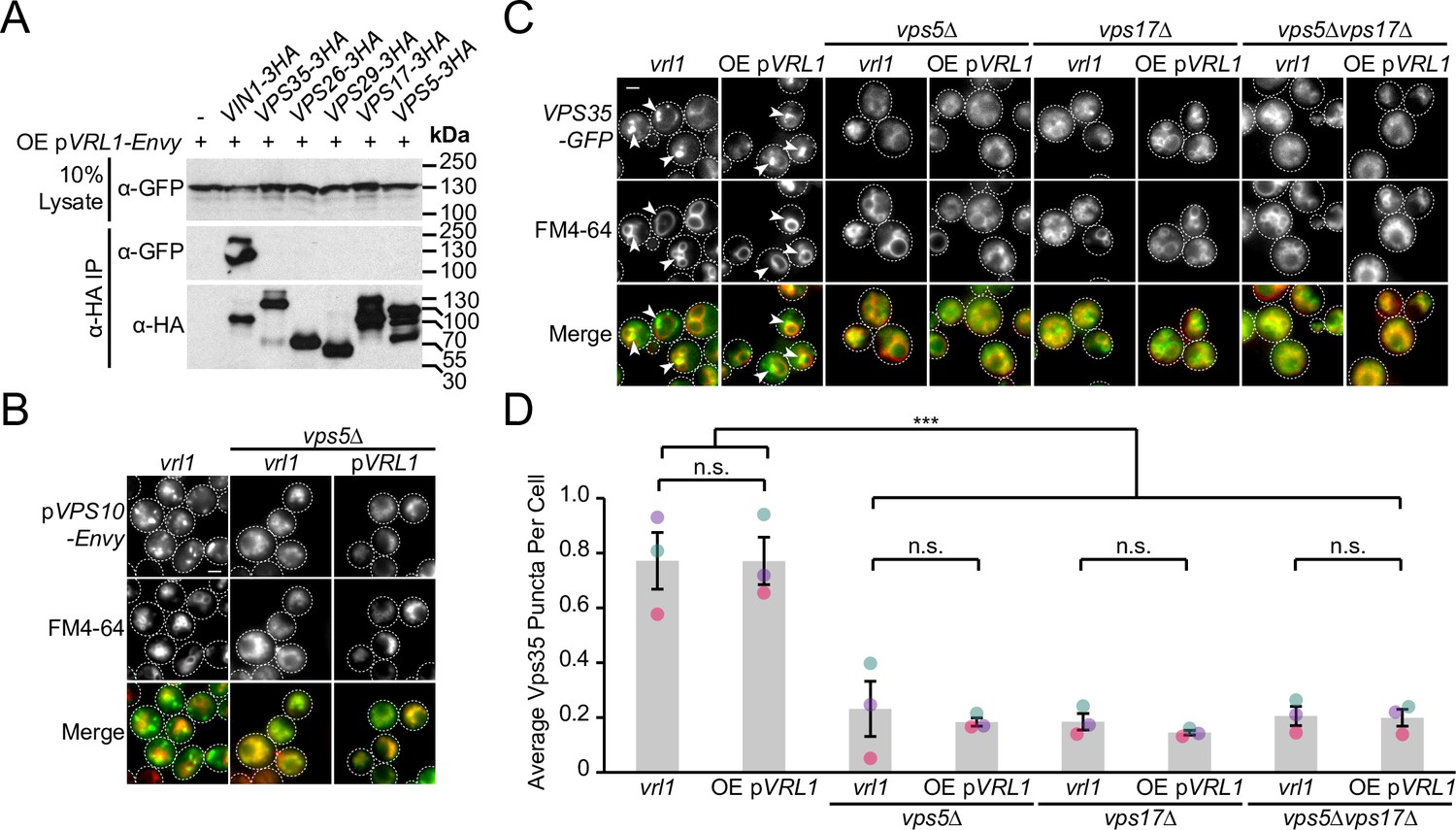
Vrl1 does not form a novel retromer-like complex.
(A) Vrl1 does not interact strongly with subunits of retromer by CoIP. (B) Expression of VRL1 does not rescue loss of endosomal Vps10-Envy in a vps5∆ strain. (C) Expression of VRL1 does not rescue loss of endosomal Vps35-GFP or vacuolar morphology defects in retromer SNX-BAR deletion strains. (D) Quantification of Vps35-GFP puncta per cell in C. One-way ANOVA with Tukey’s multiple comparison test; n=3, cells/strain/replicate ≥1243; not significant, n.s.=p > 0.05, ***=p < 0.001. Scale bars, 2 µm. Error bars report SEM. OE, over-expressed.
-
Figure 3—figure supplement 3—source data 1
Data associated with Figure 3—figure supplement 3D.
- https://cdn.elifesciences.org/articles/77035/elife-77035-fig3-figsupp3-data1-v2.xlsx
-
Figure 3—figure supplement 3—source data 2
Uncropped blot data associated with Figure 3—figure supplement 3A.
- https://cdn.elifesciences.org/articles/77035/elife-77035-fig3-figsupp3-data2-v2.zip
Figure 4 with 3 supplements

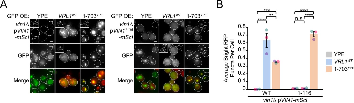
The Vrl1 AnkRD recognizes a small region of the Vin1 N-terminus.
(A) Schematic of constructs used in C, D. Full-length Vps5 was not tested but is shown for comparison. (B) Diagram of chimeric Vrl1 recruitment assay used to test for interactions with the unstructured N-terminus of either Vps5 (Vps51-276) or Vin1 (Vin11-116). (C) The AnkRD-containing Vrl1(1-703)YPE chimera recruits the N-terminus of Vin1, but not Vps5. Insets are scaled to match other images in the same channel (see Materials and methods for details). (D) Quantification of RFP puncta per cell in C. One-way ANOVA with Tukey’s multiple comparison test; n=3, cells/strain/replicate ≥902; not significant, n.s.=p > 0.05, **=p < 0.01, ***=p < 0.001, ****=p < 0.0001. (E) Schematic of Vin1 N-terminal fragments used to map the Vrl1 recruitment site. (F) The AnkRD-containing Vrl1(1-703)YPE chimera recruits a small fragment of the Vin1 N-terminus. Insets are scaled to match other images in the same channel. (G) Quantification of Vin1-mScI puncta per cell in F. Two-tailed equal variance t tests; n=3, cells/strain/replicate ≥294; not significant, n.s.=p > 0.05, *=p < 0.05, **=p < 0.01. Scale bars, 2 µm. Error bars report SEM. OE, over-expressed. FL, full-length. WT, wild type. YPE, Ypt35(PX)-Envy.
-
Figure 4—source data 1
Data associated with Figure 4D.
- https://cdn.elifesciences.org/articles/77035/elife-77035-fig4-data1-v2.xlsx
-
Figure 4—source data 2
Data associated with Figure 4G.
- https://cdn.elifesciences.org/articles/77035/elife-77035-fig4-data2-v2.xlsx
Figure 4—figure supplement 1
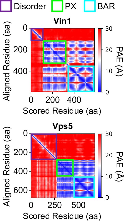
The N-terminal regions of Vin1 and Vps5 are predicted to be disordered.
A lack of off-diagonal signal in the ColabFold-generated predicted alignment error (PAE; Jumper et al., 2021; Mirdita et al., 2022) plots of Vin1 and Vps5 indicates a shared lack of structure in the N-terminus of either protein. On-diagonal signal for the PX and BAR domains provides a point of comparison for how structured domains appear in prediction PAE plots. aa, amino acids.
Figure 4—figure supplement 2
The Vin1 PX-BAR region is indispensable for Vrl1 localization.
(A) The Vin1 N-terminus is not sufficient to localize over-expressed Vrl1-Envy to membranes in a VIN1 deletion strain. The Vrl1(1-703)YPE chimera recruits the Vin1 N-terminal prey construct to puncta in strains lacking the chromosomal copy of VIN1. Insets are scaled to match other images in the same channel (see Materials and methods for details). (B) Quantification of RFP puncta per cell in A. One-way ANOVA with Tukey’s multiple comparison test; n=3, cells/strain/replicate ≥1068; not significant, n.s.=p > 0.05, **=p < 0.01, ***=p < 0.001, ****=p < 0.0001. Scale bars, 2 µm. Error bars report SEM. WT, wild type. YPE, Ypt35(PX)-Envy.
-
Figure 4—figure supplement 2—source data 1
Data associated with Figure 4—figure supplement 2B.
- https://cdn.elifesciences.org/articles/77035/elife-77035-fig4-figsupp2-data1-v2.xlsx
Figure 4—figure supplement 3
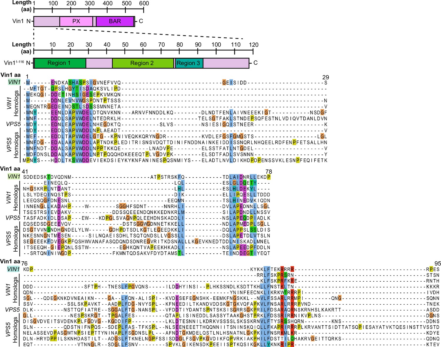
The Vin1 N-terminus has three conserved regions in fungal homologs.
Sequence alignment of Vin1 and Vps5 orthologs collected using the Yeast Gene Order Browser (http://ygob.ucd.ie/). Three conserved regions were selected for expression as mScI-tagged fragments: Vin11-29 (Region 1), Vin141-78 (Region 2), and Vin176-95 (Region 3). aa, amino acids.
Figure 5 with 2 supplements

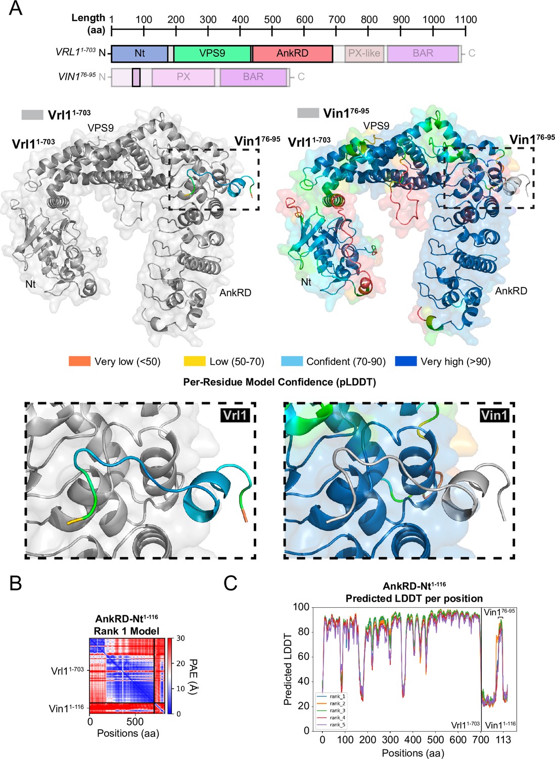
The Vrl1 AnkRD associates with the Vin1 N-terminus through electrostatic interactions.
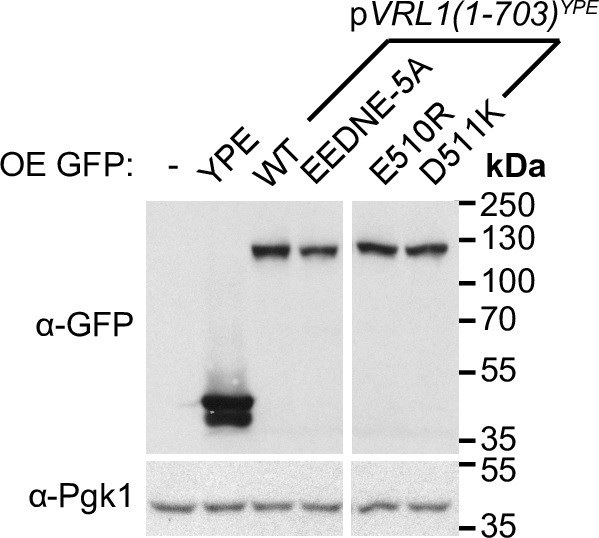
(A) Schematic of query sequences used to predict the interact between Vrl1 and the Vin1 N-terminus. Modelled regions are shown as completely opaque. (B) ColabFold-predicted interaction between the Vrl1 AnkRD and a minimal fragment of the Vin1 N-terminus (Vin176-95; pTMscore = 0.73). (C) Vrl1 sequence conservation within family Saccharomycetaceae determined by ConSurf and mapped to a surface model that was predicted by ColabFold. Strong sequence conservation can be seen at the predicted Vin176-95 interacting site and near the catalytic D373 residue. (D) Top: Vin176-95 is predicted to associate with Vrl1 through a run of basic residues. Bottom: Acidic and polar residues in the predicted Vin1-associating Vrl1 AnkRD site are among the most conserved within family Saccharomycetaceae. (E) Mutation of acidic and polar residues in the Vrl1 AnkRD reduces recruitment of the Vin1 N-terminus by the Vrl1(1-703)YPE chimera. (F) Quantification of Vin11-116-mScI puncta per cell in E. One-way ANOVA with Tukey’s multiple comparison test; n=3, cells/strain/replicate ≥863; not significant, n.s.=p > 0.05, *=p < 0.05, ***=p < 0.001, ****=p < 0.0001. Scale bars, 2 µm. Error bars report SEM. OE, over-expressed. Nt, N-terminus. WT, wild type. aa, amino acids. YPE, Ypt35(PX)-Envy.
-
Figure 5—source data 1
Data associated with Figure 5F.
- https://cdn.elifesciences.org/articles/77035/elife-77035-fig5-data1-v2.xlsx
Figure 5—figure supplement 1
Confidence measures of Vrl1 AnkRD-Vin1 N-terminus binding predictions.
(A) ColabFold-predicted interaction between the Vrl1 AnkRD and a minimal fragment of the Vin1 N-terminus (Vin176-95; pTMscore = 0.73) with pLDDT scores mapped to each residue of Vin1 (left) or Vrl1 (right). In each case, the interacting protein partner is shown in gray. (B) ColabFold-generated predicted alignment error (PAE; Jumper et al., 2021; Mirdita et al., 2022) plot for Vrl1(1-703) and the Vin1 N-terminus (Vin11-116) indicates an intermolecular interaction in the off-diagonal boxes. (C) A plot of predicted local-difference distance test (pLDDT; Jumper et al., 2021; Mirdita et al., 2022) scores for five different models of Vrl1(1-703) and Vin11-116 indicates that the minimal region of Vin1 that was recruited by the Vrl1(1-703)YPE chimera, Vin176-95, is the sole confidently predicted region. aa, amino acids. Nt, N-terminus.
Figure 5—figure supplement 2
Vrl1(1-703)YPE chimeras with AnkRD mutations are stably expressed.
AnkRD-containing Vrl1(1-703)YPE chimeras are stable by western blot when mutations are introduced to sites that are predicted to interact with the Vin1 N-terminus. Pgk1 serves as a loading control. OE, over-expressed. WT, wild type. YPE, Ypt35(PX)-Envy.
-
Figure 5—figure supplement 2—source data 1
Uncropped blot data associated with Figure 5—figure supplement 2.
- https://cdn.elifesciences.org/articles/77035/elife-77035-fig5-figsupp2-data1-v2.zip
Figure 6
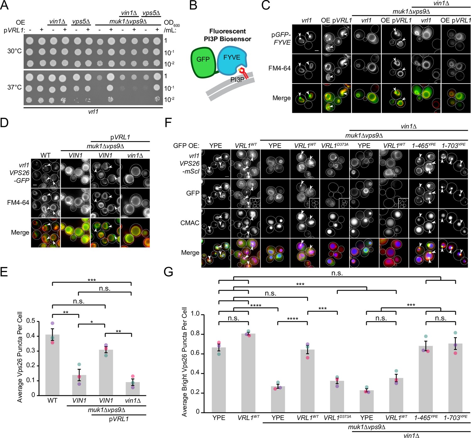
Vin1 controls Vrl1 GEF activity via membrane localization.
(A) Deletion of VIN1, but not VPS5, prevents Vrl1 from rescuing the temperature sensitivity of a strain lacking other VPS9-domain GEFs. (B) Schematic of PI3P-binding fluorescent biosensor. (C) Deletion of VIN1 prevents Vrl1 from stimulating endosomal PI3P production in a strain lacking other VPS9-domain GEFs. (D) Deletion of VIN1 prevents Vrl1 from rescuing Vps26-GFP localization in a strain lacking other VPS9-domain GEFs. (E) Quantification of Vps26-GFP puncta per cell in D. One-way ANOVA with Tukey’s multiple comparison test; n=3, cells/strain/replicate ≥1503; not significant, n.s.=p > 0.05, *=p < 0.05, **=p < 0.01, ***=p < 0.001. (F) Vin1 is dispensable for Vrl1 activity when fragments containing the N-terminus and VPS9 domain are artificially recruited by a YPE endosomal anchor. Insets are scaled to match other images in the same channel (see materials and methods for details). (G) Quantification of Vps26-mScI puncta per cell in F. One-way ANOVA with Tukey’s multiple comparison test; n=3, cells/strain/replicate ≥750; not significant, n.s.=p > 0.05, ***=p < 0.001, ****=p < 0.0001. Scale bars, 2 µm. Error bars report SEM. OE, over-expressed. WT, wild type. YPE, Ypt35(PX)-Envy.
-
Figure 6—source data 1
Data associated with Figure 6E.
- https://cdn.elifesciences.org/articles/77035/elife-77035-fig6-data1-v2.xlsx
-
Figure 6—source data 2
Data associated with Figure 6G.
- https://cdn.elifesciences.org/articles/77035/elife-77035-fig6-data2-v2.xlsx
Figure 7 with 1 supplement

The VINE complex exhibits characteristics of a membrane sorting complex.
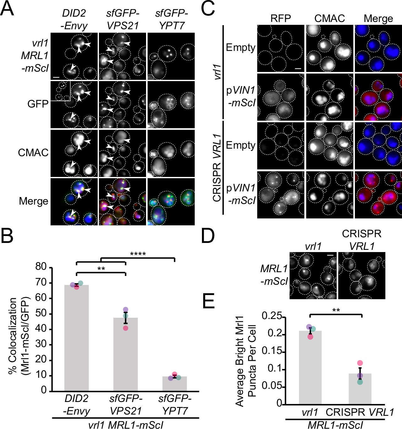
(A) Time-lapse imaging of cells over-expressing GFP-Vin1 and Vrl1 show tubules emanating from Did2-labeled endosomes. Images were uniformly enlarged using a bicubic expansion function to show detail. Solid arrowheads mark a tubule, open arrowhead marks a scission event. (B) Normalized intensity line scan analysis performed on images from A along the yellow dotted line. (C) Punctate localization of GFP-tagged Mrl1, but not other endosomal recycling cargo, is decreased in cells expressing VRL1. (D) Quantification of GFP-tagged puncta in WT and vrl1 strains in C. Two tailed Welch’s t tests; n=3, cells/strain/replicate ≥902; not significant, n.s.=p > 0.05, *=p < 0.05. (E) Mutation of the D373 residue required for VPS9 GEF activity does not prevent Vrl1 from redistributing Mrl1. (F) Quantification of Mrl1-mScI puncta per cell in E. One-way ANOVA with Tukey’s multiple comparison test; n=3, cells/strain/replicate ≥1788; not significant, n.s.=p > 0.05, *=p < 0.05. (G) Schematic of Vps10 cytosolic tail mutant and Mrl1 cytosolic tail chimera tested for VINE-mediated sorting in H, I. (H) The Mrl1 cytosolic tail is sufficient to confer VINE-mediated redistribution. (I) Percent of cells showing punctate localization of indicated GFP-tagged constructs in H. Blind scoring of GFP signal was conducted manually. One-way ANOVA with Tukey’s multiple comparison test; n=3, cells/strain/replicate ≥237; not significant, n.s.=p > 0.05, ***=p < 0.001, ****=p < 0.0001. (J) Mrl1-mScI puncta are reduced in a snx4∆ strain. (K) Quantification of Mrl1-mScI puncta per cell in J. One-way ANOVA with Tukey’s multiple comparison test; n=3, cells/strain/replicate ≥1036; not significant, n.s.=p > 0.05, *=p < 0.05, **=p < 0.01, ***=p < 0.001, ****=p < 0.0001. (L) Model for VINE activity and redistribution of Mrl1. VINE promotes its own recruitment to endosomes through a positive feedback loop involving Vrl1 GEF activity and local PI3P production. VINE-coated tubules then recycle cargo, such as Mrl1, from endosomes. VINE may target Mrl1 to the Golgi for subsequent delivery to the vacuolar membrane by the AP-3 complex. Mrl1 is then returned to the endosome by Snx4-containing complexes. See text for details. Scale bars, 2 µm. Error bars report SEM. OE, over-expressed. TM, transmembrane. WT, wild type.
-
Figure 7—source data 1
Data associated with Figure 7B.
- https://cdn.elifesciences.org/articles/77035/elife-77035-fig7-data1-v2.xlsx
-
Figure 7—source data 2
Data associated with Figure 7D.
- https://cdn.elifesciences.org/articles/77035/elife-77035-fig7-data2-v2.xlsx
-
Figure 7—source data 3
Data associated with Figure 7F.
- https://cdn.elifesciences.org/articles/77035/elife-77035-fig7-data3-v2.xlsx
-
Figure 7—source data 4
Data associated with Figure 7I.
- https://cdn.elifesciences.org/articles/77035/elife-77035-fig7-data4-v2.xlsx
-
Figure 7—source data 5
Data associated with Figure 7K.
- https://cdn.elifesciences.org/articles/77035/elife-77035-fig7-data5-v2.xlsx
Figure 7—figure supplement 1
Vrl1 redistributes Mrl1 puncta from endosomes.
(A) Mrl1-mScI puncta colocalize with Did2-Envy and sfGFP-Vps21-labeled endosomes, but not with sfGFP-Ypt7 puncta that label vacuolar sites. Insets are scaled to match other images in the same channel (see Materials and methods for details). (B) Quantification of colocalization as the percentage of Mrl1 puncta overlapping GFP puncta in A. One-way ANOVA with Tukey’s multiple comparison test; n=3, cells/strain/replicate ≥2286; **=p < 0.01, ****=p < 0.0001. (C) Localization of Vin1-mScI in BY4741 (vrl1) and an isogenic strain with the vrl1 mutation corrected at the endogenous VRL1 locus using CRISPR-Cas9 gene editing technology. Vin1-mScI localizes to puncta in a CRISPR-corrected VRL1 strain. (D) Mrl1-mScI is redistributed from puncta in the CRISPR-corrected VRL1 strain. (E) Quantification of large, bright Mrl1-mScI puncta per cell in D. Two tailed equal variance t test; n=3, cells/strain/replicate ≥1183; **=p < 0.01. Scale bars, 2 µm. Error bars report SEM.
-
Figure 7—figure supplement 1—source data 1
Data associated with Figure 7—figure supplement 1B.
- https://cdn.elifesciences.org/articles/77035/elife-77035-fig7-figsupp1-data1-v2.xlsx
-
Figure 7—figure supplement 1—source data 2
Data associated with Figure 7—figure supplement 1E.
- https://cdn.elifesciences.org/articles/77035/elife-77035-fig7-figsupp1-data2-v2.xlsx
Author response image 1
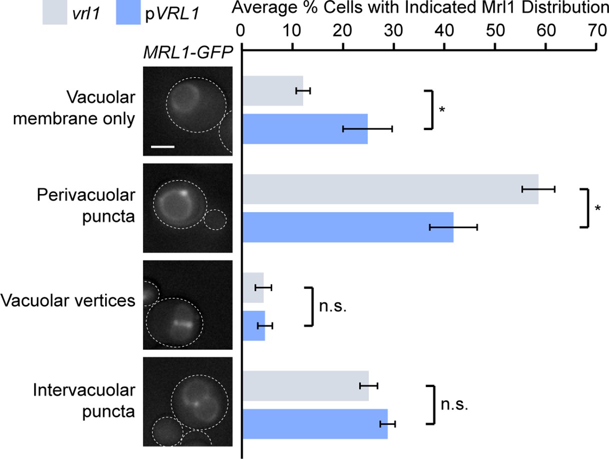
Vrl1 specifically redistributes Mrl1 from endosomes.
Quantification of Mrl1-GFP localization in vrl1 and VRL1-expressing strains. Blind scoring of GFP signal was conducted manually. Unpaired equal variance t tests; n = 3, cells/strain/replicate ≥ 465; not significant, n.s. = p > 0.05, * = p < 0.05. Scale bar, 2 µm. Error bars report standard error of the mean (SEM).
Tables
Key resources table
| Reagent type (species) or resource | Designation | Source or reference | Identifiers | Additional information |
|---|---|---|---|---|
| Antibody | Anti-HA (Mouse monoclonal) | Sigma-Aldrich | H9658; HA-7 | WB (1:1000) |
| Antibody | Anti-HA (Mouse monoclonal) | Covance | MMS-101R; HA.11 | WB (1:1000) |
| Antibody | Anti-GFP (Mouse monoclonal) | Roche | 11-814-460-001 | WB (1:1000) |
| Antibody | Anti-Pgk1 (Mouse monoclonal) | Invitrogen | AB_2532235; 22C5D8 | WB (1:1000) |
| Antibody | Anti-GFP (Rabbit polyclonal) | Eusera | EU2 | CoIP |
| Antibody | Anti-HA (Rabbit polyclonal) | AbCam | Ab9110 | CoIP |
| Antibody | HRP-Anti-Mouse (Goat polyclonal) | Jackson | 115-035-146 | WB (1:20,000) |
| Other | nProtein A Sepharose 4 Fast Flow | Cytiva | 17528004 | CoIP |
| Other | Amersham Hyperfilm | GE Healthcare | 28906839 | Chemiluminescent film |
| Other | Amersham ECL | Cytiva | GERPN2209 | Chemiluminescent reagent |
| Other | Amersham ECL Prime | Cytiva | GERPN2232 | Chemiluminescent reagent |
| Other | Yeast/Fungal ProteaseArrest | GBiosciences | 786–435 | Yeast protease inhibitor; 100 X |
| Other | Concanavalin A | Sigma-Aldrich | C2010 | Yeast live-cell imaging preparation |
| Other | FM4-64 | Invitrogen | T3166 | Vacuolar Rim Stain; 4 μM |
| Other | CMAC | Setareh Biotech | 6627 | Vacuole Stain; 100 µM |
| Chemical compound, drug | Methotrexate | Enzo Life Sciences | ALX-440–045 G001 | DHFR inhibitor; 200 μg/mL, DMSO |
| Software, algorithm | CellProfiler | Lamprecht et al., 2007 | Yeast colony array image analysis | |
| Software, algorithm | MetaMorph | MDS Analytical Technologies | Version 7.8 | Automated image analysis |
| Software, algorithm | GraphPad Prism | GraphPad Software | Version 9.1.0 | Statistical analysis |
| Software, algorithm | ImageJ | NIH | Band densitometry | |
| Software, algorithm | ColabFold AlphaFold2 Advanced | Jumper et al., 2021; Mirdita et al., 2022 | Protein structure and binding prediction software |
Additional files
-
Supplementary file 1
Vrl1 DHFR Interactors.
List of Z-scores from the Vrl1 DHFR screen.
- https://cdn.elifesciences.org/articles/77035/elife-77035-supp1-v2.xlsx
-
Supplementary file 2
Vrl1 DHFR Ontology Enrichment.
List of enriched ontology terms for Vrl1 DHFR interactors (Z>2).
- https://cdn.elifesciences.org/articles/77035/elife-77035-supp2-v2.xlsx
-
Supplementary file 3
Yeast SNX-BAR Dimer Predictions.
Results of pairwise yeast SNX-BAR prediction matrix generated using ColabFold.
- https://cdn.elifesciences.org/articles/77035/elife-77035-supp3-v2.xlsx
-
Supplementary file 4
List of Saccharomyces cerevisiae strains used in this study.
- https://cdn.elifesciences.org/articles/77035/elife-77035-supp4-v2.xlsx
-
Supplementary file 5
List of plasmids used in this study.
- https://cdn.elifesciences.org/articles/77035/elife-77035-supp5-v2.xlsx
-
Transparent reporting form
- https://cdn.elifesciences.org/articles/77035/elife-77035-transrepform1-v2.pdf
Download links
A two-part list of links to download the article, or parts of the article, in various formats.
Downloads (link to download the article as PDF)
Open citations (links to open the citations from this article in various online reference manager services)
Cite this article (links to download the citations from this article in formats compatible with various reference manager tools)
The VINE complex is an endosomal VPS9-domain GEF and SNX-BAR coat
eLife 11:e77035.
https://doi.org/10.7554/eLife.77035


