Lung evolution in vertebrates and the water-to-land transition
- Departamento de Zoologia-IBRAG, Universidade do Estado do Rio de Janeiro, Brazil
- Department of Earth and Planetary Science, Graduate School of Science, The University of Tokyo, Japan
- Department of Anatomy, The Jikei University School of Medicine, Japan
- Kitakyushu Museum of Natural History and Human History, 2-4-1 Higashida, Yahatahigashi-ku, Kitakyushu, Japan
- Institute of Earth Sciences, University of Lausanne, Switzerland
- Université Paris-Saclay, CNRS, ministère de la Culture, UVSQ, MNHN, Institut photonique d’analyse non-destructive européen des matériaux anciens, France
- Department of Anatomy, Iwate Medical University School of Medicine, Japan
- Kanagawa Prefectural Museum of Natural History, Japan
- Atmosphere and Ocean Research Institute, The University of Tokyo, Japan
- Seikei Education and Research Center for Sustainable Development, Japan
- Synchrotron SOLEIL, L’orme des Merisiers Saint-Aubin, France
- Japan Synchrotron Radiation Research Institute (JASRI/SPring-8), Japan
Figures
Figure 1 with 2 supplements

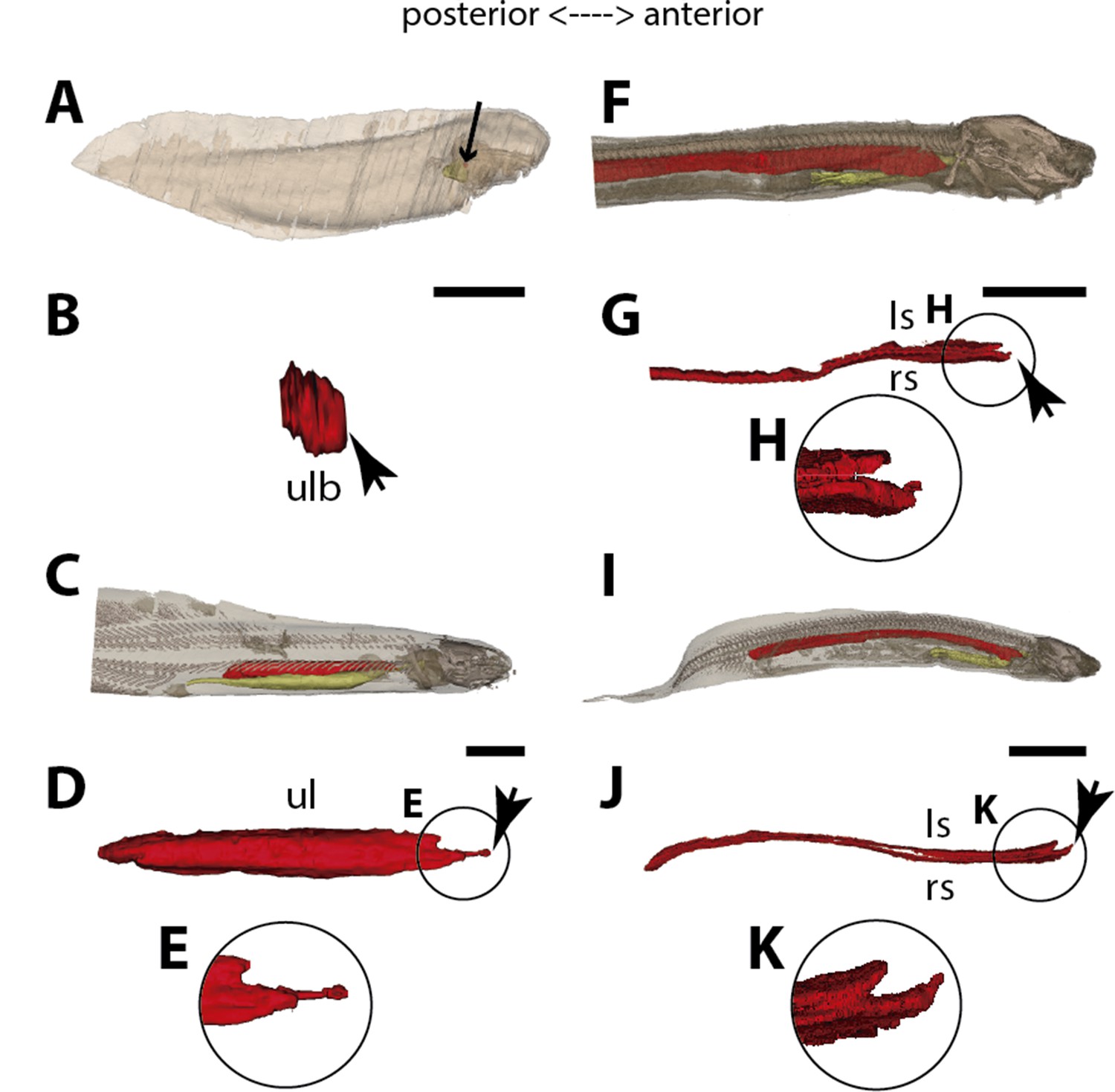
Three-dimensional reconstructions of the pulmonary complex of Polypterus senegalus.
(A) Early embryo (9.3mm) total length (TL) in right lateral view, (B) isolated lung of the early embryo in dorsal view, (C) juvenile (45mm TL) in right lateral view, (D) isolated lung of the juvenile in dorsal view, (E) close-up of (D) highlighting the lung in ventral view and pointing out the region of the independent and secondary connection of the left sac to the right one by a lateral opening. Yellow, foregut including the stomach; red, lung. Black arrow in (A) pointing to the lung. Arrowheads in (B) pointing to the lung connection to the foregut and in (D) pointing to the pneumatic duct connection to the foregut. Black arrow in (E) pointing to the independent connection. Ls, left sac; rs, right sac; ulb, unpaired lung bud. Scale bars, 5.0mm (A); 0.075mm (B); 5.0mm (C, D); 1.0mm (E).
Figure 1—figure supplement 1
Sections of synchrotron x-ray microtomography of a juvenile of Polypterus senegalus (23 mm total length; TL).
(A) Unpaired lung origin. (B) Right sac arising from the foregut. (C) Left sac arising from an independent and lateral connection to the right sac. (D) Right and left sacs. Yellow, foregut; red, lung. Orange arrow, opened connection between foregut and lung. Fg, foregut; ls, left sac; rs, right sac. Scale bars, 0.5 mm (A–D).
Figure 1—figure supplement 2
Three-dimensional reconstructions of the pulmonary complex of Polypterus senegalus.
(A) Virtual section of the juvenile (45 mm total length; TL) in anterior view, evidencing the esophagus and the lung in 3D. (B) Isolated right lung of the juvenile in lateral view, evidencing the independent and secondary connection of the left sac to the right one by a lateral opening. Yellow, foregut including the stomach; red, right sac; blue, left sac, dashed line, independent and secondary connection of the left sac to the right one. Scale bars, 1.0 mm (A, B).
Figure 2
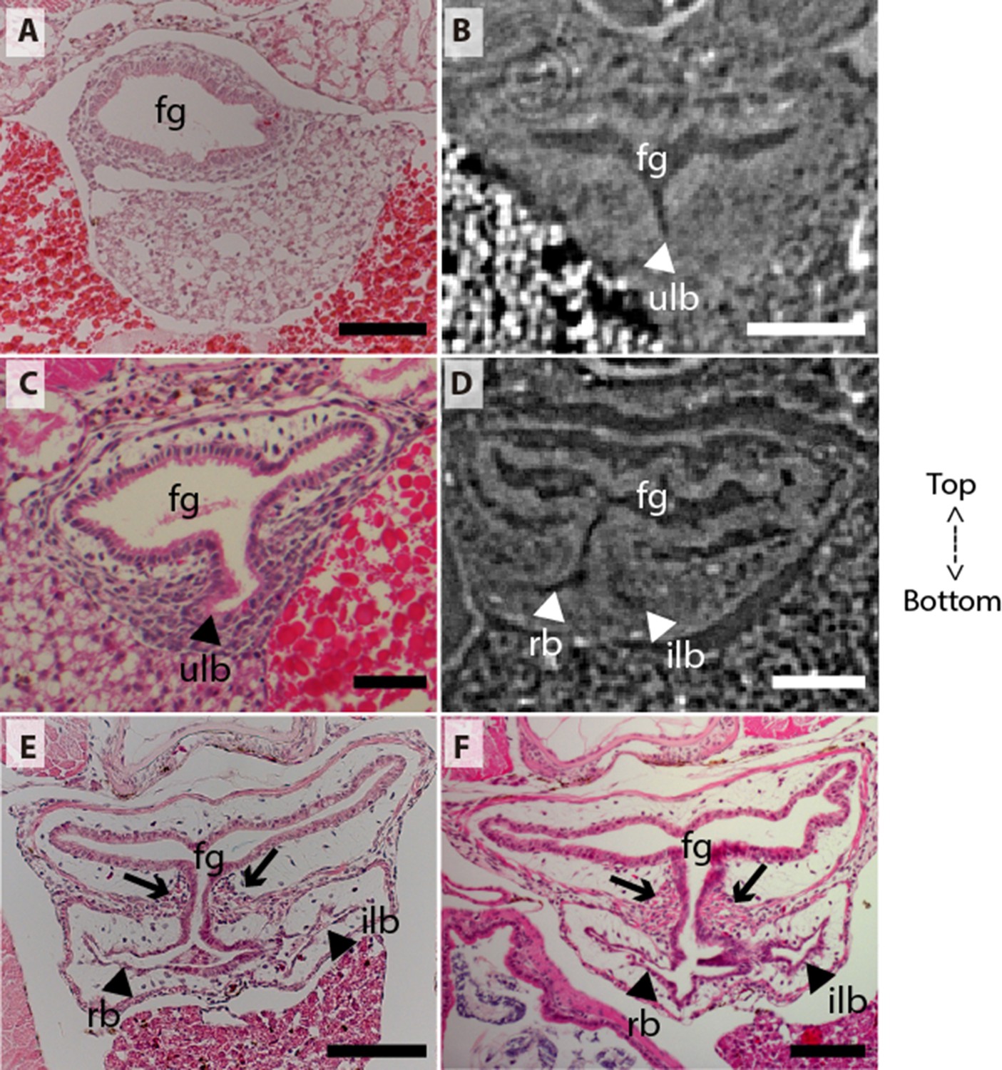
Coronal sections of the unpaired lung in the living actinopterygian fish Polypterus senegalus.
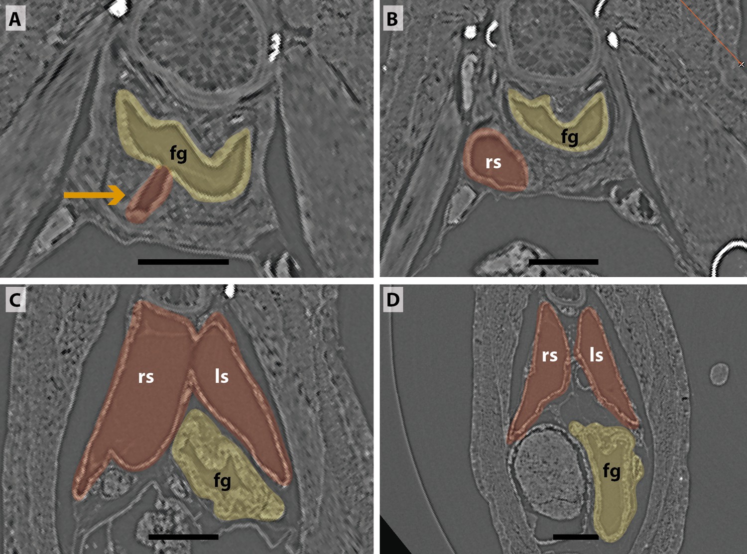
(A) No lung bud in 8.0mm total length (TL) specimen, (B) origin of an unpaired lung bud in 8.5mm TL specimen, (C) unpaired lung bud in 9.1mm TL specimen, (D) first register of an independent and lateral second lung bud in 12mm TL specimen, (E, F) independent and lateral second lung bud arising from the principal tube in 15.5mm TL and 18mm TL specimens. (A, C, E–F) Histological thin-sections. (B, D) Sections of synchrotron x-ray microtomography of the early embryo. Black and white head arrows pointing to the lumen of the unpaired lung buds; arrows pointing to the undifferentiated cells surrounding the glottis. Fg, foregut; ilb, independent lateral bud; rb, right bud; ulb, unpaired lung bud. Scale bars, 0.2mm (A, E); 0.1mm (B, D, F); 0.05mm (C).
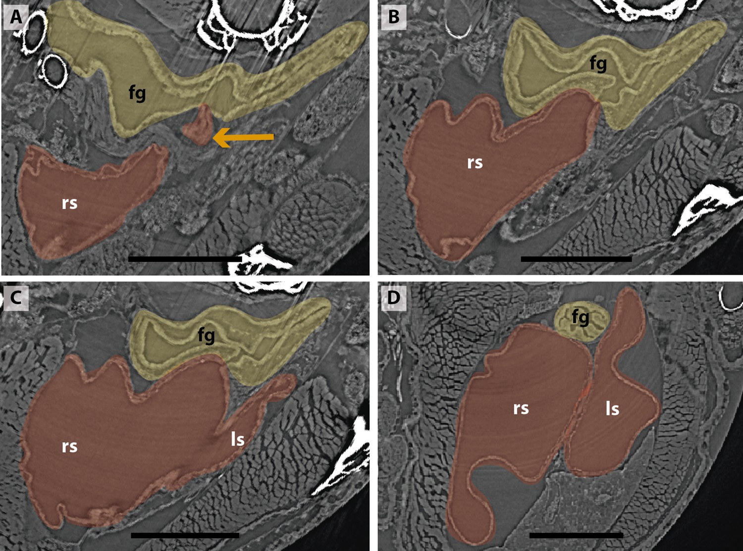
Figure 3

Three-dimensional reconstructions of the pulmonary complex of Latimeria chalumnae.
(A) Early embryo of Latimeria chalumane (45mm total length; TL) in right lateral view (Cupello et al., 2015), (B) isolated unpaired lung of the early embryo in dorsal view, (C) adult specimen of Latimeria chalumnae (1300mm TL) in right lateral view (Cupello et al., 2015), (D) isolated unpaired lung of the adult specimen in dorsal view. Yellow, foregut including the stomach; red, lung. Arrowheads in (B) and (D) pointing to the lung connection to the foregut. Black arrow in (C) pointing to the lung. Ul, unpaired lung bud in (B) and unpaired lung in (D). Scale bars, 5.0mm (A); 5.0mm (B); 200.0mm (C); 40mm (D). Modified from Cupello et al., 2015.
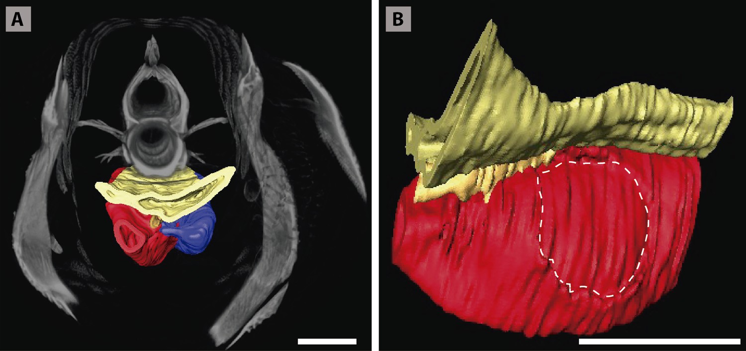
Figure 4 with 3 supplements
Three-dimensional reconstructions of the pulmonary complex of two species of lungfishes.
(A) Early embryo of Neoceratodus forsteri (13.5mm total length; TL) in right lateral view, (B) isolated unpaired lung of the early embryo in dorsal view, (C) adult specimen of Neoceratodus forsteri (200mm TL) in right lateral view, (D) isolated unpaired lung of the adult specimen in dorsal view, (E) close-up of the lung unpaired connection to the foregut in (D), (F) larva of Lepidosiren paradoxa (46mm TL) in lateral view, (G) isolated lung of the larval specimen in dorsal view, (H) close-up of the lung unpaired connection to the foregut in (G), (I) juvenile of Lepidosiren paradoxa young adult (68mm TL) in lateral view, (J) isolated lung of the juvenile specimen in dorsal view, (K) close-up of the lung unpaired connection to the foregut in (J).Yellow, foregut including the stomach; red, lung. Black arrow in (A) pointing to the lung. Arrowheads in (B), pointing to the lung connection to the foregut and in (D), (G) and (J) pointing the pneumatic duct connection to the foregut. Ls, left sac; rs, right sac; ul, unpaired lung; ulb, unpaired lung bud. Scale bars, 2.5mm (A); 0.1mm (B); 20mm (C); 10mm (D, I); 7.2 mm (J); 5.0mm (F, G).
Figure 4—figure supplement 1
Sections of synchrotron x-ray microtomography of a larva of Neoceratodus forsteri (19 mm total length; TL).
(A) Unpaired lung origin. (B) Unique sac arising from the foregut. (C, D) Unique sac developing. Yellow, foregut; red, lung. Orange arrow, opened connection between foregut and lung. fg, foregut; us, unique sac. Scale bars, 0.5 mm (A–D).
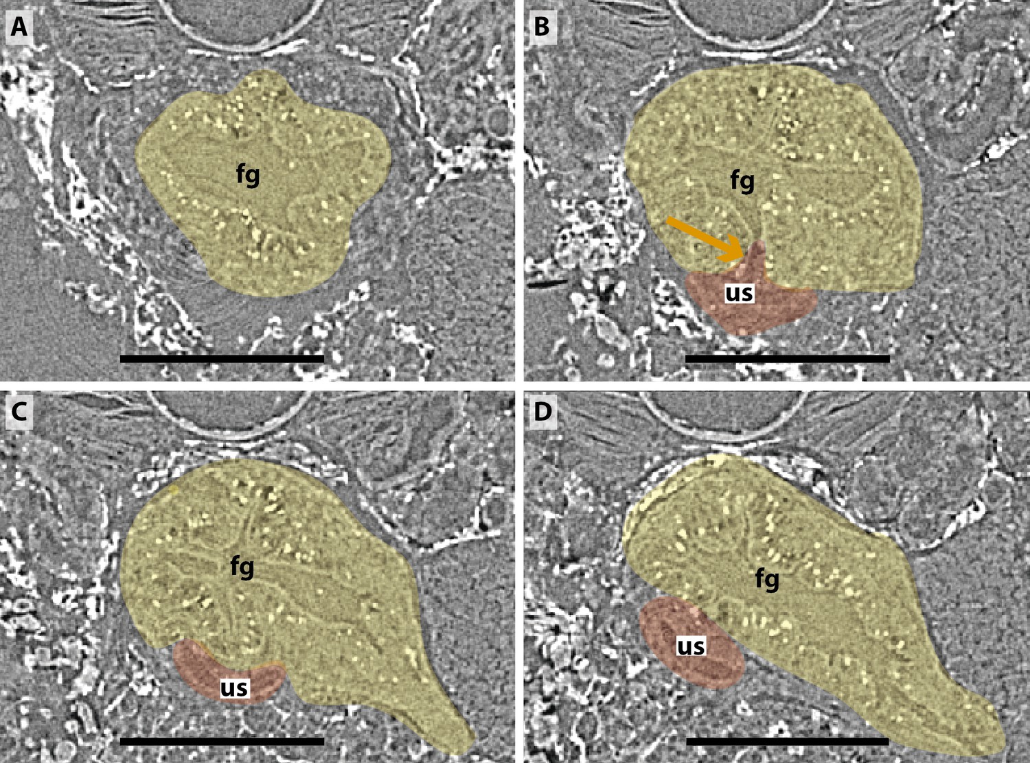
Figure 4—figure supplement 2
Sections of synchrotron x-ray microtomography of a juvenile of Lepidosiren paradoxa (68mm total length; TL).
(A) Unpaired lung origin. (B) Right sac arising from the foregut. (C) Left sac arising from an independent and lateral connection to the right sac. (D) Right and left sacs. Yellow, foregut; red, lung. Orange arrow, opened connection between foregut and lung. Fg, foregut; ls, left sac; rs, right sac. Scale bars, 0.5mm (A–D).
Figure 4—figure supplement 3
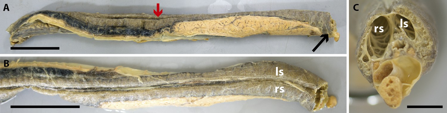
Dissection of the lung of an adult Lepidosiren paradoxa (400mm total length; TL).
Red arrow, lung. Black arrow, ventral insertion of the right sac. Ls, left sac; rs, right sac. Scale bars, 50mm (A, B); 10mm (C).
Figure 5 with 1 supplement
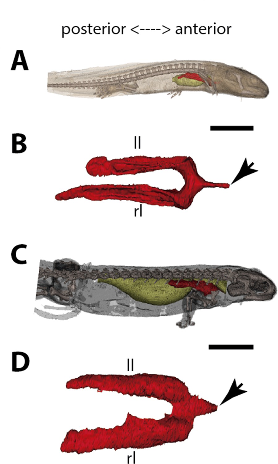
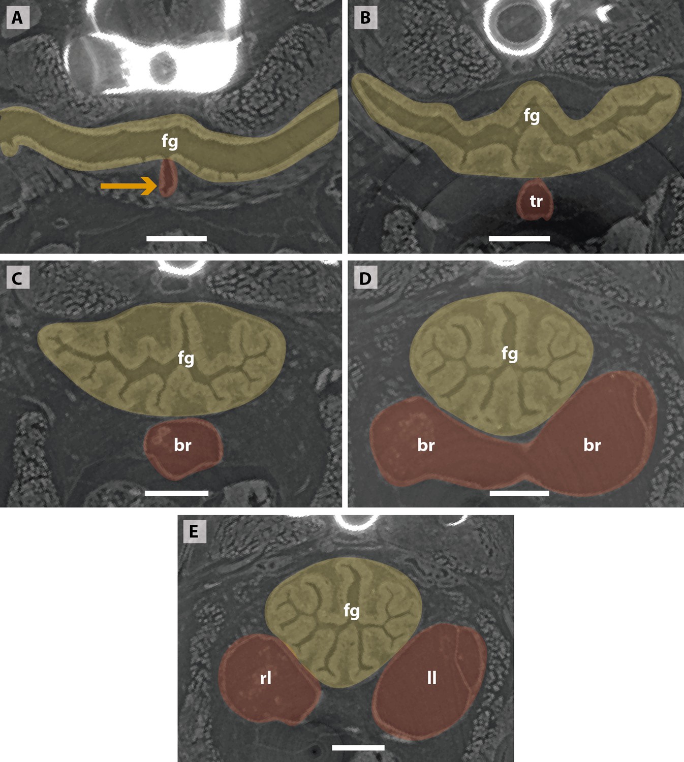
Three-dimensional reconstructions of the pulmonary complex of Salamandra salamandra.
(A) Early larva of Salamandra salamandra (35.5mm total length; TL) in right lateral view, (B) isolated paired lung of the larva embryo in dorsal view, (C) juvenile of Salamandra salamandra (81.85mm TL) in right lateral view, (D) isolated paired lung of the juvenile specimen in dorsal view. Yellow, foregut including the stomach; red, lung. Arrowheads in (B) and (D) pointing to the trachea connection to the foregut. Ll, left lung; rl, right lung. Scale bars, 5.0mm (A); 3.125mm (B); 10mm (C); 6.25cm (D).
Figure 5—figure supplement 1
Sections of synchrotron x-ray microtomography of a larva of Salamandra salamandra (42.8mm total length; TL).
(A, B) Trachea arising. (C, D) Fist order bronchioles. (E) Right and left lungs arising simultaneously and symmetrically. Yellow, foregut; red, lung. Orange arrow, opened connection from the foregut. Br, braonchile; fg, foregut; ll, left lung; rl, right lung; tr, trachea. Scale bars, 0.5mm (A–D).
Figure 6
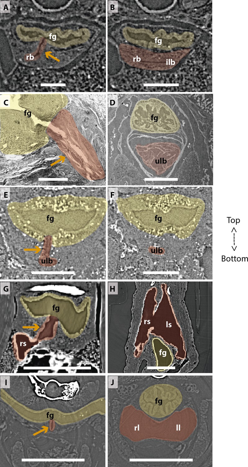
Comparison of sections showing the difference in lung origin and connection between unpaired (A–H) and true paired lungs (I, J).
(A, B) Virtual section of Polypterus senegalus (12mm total length; TL), (C) histological thin section of Latimeria chalumnae (127cm) (Cupello et al., 2017a), (D) virtual section of L. chalumnae (40mm TL; modified from Cupello et al., 2017a), (E, F) virtual section of Neoceratodus forsteri (16mm TL), (G, H) virtual section of Lepidosiren paradoxa (46mm TL), (I, J) virtual section of Salamandra salamandra (35.5mm TL). Yellow, foregut including the stomach; red, lung. Orange arrows, opened connection between the foregut and the lung. Fg, foregut; ll, left lung; ls, left sac; rb, right bud; ilb, independent lung bud; rl, right lung; rs, right sac; ulb, unpaired lung bud. Scale bars, 0.25mm (A, B); 3.0mm (C); 1.0mm (D); 0.1mm (E, F); 0.5mm (G, H); 1.25mm (I, J).
Figure 7
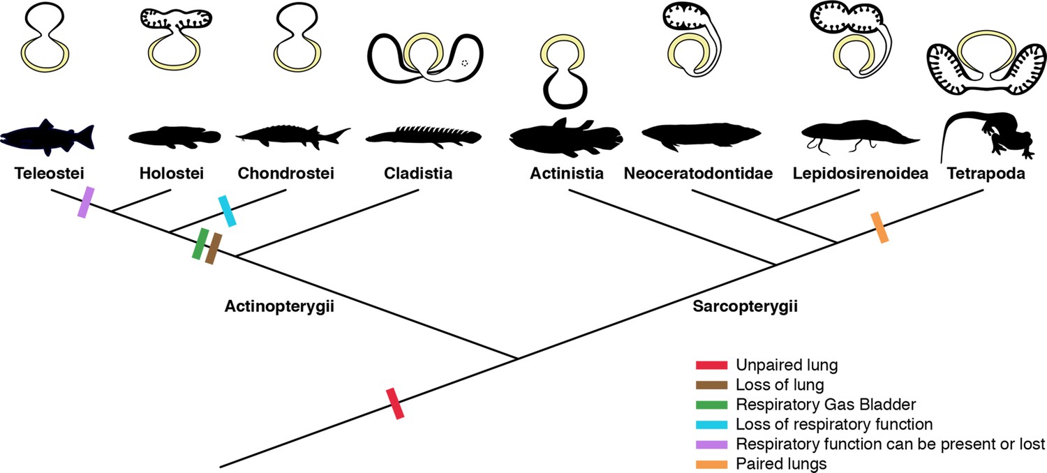
Schematic figure reconstructing the evolutionary history of vertebrate lungs.
All living actinopterygian and sarcopterygian fishes have unpaired lungs. True paired lungs are a synapomorphy of tetrapods. Dashed circle in Cladistia lung pointing to the secondary and independent opening to a left sac, at the lung level. Modified from Liem, 1988. This figure was made with free silhouettes from PhyloPic.
Additional files
Download links
A two-part list of links to download the article, or parts of the article, in various formats.
Downloads (link to download the article as PDF)
Open citations (links to open the citations from this article in various online reference manager services)
Cite this article (links to download the citations from this article in formats compatible with various reference manager tools)
Lung evolution in vertebrates and the water-to-land transition
eLife 11:e77156.
https://doi.org/10.7554/eLife.77156


