RNA sequence to structure analysis from comprehensive pairwise mutagenesis of multiple self-cleaving ribozymes
- Biomolecular Sciences Graduate Programs, Boise State University, United States
- Computing PhD Program, Boise State University, United States
- Department of Biological Science, Boise State University, United States
Figures
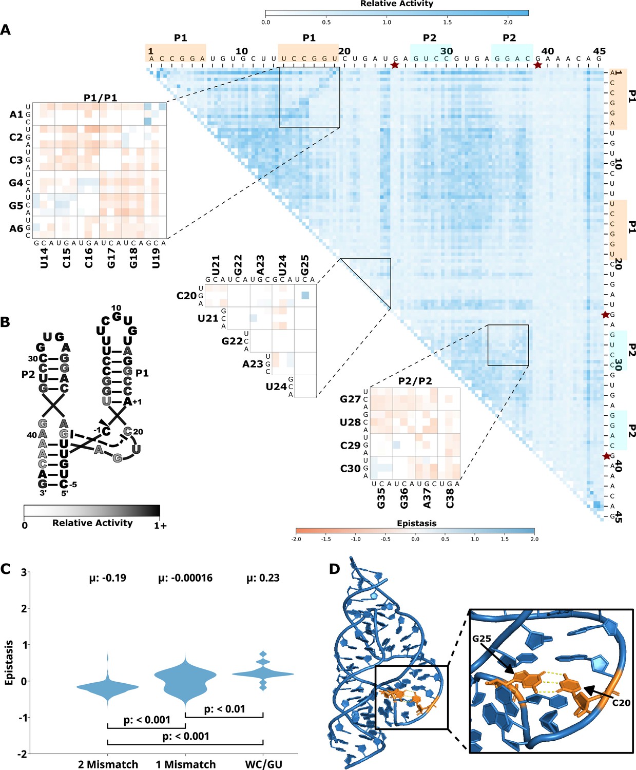
Figure 1
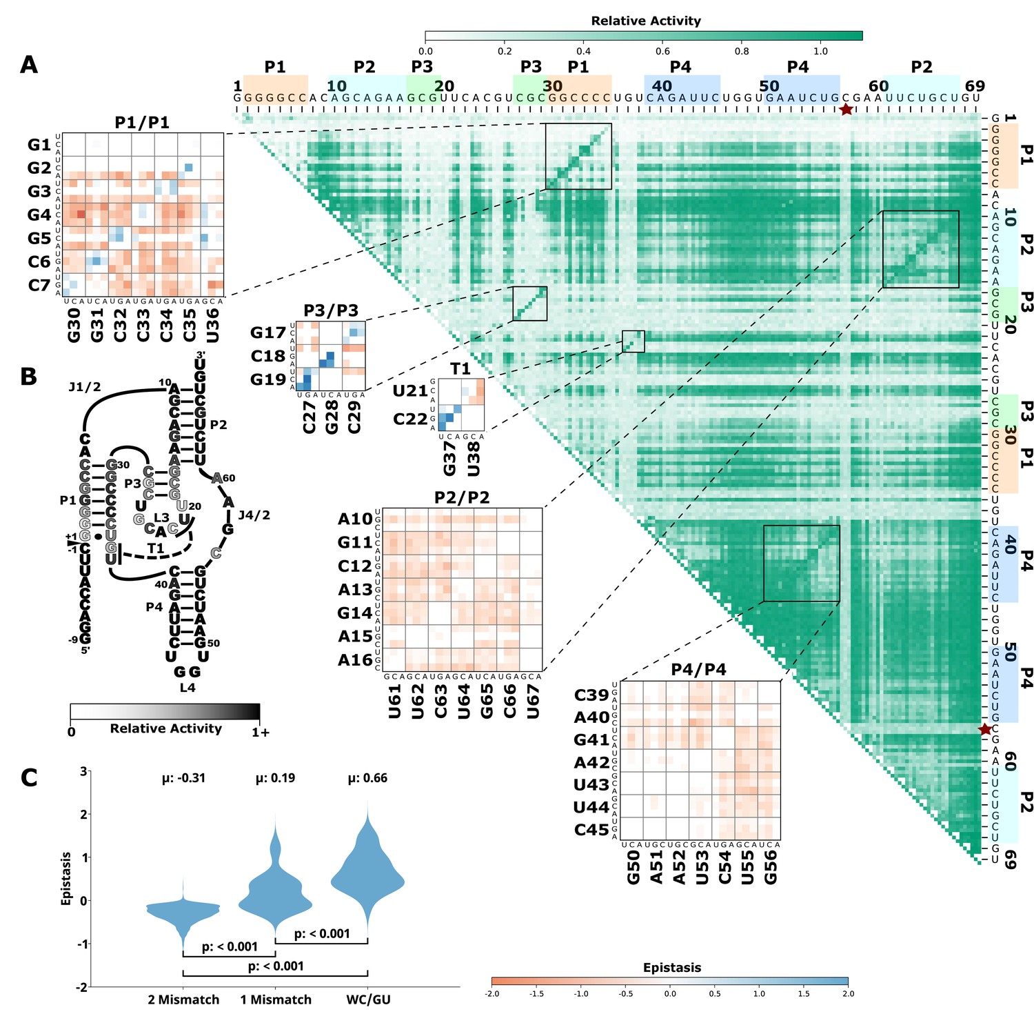
Effects of mutations and pairwise epistasis in a CPEB3 ribozyme.
(A) Relative activity heatmap depicting all possible pairwise effects of mutations on the cleavage activity of a mammalian CPEB3 ribozyme. Base-paired regions P1, P2, P3, P4, and T1 are highlighted and color coordinated along the axes, and surrounded by black squares within the heatmap. Pairwise epistasis interactions observed for each paired regions are each shown as expanded insets for easy identification of the specific epistatic effects measured for each pair of mutations. Instances of positive epistasis are shaded blue, and negative epistasis is shaded red, with higher color intensity indicating a greater magnitude of epistasis. Catalytic residues are indicated by stars along the axes (A is reproduced from Figure 1B from Beck et al., 2022). (B) Secondary structure of the CPEB3 ribozyme used in this study. Each nucleotide is shaded to indicate the average relative cleavage activity of all single mutations at that position. (C) Distributions of epistasis values in the paired regions of the CPEB3 ribozyme. Data were categorized as double mutations that result in two mismatches (2 Mismatch), a single mismatch (1 Mismatch), or no mismatches because of a new Watson-Crick base pair or GU wobble results (WC/GU).
Figure 2
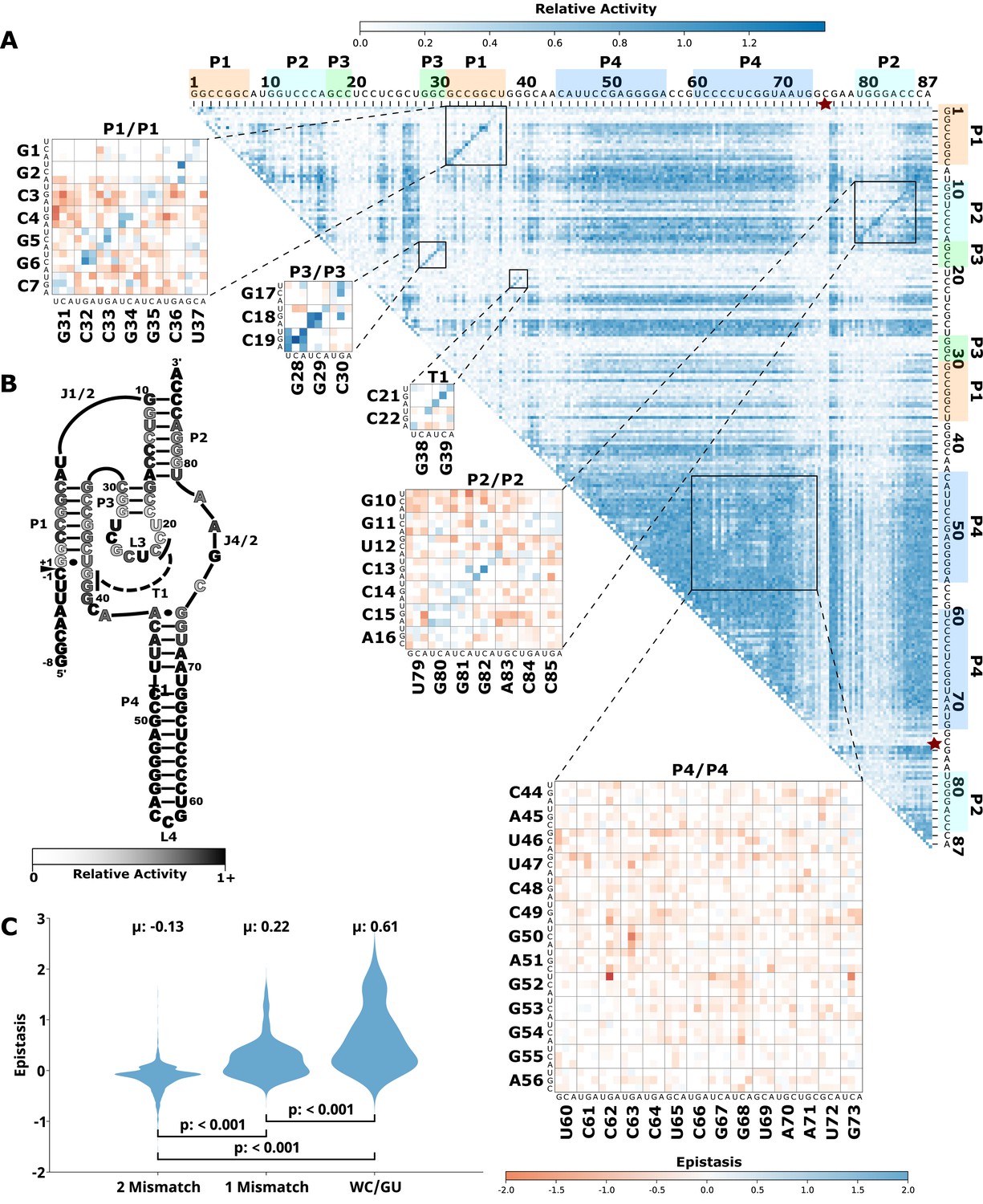
Effects of mutations and pairwise epistasis in a HDV self-cleaving ribozyme.
(A) Relative activity heatmap depicting all possible pairwise effects of mutations on the cleavage activity of an HDV ribozyme. Base-paired regions P1, P2, P3, P4, and T1 are highlighted and color coordinated along the axes, and surrounded by black squares within the heatmap. Pairwise epistasis interactions observed for each paired regions are each shown as expanded insets for easy identification of the specific epistatic effects measured for each pair of mutations. Instances of positive epistasis are shaded blue, and negative epistasis is shaded red, with higher color intensity indicating a greater magnitude of epistasis. Catalytic residues are indicated by stars along the axes. (B) Secondary structure of the HDV ribozyme used in this study. Each nucleotide is shaded to indicate the average relative cleavage activity of all single mutations at that position. (C) Distributions of epistasis values in the paired regions of the HDV ribozyme. Data were categorized as double mutations that result in two mismatches (2 Mismatch), a single mismatch (1 Mismatch), or no mismatches because of a new Watson-Crick base pair or GU wobble results (WC/GU). HDV, hepatitis delta virus.
Figure 3
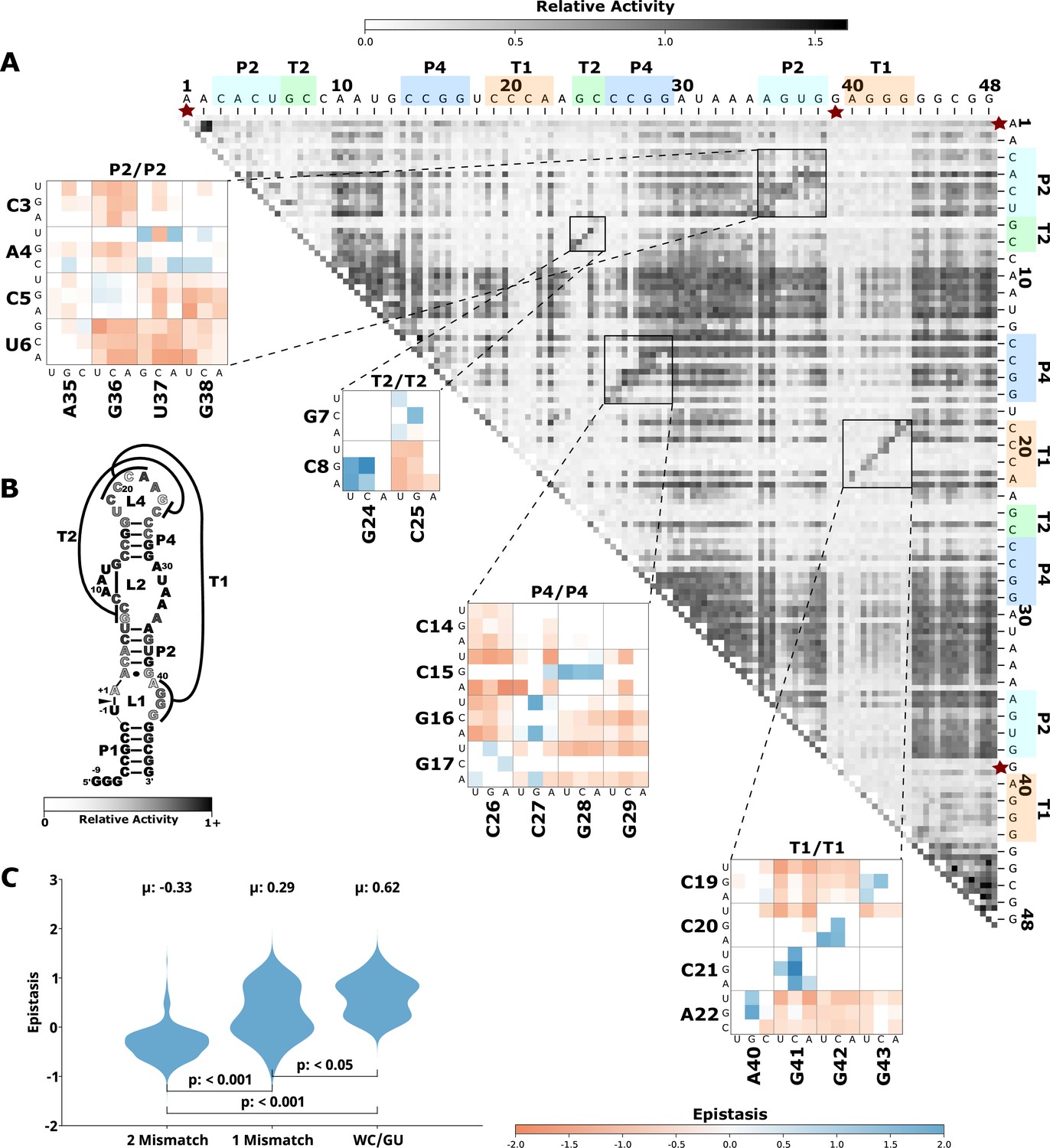
Effects of mutations and pairwise epistasis in a twister self-cleaving ribozyme.
(A) Relative activity heatmap depicting all possible pairwise effects of mutations on the cleavage activity of a twister ribozyme. Base-paired regions P2, P4, T1, and T2 are highlighted and color coordinated along the axes, and surrounded by black squares within the heatmap. Pairwise epistasis interactions observed for each paired region are each shown as expanded insets for easy identification of the specific epistatic effects measured for each pair of mutations. Instances of positive epistasis are shaded blue, and negative epistasis is shaded red, with higher color intensity indicating a greater magnitude of epistasis. Catalytic residues are indicated by stars along the axes. (B) Secondary structure of the twister ribozyme used in this study. Each nucleotide is shaded to indicate the average relative cleavage activity of all single mutations at that position. (C) Distributions of epistasis values in the paired regions of the twister ribozyme. Data were categorized as double mutations that result in two mismatches (2 Mismatch), a single mismatch (1 Mismatch), or no mismatches because of a new Watson-Crick base pair or GU wobble results (WC/GU).
Figure 4 with 1 supplement
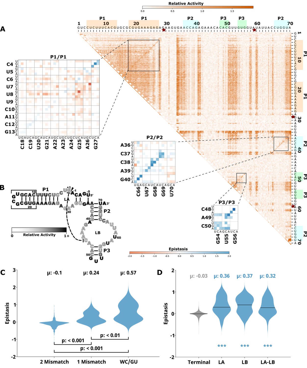
Effects of mutations and pairwise epistasis in a hairpin self-cleaving ribozyme.
(A) Relative activity heatmap depicting all possible pairwise effects of mutations on the cleavage activity of a hairpin ribozyme. Base-paired regions P1, P2, and P3 are highlighted and color coordinated along the axes, and surrounded by black squares within the heatmap. Pairwise epistasis interactions observed for each paired region are each shown as expanded insets for easy identification of the specific epistatic effects measured for each pair of mutations. Instances of positive epistasis are shaded blue, and negative epistasis is shaded red, with higher color intensity indicating a greater magnitude of epistasis. Catalytic residues are indicated by stars along the axes. (B) Secondary structure of the hairpin ribozyme used in this study. Each nucleotide is shaded to indicate the average relative cleavage activity of all single mutations at that position. (C) Distributions of epistasis values in the paired regions of the hairpin ribozyme. Data were categorized as double mutations that result in two mismatches (2 Mismatch), a single mismatch (1 Mismatch), or no mismatches because of a new Watson-Crick base pair or GU wobble results (WC/GU). (D) The distributions of epistasis values in all terminal stem loops across all five ribozymes, and epistasis observed within loop A, loop B, and between loop A and loop B in the hairpin ribozyme.
Figure 4—figure supplement 1
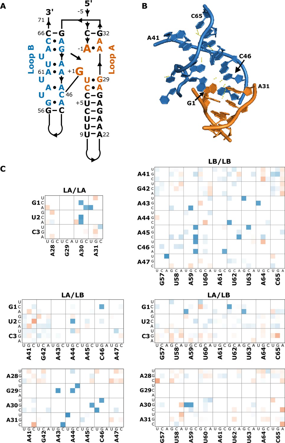
Epistasis in the internal loops of a hairpin ribozyme.
(A) Secondary structure of the loop domains in hairpin. Loop B nucleotides are shaded blue, and loop A nucleotides are shaded in orange. Canonical base pairs are connected by lines, non-canonical pairs are indicated by connecting dots. (B) Crystal structure of a hairpin ribozyme (PDB 1M5K) showing loop A and loop B domain interactions. Loop B is shaded in blue, and loop A is shaded in orange. Hydrogen bonds are shown as dashed yellow lines.
Figure 5 with 1 supplement
Effects of mutations and pairwise epistasis in a hammerhead self-cleaving ribozyme.
(A) Relative activity heatmap depicting all possible pairwise effects of mutations on the cleavage activity of a hammerhead ribozyme. Base-paired regions, P1 and P2, are highlighted and color coordinated along the axes, and surrounded by black squares within the heatmap. Pairwise epistasis interactions observed for each paired region are each shown as expanded insets for easy identification of the specific epistatic effects measured for each pair of mutations. Instances of positive epistasis are shaded blue, and negative epistasis is shaded red, with higher color intensity indicating a greater magnitude of epistasis. Catalytic residues are indicated by stars along the axes. (B) Secondary structure of the hammerhead ribozyme used in this study. Each nucleotide is shaded to indicate the average relative cleavage activity of all single mutations at that position. (C) Distributions of epistasis values in the paired regions of the hammerhead ribozyme. Data were categorized as double mutations that result in two mismatches (2 Mismatch), a single mismatch (1 Mismatch), or no mismatches because of a new Watson-Crick base pair or GU wobble results (WC/GU). (D) Crystal structure of a hammerhead ribozyme (3ZD5) with C20 and G25 indicated (orange) and hydrogen bonds between the nucleotides shown as yellow dashed lines.
Figure 5—figure supplement 1

Distribution of pairwise epistasis observed between the loops of P1 and P2 in the hammerhead ribozyme.
Figure 6 with 3 supplements
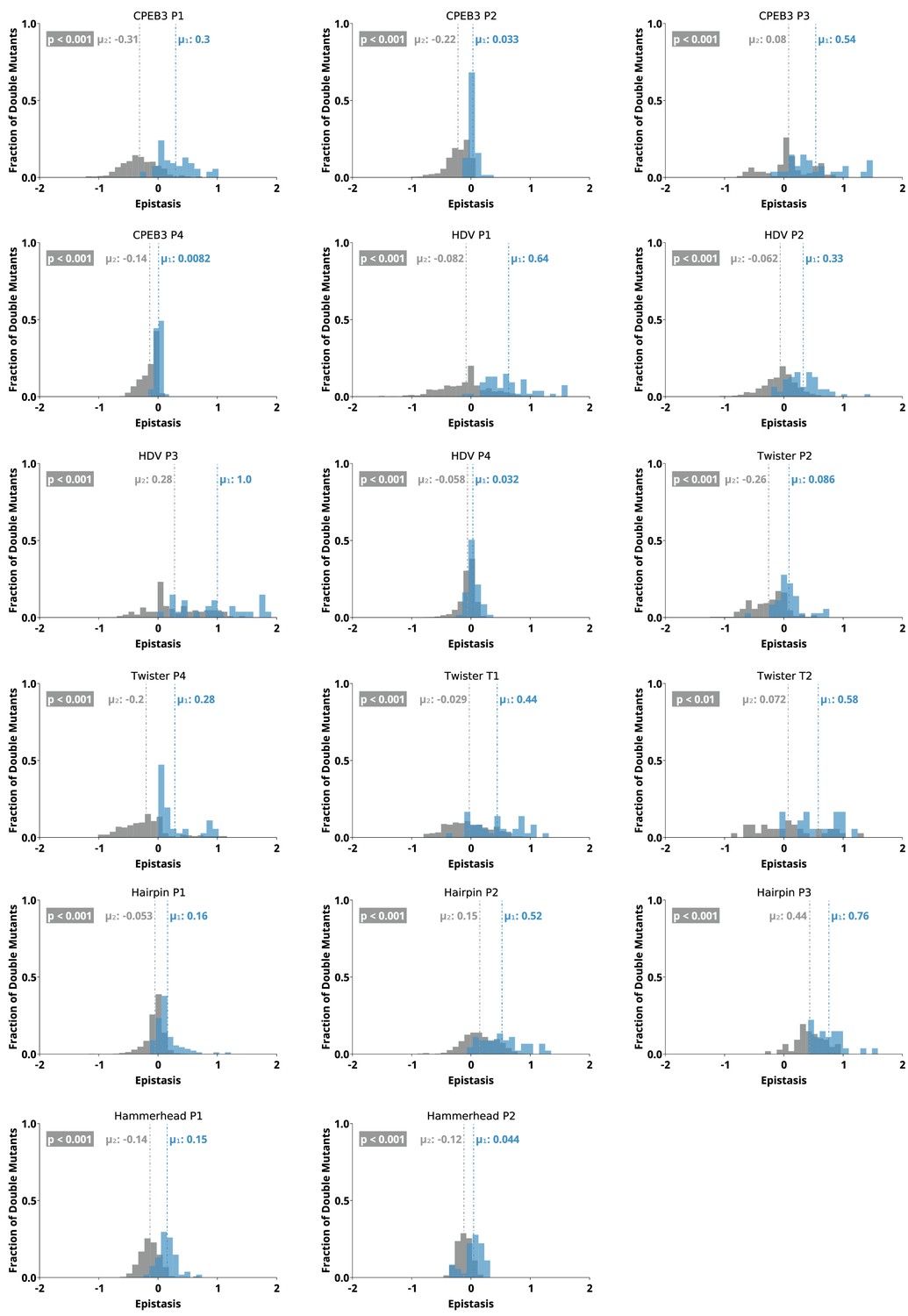
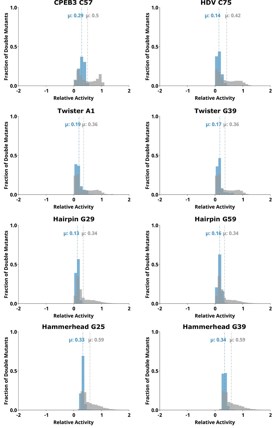
Distributions of epistasis values calculated for individual paired regions in all five ribozymes.
For each region, epistasis values were separated into double mutants that restore a Watson-Crick base pair (‘on-diagonal’, blue) and all other double mutants (‘off-diagonal’, gray). The mean of each distribution (µ) is reported and indicated by the dashed line. The p value is the probability that values were drawn from the same distribution by chance (Mann-Whitney U test).
Figure 6—figure supplement 1
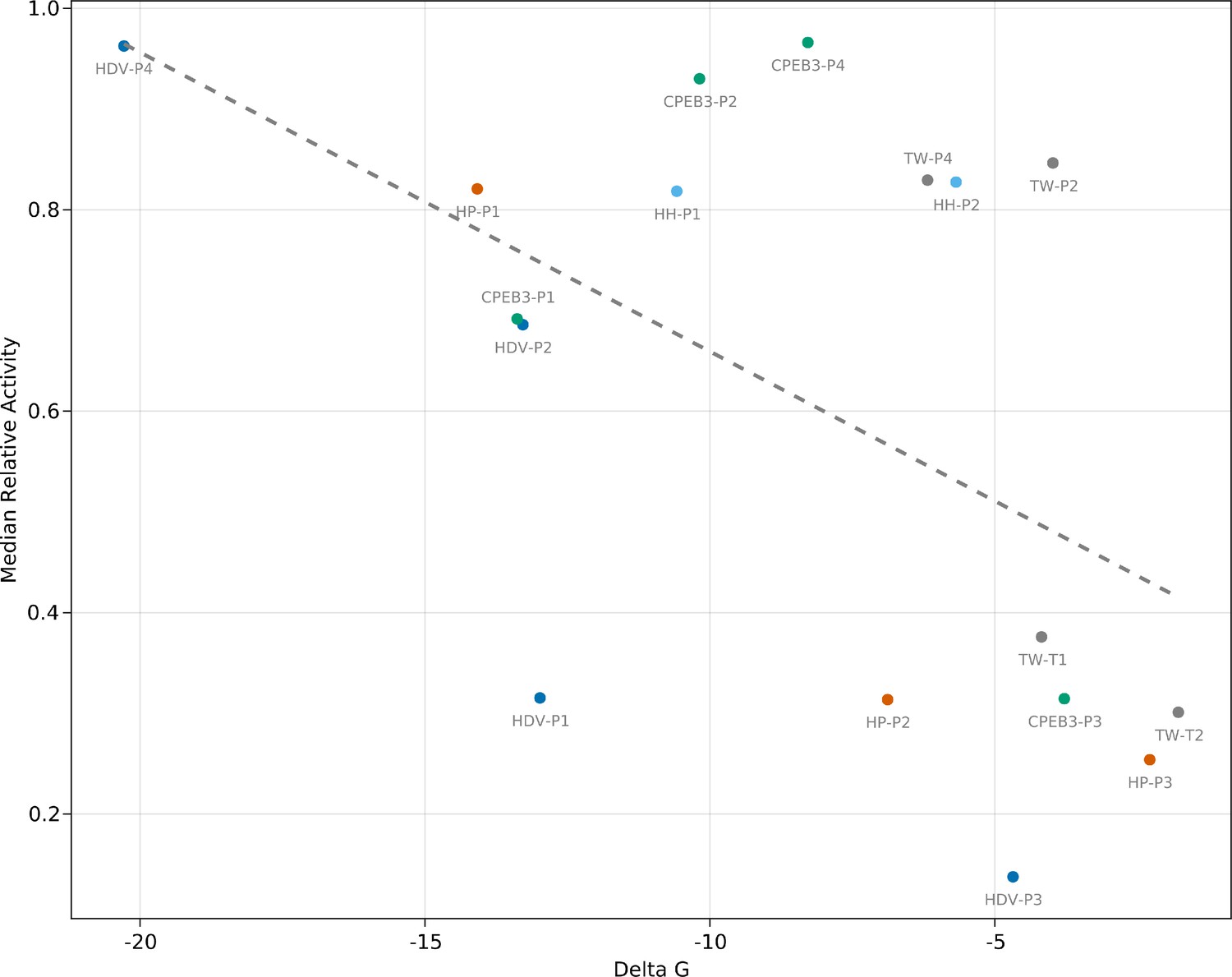
Correlation between mutational effects and RNA helix stability.
Relationship between the Gibbs free energy (ΔG) of each base-paired region belonging to the hairpin, hammerhead, CPEB3, HDV, and twister ribozymes, and the median relative activity of all single mutants within each base-paired region (Pearson correlation=–0.53, p=0.029, R2=0.27).
Figure 6—figure supplement 2
Relative activity (RA) values for sequences with mutations at catalytic and non-catalytic nucleotide positions.
The distribution of RA values for sequences with pairs of mutations where one mutation is a catalytic nucleotide is shown in blue. The specific catalytic nucleotide is indicated in the title above each plot. The distributions for all pairs of mutations for each ribozyme that do not involve a catalytic nucleotide are shown in gray. The mean values (µ) for each distribution are marked by dashed lines.
Figure 6—figure supplement 3

Histogram of the distributions of read counts (read depth) for the single and double mutants matching to each ribozyme analyzed in this study (CPEB3, HDV, twister, hairpin, and hammerhead).
Tables
Table 1
Summary of the lengths of each self-cleaving ribozyme used in this study, the number of single and double mutants whose cleavage activity was analyzed, and the average fraction cleaved observed for all single and double mutants.
| Ribozyme name | Ribozyme length | Possible single Mmutants | Possible double mutants | Total mapped reads | Wild-type fraction cleaved | Single mutant average fraction cleaved | Double mutant average fraction cleaved |
|---|---|---|---|---|---|---|---|
| CPEB3 | 69 | 207 | 21,114 | 9,238,603 | 0.90 | 0.69 | 0.44 |
| HDV | 87 | 261 | 33,669 | 3,316,380 | 0.60 | 0.40 | 0.25 |
| Twister | 48 | 144 | 10,152 | 7,762,863 | 0.60 | 0.41 | 0.21 |
| Hairpin | 71 | 213 | 22,365 | 5,067,216 | 0.52 | 0.29 | 0.17 |
| Hammerhead | 45 | 135 | 8,910 | 8,054,498 | 0.34 | 0.27 | 0.19 |
Additional files
-
Supplementary file 1
Oligonucleotides used in this study.
- https://cdn.elifesciences.org/articles/80360/elife-80360-supp1-v2.docx
-
MDAR checklist
- https://cdn.elifesciences.org/articles/80360/elife-80360-mdarchecklist1-v2.docx
Download links
A two-part list of links to download the article, or parts of the article, in various formats.
Downloads (link to download the article as PDF)
Open citations (links to open the citations from this article in various online reference manager services)
Cite this article (links to download the citations from this article in formats compatible with various reference manager tools)
RNA sequence to structure analysis from comprehensive pairwise mutagenesis of multiple self-cleaving ribozymes
eLife 12:e80360.
https://doi.org/10.7554/eLife.80360


