Molecular and spatial profiling of the paraventricular nucleus of the thalamus
- National Institute of Mental Health, United States
- Department of Neuroscience, Brown University, United States
- National Institute on Alcohol Abuse and Alcoholism, United States
- National Institute of Child Health and Human Development, United States
Figures
Figure 1 with 2 supplements
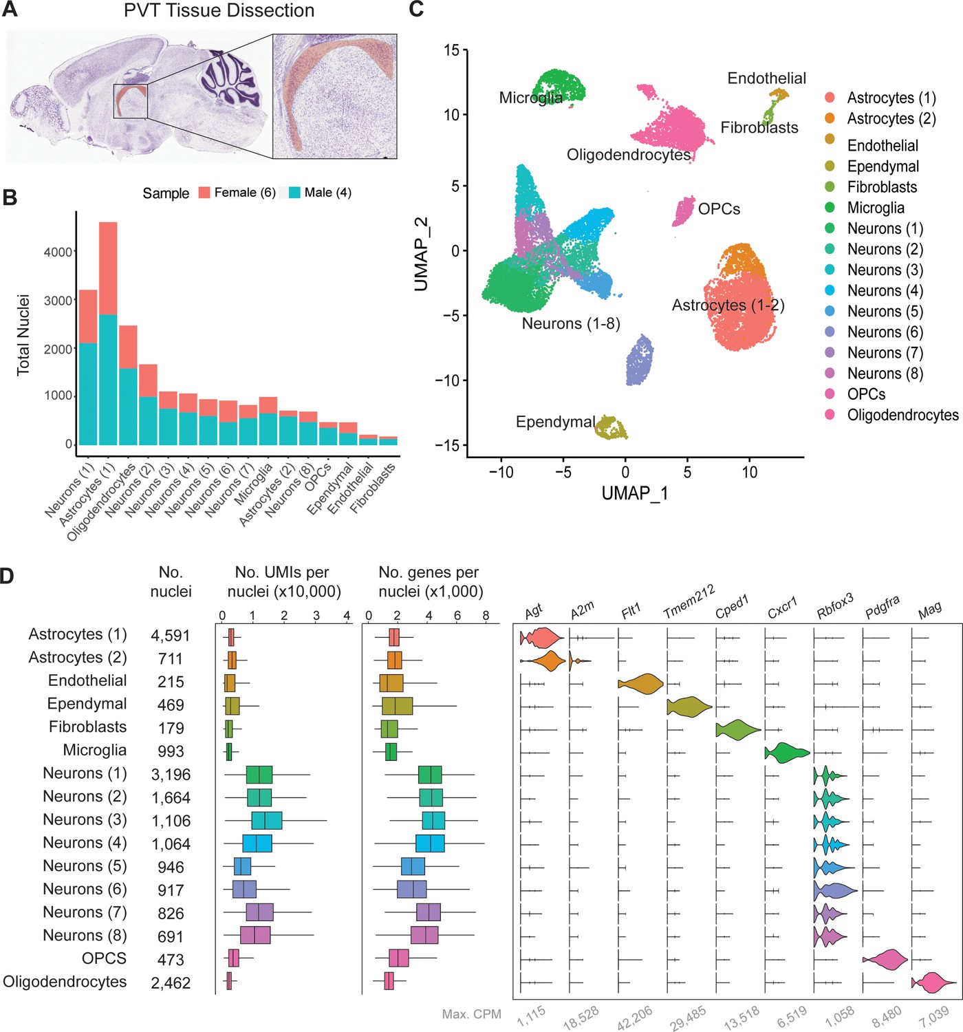
Single-nucleus RNA-sequencing in and around the mouse paraventricular nucleus of the thalamus (PVT).
(a) Sagittal view of the PVT (red) illustrating the dissection target location. (b) Distribution of nuclei from four samples across all cell types. (c) The uniform manifold approximation and projection (UMAP) plot of all 13,220 nuclei from the combined dataset shows 14 cell clusters. (d) Cell type classification is based on the expression of marker genes in all 14 clusters. Left: box plot of UMI number in each cell cluster. Middle: box plot of genes detected per cell in each cell cluster. Right: violin plot showing expression profile of marker genes in 14 cell clusters. Max. CPM, maximum counts per million reads. Box plot legend: box is defined by 25th and 75th percentiles, whiskers are determined by 5th and 95th percentiles, and the mean is depicted by the square symbol.
Figure 1—figure supplement 1
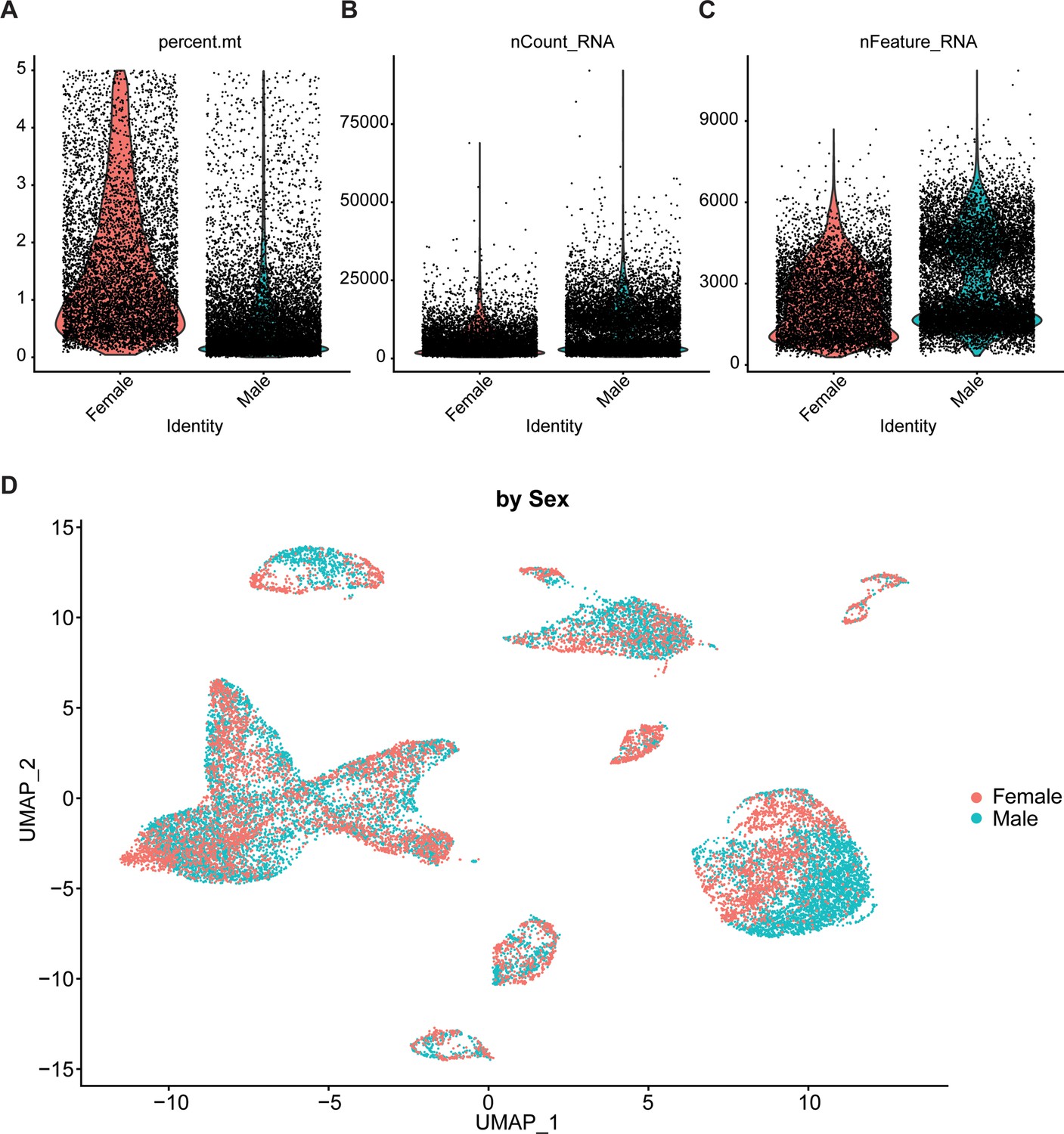
Quality control statistics and uniform manifold approximation and projection (UMAP) by sample.
(a–c) Violin plots depicting the percent mitochondrial RNA (a), number of UMIs (b), and the number of genes (c) detected by sample. (d) The UMAP plot of the same nuclei from Figure 1c is labeled by the sample of origin.
Figure 1—figure supplement 2
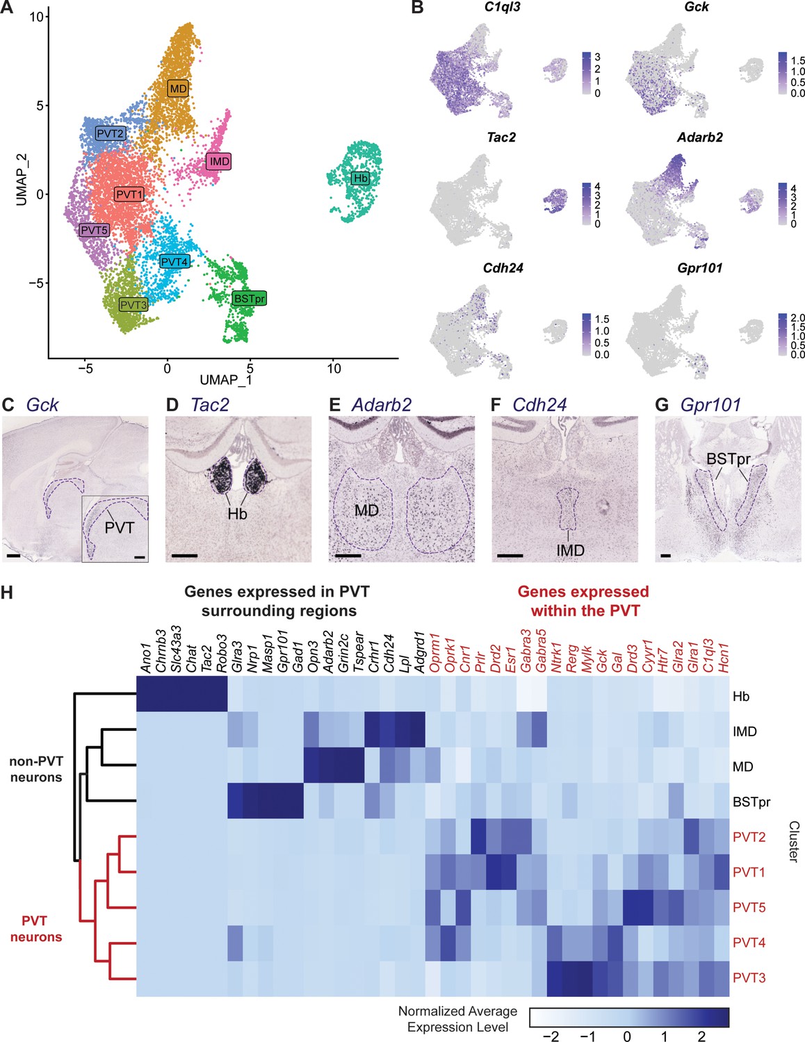
Clustering analysis and classification of all neurons.
(a) The uniform manifold approximation and projection (UMAP) plot of 9845 neuronal nuclei shows nine clusters. (b) Feature plots of representative gene expression of markers from different clusters. (c) In situ hybridization of Gck (ABA Experiment #68269269), scale bar 500 μm, outline depicts the PVT. (d) In situ hybridization of Tac2 (ABA Experiment #72339556), scale bar 300 μm, outline depicts the Hb. (e) In situ hybridization of Adarb2 (ABA Experiment #73925721), scale bar 300 μm, outline depicts the MD. (f) In situ hybridization of Cdh24 (ABA Experiment #70231307), scale bar 300 μm, outline depicts the IMD. (g) In situ hybridization of Gpr101 (ABA Experiment #79591639), scale bar 300 μm, outline depicts the BSTpr. (h) Heatmap of normalized average gene expression of PVT-enriched genes (red) and PVT-depleted genes (black). ABA, Allen Brain Atlas.
Figure 2 with 1 supplement
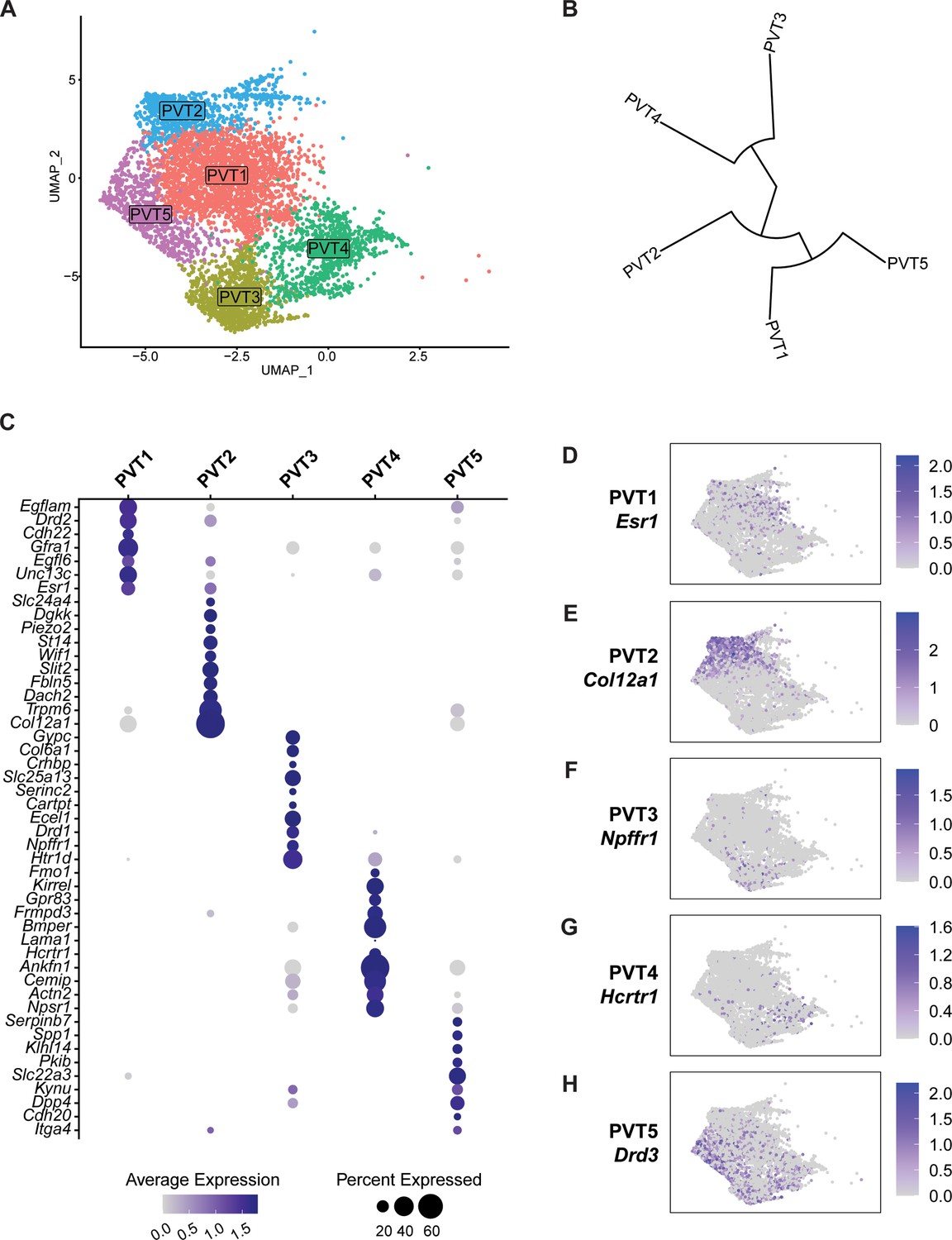
Five transcriptionally distinct neuronal subtypes are found in the paraventricular nucleus of the thalamus (PVT).
(a) The uniform manifold approximation and projection (UMAP) plot of 5737 PVT neuronal nuclei shows five clusters. (b) Phylogenetic tree depicting cluster relationships based on the distance between clusters in gene expression space. (c) Dot plot of top gene marker average expression across PVT clusters selected based on pct. ratio value. (d-h) Feature plots of top marker genes (d) Esr1, (e) Col12a1, (f) Npffr1, (g) Hcrtr1, and (h) Drd3 for each PVT subtype.
Figure 2—figure supplement 1
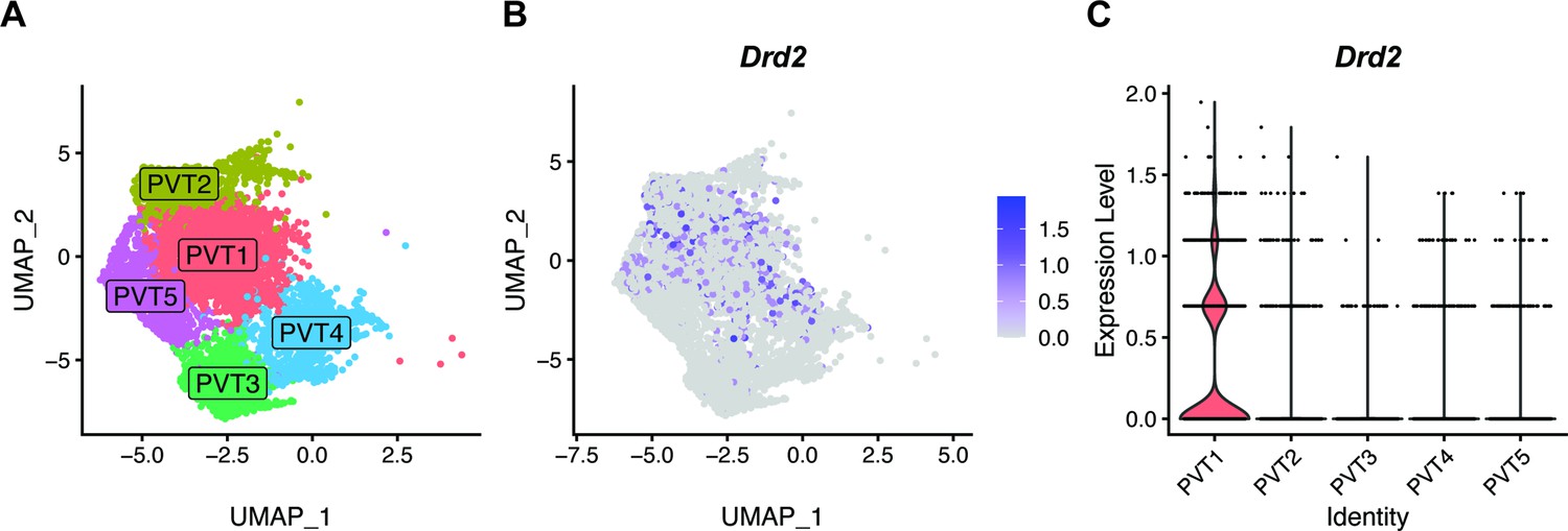
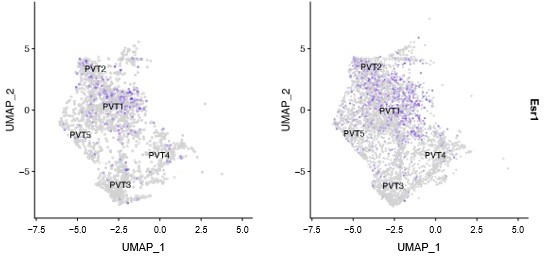
Drd2 expression is found in PVT1 and PVT2.
(a) The uniform manifold approximation and projection (UMAP) plot of 4067 paraventricular nucleus of the thalamus (PVT) neuronal nuclei shows five clusters. (b) Feature plot of Drd2 expression across all PVT neurons. (c) Violin plot of Drd2 expression level across five PVT clusters.
Figure 3 with 3 supplements
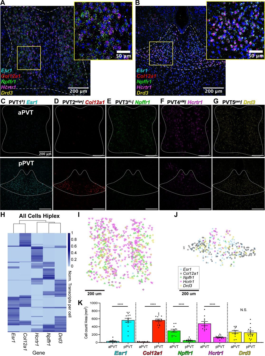
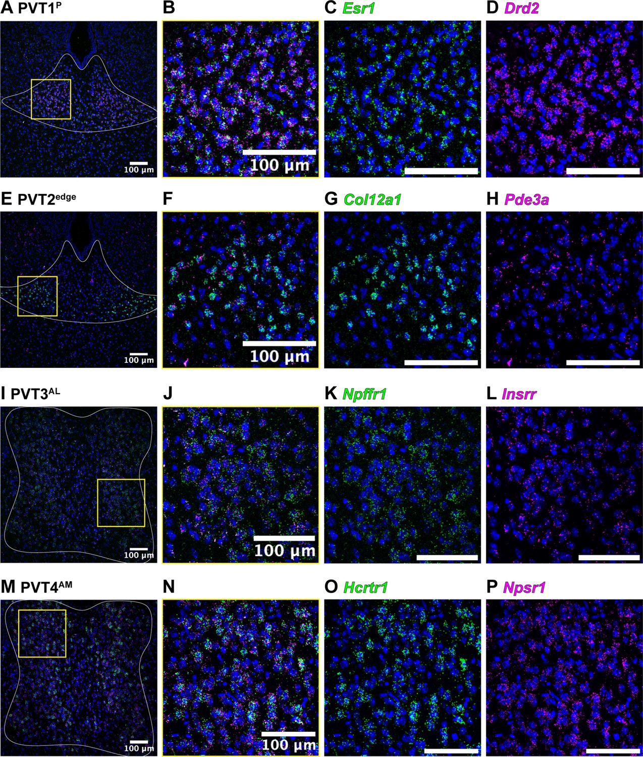
Mapping of five paraventricular nucleus of the thalamus (PVT) subtypes reveals a gradient-like segregated distribution.
(a) RNA in situ hybridization in anterior PVT (white dotted outline) with closeup insert (right, yellow square) labeling one gene marker from each subtype: Esr1 (light blue), Col12a1 (red), Npffr1 (green), Hcrtr1 (magenta), and Drd3 (yellow). (b) RNA in situ hybridization in posterior PVT (white dotted outline) with closeup insert (right, yellow square) labeling one gene marker from each subtype: Esr1 (blue), Col12a1 (red), Npffr1 (green), Hcrtr1 (magenta), and Drd3 (yellow). (c–g) RNA in situ hybridization showing aPVT (top) and pPVT (bottom) of (c) Esr1 from PVT1 subtype, (d) Col12a1 from PVT2 subtype, (e) Npffr1 from PVT3 subtype, (f) Hcrtr1 from PVT4 subtype, (g) Drd3 from PVT5 subtype. DAPI (blue). Scale bar 200 μm; all images are 20 X representative confocal images with brightness and contrast adjusted depicting expression patterns found in all sections from N=3 animals. (h) Heatmap of gene expression matrix with rows showing normalized transcripts per cell and columns showing gene markers from each PVT subtype; Left dendrogram displays hierarchical clustering by cell, top dendrogram displays hierarchical clustering by gene marker, and right bar shows heatmap legend. (i–j) Coordinates of positive cells from aPVT (i) and pPVT (j) with Esr1 (blue square), Col12a1 (black circle), Npffr1 (green triangle), Hcrtr1 (magenta downward triangle), Drd3 (yellow circle) shown in the legend. (k) Bar graphs of the number of positive cells over the area (mm2) per section of Esr1 (blue; aPVT: 30.71±11.73; pPVT: 179.78±48.05; ****p=0.0000000015, two-sided Paired sample t-test), Col12a1 (black; aPVT: 15.85±2.95; pPVT: 563.13±30.37; ****p=0.000000000000071, two-sided Paired sample t-test), Npffr1 (green; aPVT: 297.64±37.93; pPVT: 51.03±9.13; ****p=0.00000035), Hcrtr1 (magenta; aPVT: 478.53±52.05; pPVT: 133.62±10.76; ****p=0.00000021), and Drd3 (yellow; aPVT: 265.97±38.52; pPVT: 250.43±41.18; N.S., not significant, p=0.79) in aPVT and pPVT. Data from N=3 animals are shown as mean ± SEM. Each data point represents one section.
Figure 3—figure supplement 1
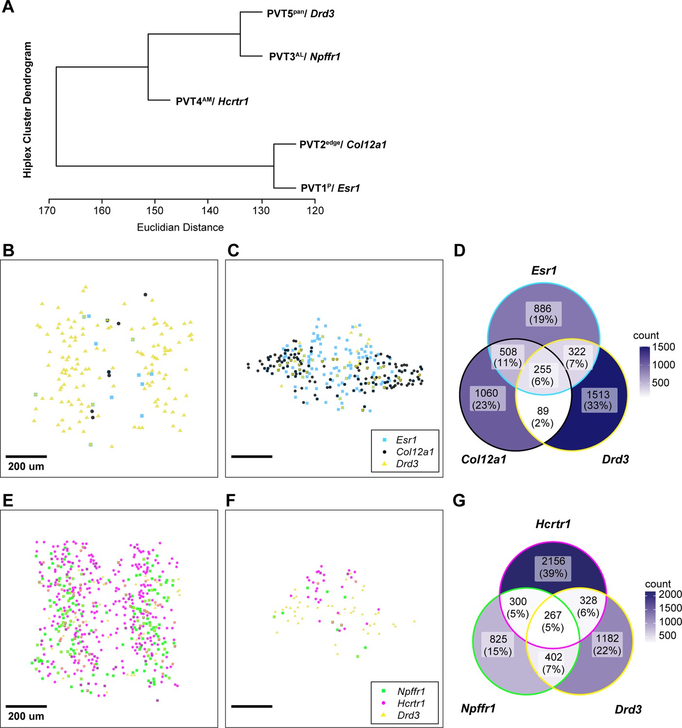
Spatial distribution and overlap across anterior- or posterior- and pan-PVT subtypes.
(a) Dendrogram depicting Euclidian distance between cluster types based on gene transcript expression. (b–c) Location of positive cells expressing Esr1 (blue square), Col12a1 (black circle), and Drd3 (yellow triangle) in aPVT (b) or pPVT (c). (d) Venn diagram of number and percent of Esr1-, Col12a1-, or Drd3-positive cells. (e–f) Location of positive cells expressing Npffr1 (green square), Hcrtr1 (magenta circle), or Drd3 (yellow triangle) in aPVT (e) or pPVT (f). (g) Venn diagram of number and percent of Npffr1-, Hcrtr1-, or Drd3-positive cells. Venn Diagram legends, color intensity correlates to cell count.
Figure 3—figure supplement 2
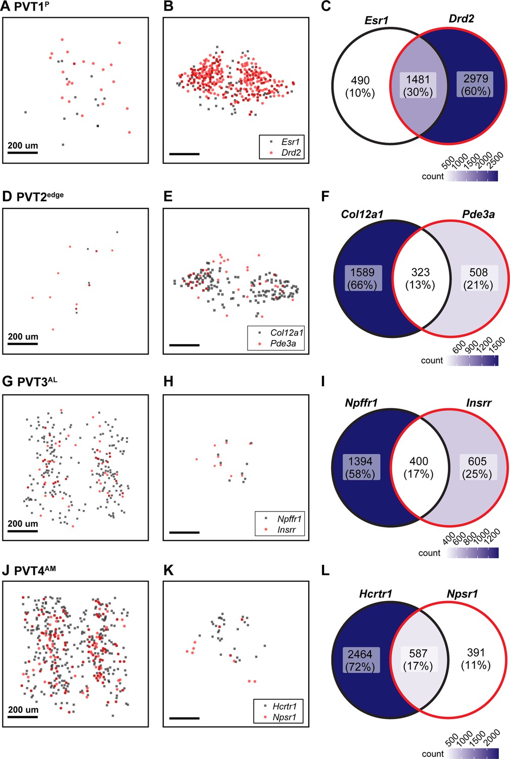
Overlap in gene expression among top markers from the same cluster.
(a–b) Location of Esr1 (black) or Drd2 (red) positive cells in aPVT (a) or pPVT (b) from PVT1 subtype. (c) Venn diagram of cell count and percent overlap between Esr1- and Drd2-positive cells. (d-e) Location of Col12a1 (black) or Pde3a (red) positive cells in aPVT (a) or pPVT (b) from PVT2 subtype. (f) Venn diagram of cell count and percent overlap between Col12a1- and Pde3a-positive cells. (g–i) Location of Npffr1 (black) or Insrr (red) positive cells in aPVT (a) or pPVT (b) from PVT3 subtype. (f) Venn diagram of cell count and percent overlap between Npffr1- and Insrr-positive cells. (j–k) Location of Hcrtr1 (black) or Npsr1 (red) positive cells in aPVT (a) or pPVT (b) from PVT4 subtype. (f) Venn diagram of cell count and percent overlap between Hcrtr1- and Npsr1-positive cells. Venn Diagram legends, color intensity correlates to cell count. Black scale bar indicates 200 μm.
Figure 3—figure supplement 3
In situ hybridization of top markers from the same cluster.
(a) In situ hybridization of Esr1 (green), Drd2 (magenta), and DAPI (blue) in pPVT (white outline). (b–d) Yellow square insert from (a). (e) In situ hybridization of Col12a1 (green), Pde3a (magenta), and DAPI (blue) in pPVT (white outline). (f–d) Yellow square insert from (e). (i) In situ hybridization of Npffr1 (green), Insrr (magenta), and DAPI (blue) in aPVT (white outline). (j–l) Yellow square insert from (i). (m) In situ hybridization of Hcrtr1 (green), Npsr1 (magenta), and DAPI (blue) in aPVT (white outline). (n–p) Yellow square insert from (m). All images are 20 X representative confocal images with brightness and contrast adjusted depicting expression patterns found in all sections from N=3 animals. White scale bar indicates 100 μm.
Figure 4
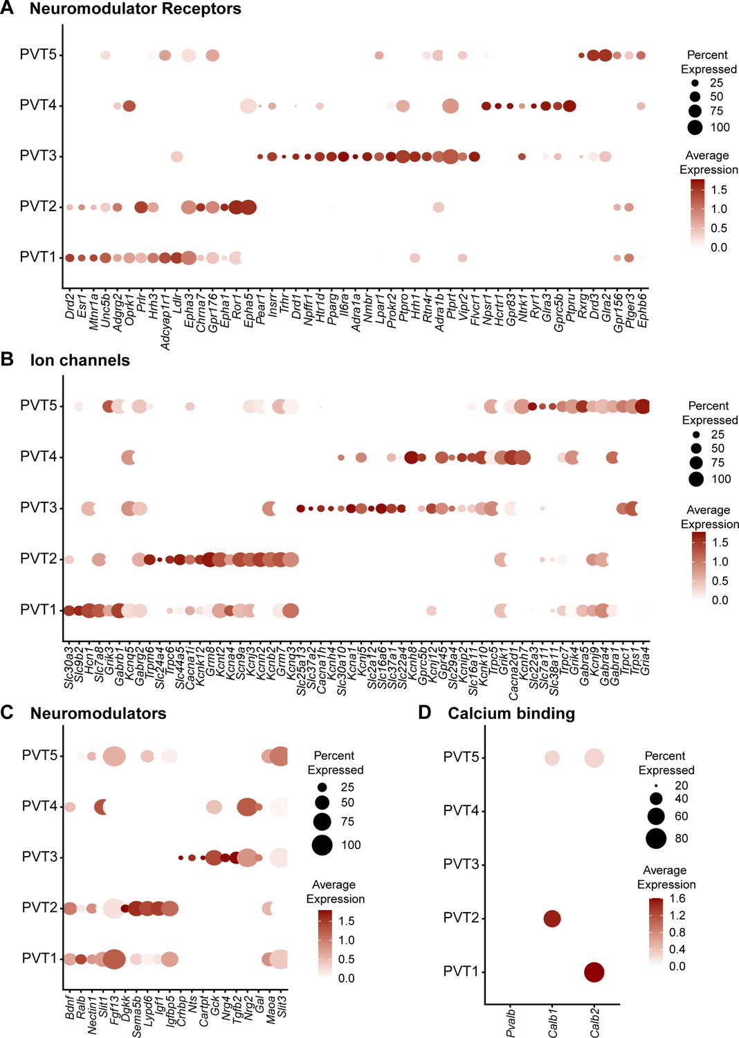
Five paraventricular nucleus of the thalamus (PVT) subtypes have diverse neuromodulator receptor, neuromodulator, and ion channel expression.
(a) Dot plot depicting neuromodulator receptor gene expression across five PVT subtypes. (b) Dot plot depicting ion channel gene expression across five PVT subtypes. (c) Dot plot depicting neuromodulator gene expression across five PVT subtypes. (d) Dot plot depicting calcium binding gene (column) expression across five PVT subtypes. Legend: top, percent of nuclei in each cluster expressing a given gene; bottom, color intensity corresponding to average gene expression level.
Figure 5 with 4 supplements
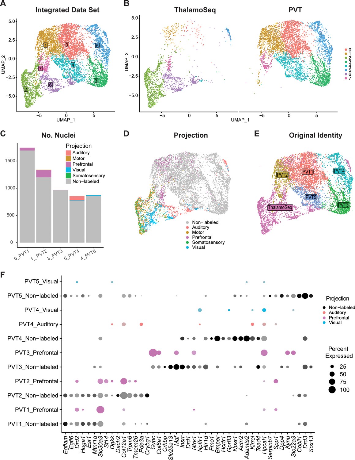
Cross-validation with ThalamoSeq dataset reveals overlap between paraventricular nucleus of the thalamus (PVT) subtypes and thalamic cortical projectors.
(a) The uniform manifold approximation and projection (UMAP) plot of 7709 cells or nuclei from combined datasets annotated by cluster identity. (b) The same cells/nuclei are separated by the study of origin: ThalamoSeq (left) and present study (right) (c) Proportion of nuclei from each cortical projection target in the clusters that represent the molecular PVT subtypes from the integrated dataset: 0=PVT1, 1=PVT2, 3=PVT3, 5=PVT4, 4=PVT5. (d) The same cells/nuclei are colored by the cortical projection target. (e) The same cells/nuclei are colored by the original identity. (f) Dot plot of PVT representing clusters (0,1,3, 5, 4) in the integrated dataset depicting expression of genes obtained from the conserved marker analysis (Supplementary file 1) split by projection identity.
Figure 5—figure supplement 1
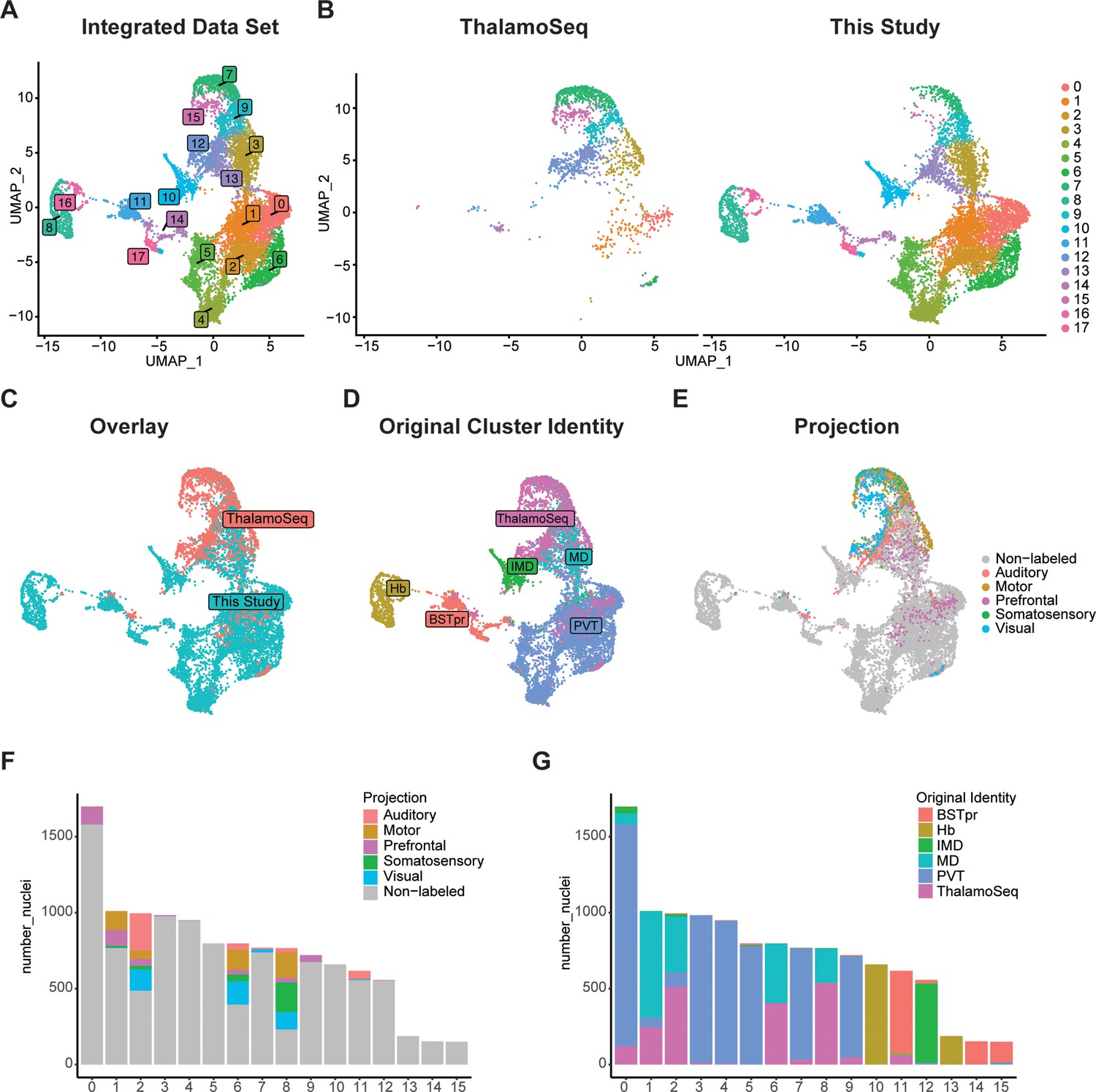
RPCA-based integration with ThalamoSeq dataset and all neurons from the present study reveals mostly non-overlapping transcriptomes.
(a) The uniform manifold approximation and projection (UMAP) plot of 11,817 cells or nuclei from combined datasets annotated by cluster identity. (b) The same cells/nuclei are separated by the study of origin: ThalamoSeq (left) and present study (right) and overlayed in (c). (d) The same cells/nuclei are colored by the original identity. (e) The same cells/nuclei are colored by the cortical projection target. (f) Proportion of nuclei in each cluster labeled by cortical projection target. (g) Proportion of nuclei in each cluster labeled by original identity.
Figure 5—figure supplement 2
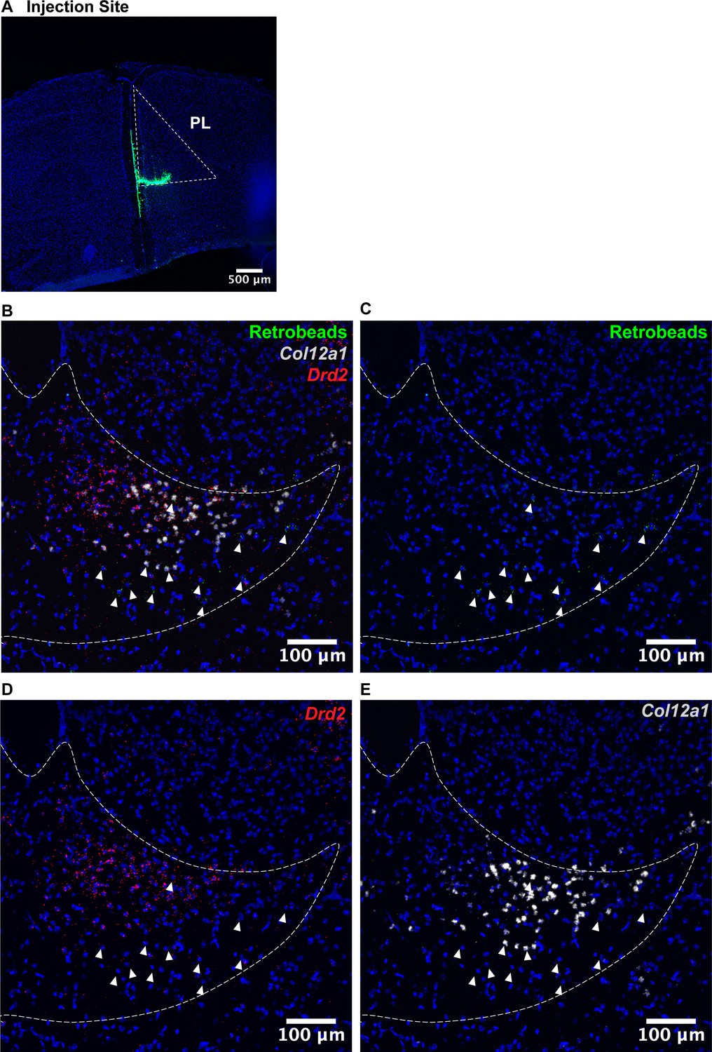
PL-projecting pPVT neurons are localized to the ventroposterior edge of the paraventricular nucleus of the thalamus (PVT) and partially overlap with Col12a1.
(a) Representative image of the injection site of green fluorescent retrobeads in the prelimbic cortex (PL). (b–e) Representative image of the localization of retrobeads (green) and Col12a1 mRNA (light grey) and Drd2 mRNA (red) in the posterior PVT. N=2.
Figure 5—figure supplement 3
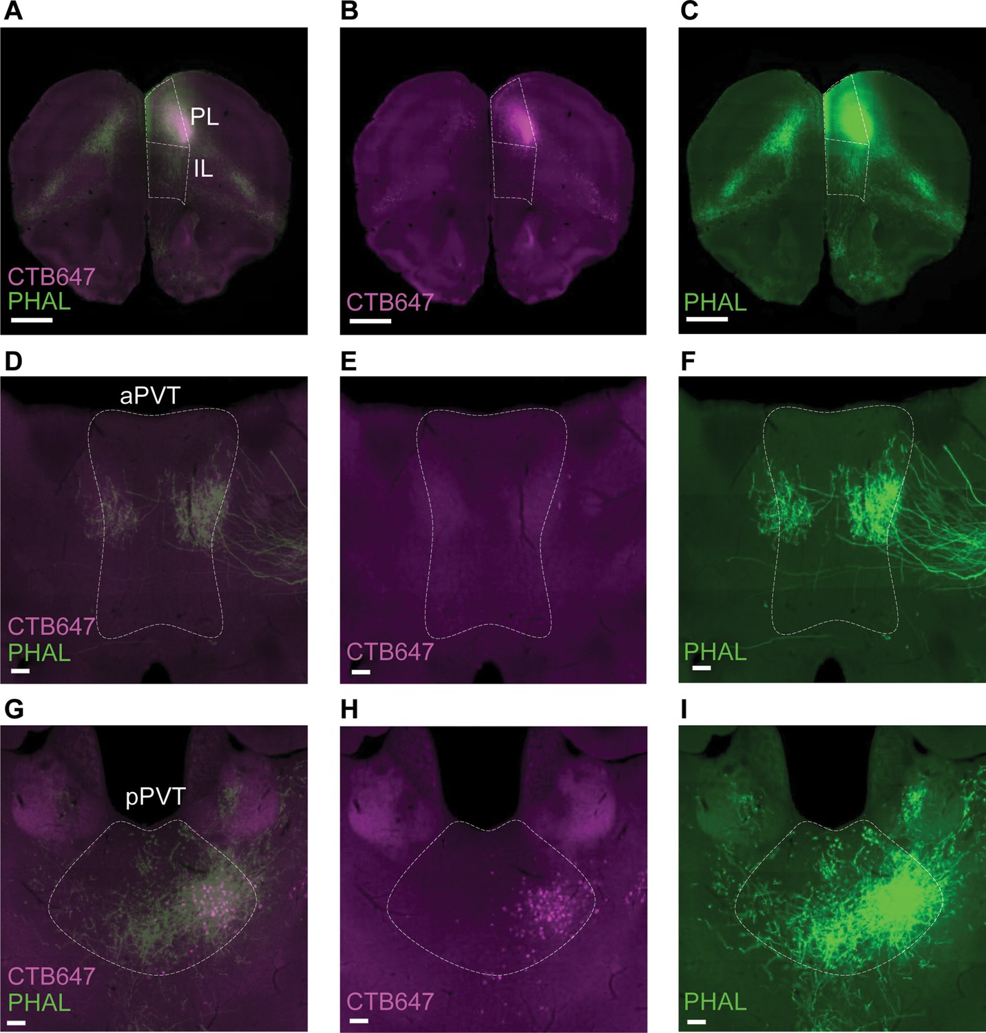
Dual anterograde and retrograde tracing from the prelimbic cortex (Mouse Connectome Project Experiment SW120125-02A).
(a–c) Representative image of the injection site in the prelimbic cortex (PL) of retrograde tracer CTB647 (magenta) and anterograde tracer PHAL (green); Scale bar 1 mm. (d-f) Representative image of retrogradely labeled cells (magenta) and anterogradely labeled fibers (green) in the aPVT; Scale bar 100 μm. (g–i) Representative image of retrogradely labeled cells (magenta) and anterogradely labeled fibers (green) in the pPVT; Scale bar 100 μm. Legend: IL, infralimbic cortex; https://www.mouseconnectome.org/.
Figure 5—figure supplement 4
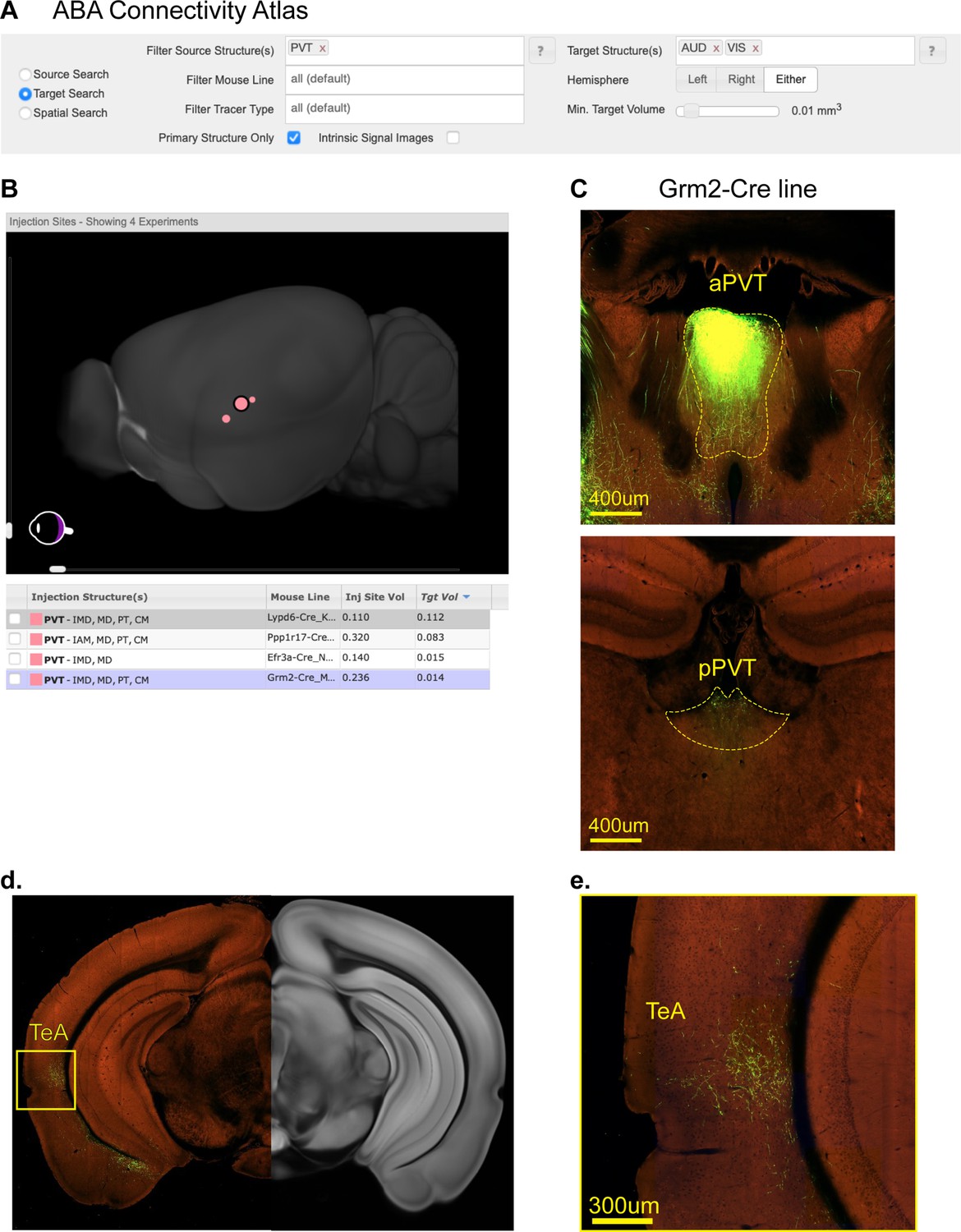
Projections from aPVT to Auditory and Visual cortical areas.
(a) Schematic of Target Search tool used to identify experiments where projections from the PVT to Auditory (AUD) and Visual (VIS) areas were detected (Mouse Brain Connectivity Atlas of the Allen Brian Institute, https://connectivity.brain-map.org). (b) Four experiments were identified from the target search. (c) Representative image from Experiment #183225830 depicting the injection site in the aPVT (top) and pPVT (bottom). (d) Representative image of fibers located in the temporal association cortex (TeA) from Experiment #183225830 (e) Same image of the yellow insert from (d).
Author response image 1
Author response image 2
Author response image 3
Additional files
-
MDAR checklist
- https://cdn.elifesciences.org/articles/81818/elife-81818-mdarchecklist1-v2.pdf
-
Supplementary file 1
Lists of differential gene expression for snRNA-seq data and conserved marker analysis from the integrated dataset.
- https://cdn.elifesciences.org/articles/81818/elife-81818-supp1-v2.xlsx
Download links
A two-part list of links to download the article, or parts of the article, in various formats.
Downloads (link to download the article as PDF)
Open citations (links to open the citations from this article in various online reference manager services)
Cite this article (links to download the citations from this article in formats compatible with various reference manager tools)
Molecular and spatial profiling of the paraventricular nucleus of the thalamus
eLife 12:e81818.
https://doi.org/10.7554/eLife.81818


