Antibacterial T6SS effectors with a VRR-Nuc domain are structure-specific nucleases
- Departamento de Microbiologia, Instituto de Ciências Biomédicas, Universidade de São Paulo, Brazil
- Department of Biosciences, University of Birmingham, United Kingdom
- Departamento de Bioquímica, Instituto de Química, Universidade de São Paulo, Brazil
Figures
Figure 1

The S. bongori SPI-22 encodes an antibacterial T6SS.
(A) Comparison between the SPI-22 T6SS of S. bongori with the systems of C. rodentium and P. aeruginosa. The T6SS proteins forming the three subcomplexes are in colors: membrane components (orange), sheath and inner tube (blue), and baseplate and spike components (green). (B) Representation of the circular genome of S. bongori with T6SS components highlighted: the structural cluster is marked by a black line; VgrG proteins are represented by green lines; Hcps are in blue; adaptor proteins are in orange; and PAAR or PAAR-like proteins are in red. TseV1, TseV2, and TseV3 fused to PAAR-like domain are also in red, and TseV4 is in gray. (C) Bacterial competition assays between S. bongori WT, ΔtssB, or ΔtssB complemented with pFPV25.1 tssB against E. coli in LB agar incubated for 24 hr. The prey recovery rate was calculated by dividing the colony-forming unit (CFU) counts of the output by the input. Data represent the mean ± standard deviation (SD) of six independent experiments and were analyzed through comparison with WT that were normalized to 1. One-way analysis of variance (ANOVA) followed by Dunnett’s multiple comparison test. ***p < 0.01 and ns (not significant).
-
Figure 1—source data 1
CFU counts used to calculate the prey recovery rate of Figure 1C.
- https://cdn.elifesciences.org/articles/82437/elife-82437-fig1-data1-v2.zip
Figure 2 with 5 supplements
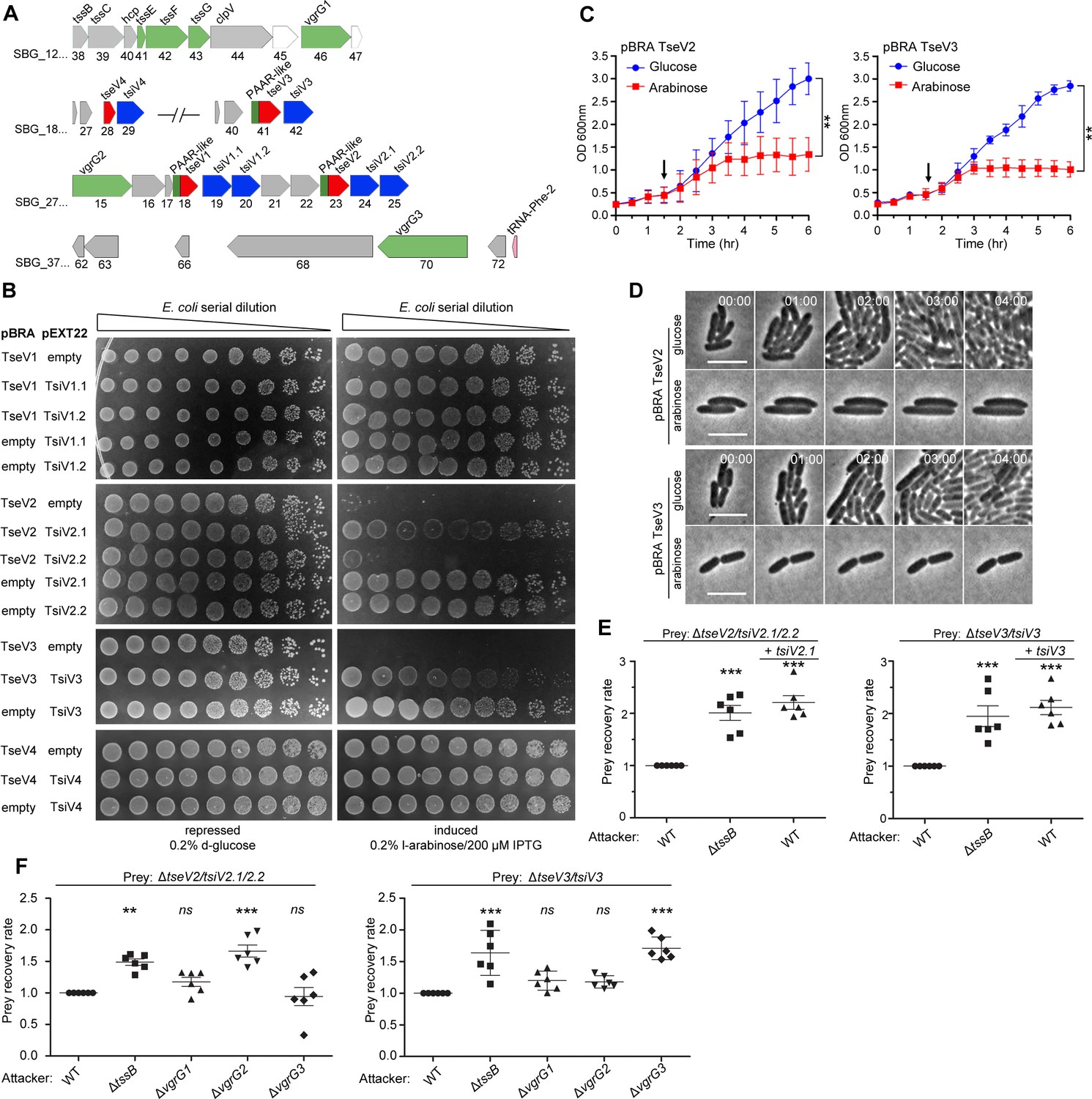
TseV2 and TseV3 are antibacterial SPI-22 T6SS effectors.
(A) Scheme of the genomic region containing VgrGs and TseV/TsiV effector/immunity pairs. VRR-Nuc domain (red), PAAR-like domain (dark green), VgrG (light green), and DUF3396-containing immunities (blue). (B) E. coli toxicity assay. Serial dilutions of E. coli containing pBRA and pEXT22 constructs, as indicated, spotted onto LB agar plates, and grown for 20 hr. Images are representative of three independent experiments. (C) Growth curve of E. coli harboring pBRA TseV2 or TseV3 before and after toxin induction by the addition of 0.2% l-arabinose (arrow). Results represent the mean ± standard deviation (SD) of three independent experiments performed in duplicate. **p < 0.01 (Student’s t-test). (D) Time-lapse microscopy of E. coli carrying either pBRA TseV2 or pBRA TseV3 grown on LB agar pads containing either 0.2% d-glucose (repressed) or 0.2% l-arabinose (induced). Scale bar: 5 µm. Timestamps in hh:mm. (E) Bacterial competition assay using S. bongori WT, ΔtssB, or ΔtssB complemented with pFPV25.1 tssB against S. bongori ΔtseV2/tsiV2.1/tsiV2.2 or ΔtseV3/tsiV3 complemented or not with pFPV25.1 tsiV2.1 or pFPV25.1 tsiV3. Strains were coincubated for 20 hr (ΔtseV2/tsiV2.1/tsiV2.2) or 6 hr (ΔtseV3/tsiV3) prior to measuring CFU counts. The prey recovery rate was calculated by dividing the CFU of the output by the input. Data represent the mean ± SD of six independent experiments and were analyzed through comparison with WT that were normalized to 1. One-way analysis of variance (ANOVA) followed by Dunnett’s multiple comparison test. **p < 0.01, and ***p < 0.001. (F) Bacterial competition assay using S. bongori WT, ΔtssB, ΔvgrG1, ΔvgrG2, or ΔvgrG3 against S. bongori ΔtseV2/tsiV2.1/tsiV2.2 or ΔtseV3/tsiV3. Strains were coincubated for 20 hr prior to measuring CFU counts. Prey recovery rate was calculated as in (E). Data represent the mean ± SD of six independent experiments. One-way ANOVA followed by Dunnett’s multiple comparison test. *p < 0.05, **p < 0.01, and ns (not significant).
-
Figure 2—source data 1
Original images of the E. coli plates shown in Figure 2B.
- https://cdn.elifesciences.org/articles/82437/elife-82437-fig2-data1-v2.zip
-
Figure 2—source data 2
OD600 nm measures of the growth curve shown in Figure 2C.
- https://cdn.elifesciences.org/articles/82437/elife-82437-fig2-data2-v2.zip
-
Figure 2—source data 3
CFU counts used to calculate the prey recovery rate of Figure 2E, F.
- https://cdn.elifesciences.org/articles/82437/elife-82437-fig2-data3-v2.zip
Figure 2—figure supplement 1
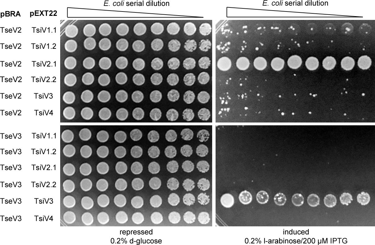
Toxicity assay in E. coli cotransformed with pBRA TseV2 or pBRA TseV3 and the six different immunity proteins.
Only one specific immunity could abrogate the toxic effect. Images are representative of three independent experiments.
Figure 2—video 1
Time-lapse microscopy of E. coli harboring pBRA TseV2 growing in media supplemented with 0.2% d-glucose.
Timestamp in hh:mm. Scale bar: 5 μm. Arrows indicate selected bacteria shown in Figure 2D.
Figure 2—video 2
Time-lapse microscopy of E. coli harboring pBRA TseV2 growing in media supplemented with 0.2% l-arabinose.
Timestamp in hh:mm. Scale bar: 5 μm. Arrows indicate selected bacteria shown in Figure 2D.
Figure 2—video 3
Time-lapse microscopy of E. coli harboring pBRA TseV3 growing in media supplemented with 0.2% d-glucose.
Timestamp in hh:mm. Scale bar: 5 μm. Arrows indicate selected bacteria shown in Figure 2D.
Figure 2—video 4
Time-lapse microscopy of E. coli harboring pBRA TseV3 growing in media supplemented with 0.2% l-arabinose.
Timestamp in hh:mm. Scale bar: 5 μm. Arrows indicate selected bacteria shown in Figure 2D.
Figure 3 with 1 supplement
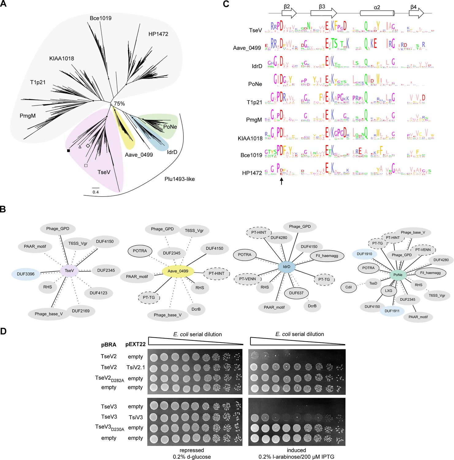
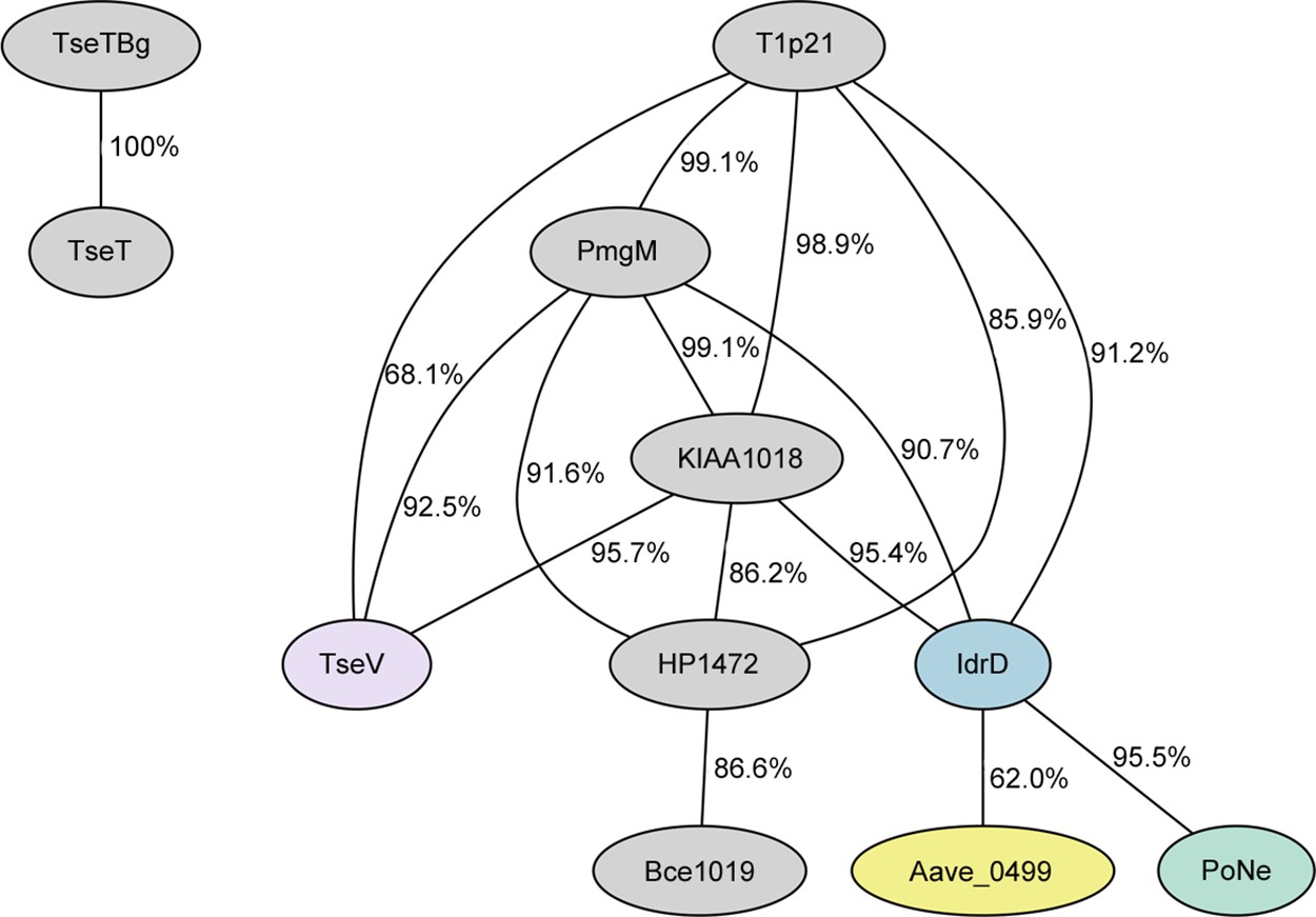
VRR-Nuc-containing effectors are evolutionarily related to enzymes involved in DNA repair.
(A) Maximum-likelihood phylogenetic tree of VRR-Nuc family members (Bce1019, PmgM, T1p21, KIAA1018, HP1472, and Plu1493) (Iyer et al., 2006) and recently reported bona fide or putative T6SS effectors belonging to the PD-(D/E)xK superfamily (TseT, PoNe, IdrD-CT, TseTBg, Aave_0499, and TseVPA). In the TseV clade (pink) the localization of TseV1 (□), TseV2 (Δ), TseV3 (■), and Plu1493 (◊) are marked. (B) Contextual network representation of domains and the genomic context of proteins belonging to Plu1493-like group (TseV, Aave_0499, IdrD, PoNe). Each circle represents a domain, which is either fused to (solid line) or encoded up- or downstream (dashed line) of the gene of interest (center). Borderless gray circles represent domains related to T6SS; bordered gray circles denote domains associated with a different bacterial secretion system; dashed nodes indicate pre-toxin domains; and light blue circles represent immunity proteins. (C) Sequence logo from the conserved β2β3α2β4 of the PD-(D/E)xK enzymatic core from all clades shown in (A). The arrow indicates conserved aspartic acid that was mutated in (D). (D) E. coli toxicity assay. Serial dilution of E. coli containing pBRA and pEXT22 constructs, as indicated, spotted onto LB agar plates and grown for 20 hr. Images are representative of three independent experiments.
-
Figure 3—source data 1
Amino acid sequence alignments used to generate the phylogenetic tree and sequence logos in Figure 3A, C.
- https://cdn.elifesciences.org/articles/82437/elife-82437-fig3-data1-v2.zip
-
Figure 3—source data 2
Original images of the E. coli plates shown in Figure 3D.
- https://cdn.elifesciences.org/articles/82437/elife-82437-fig3-data2-v2.zip
Figure 3—figure supplement 1
Comparison of the HMM (Hidden Markov Model) from each clade shown in Figure 3A.
All clades present enough similarity to be clustered together, while the homologs of P. aeruginosa TseT and B. gladioli TseTBg differ from the other models.
Figure 4 with 1 supplement
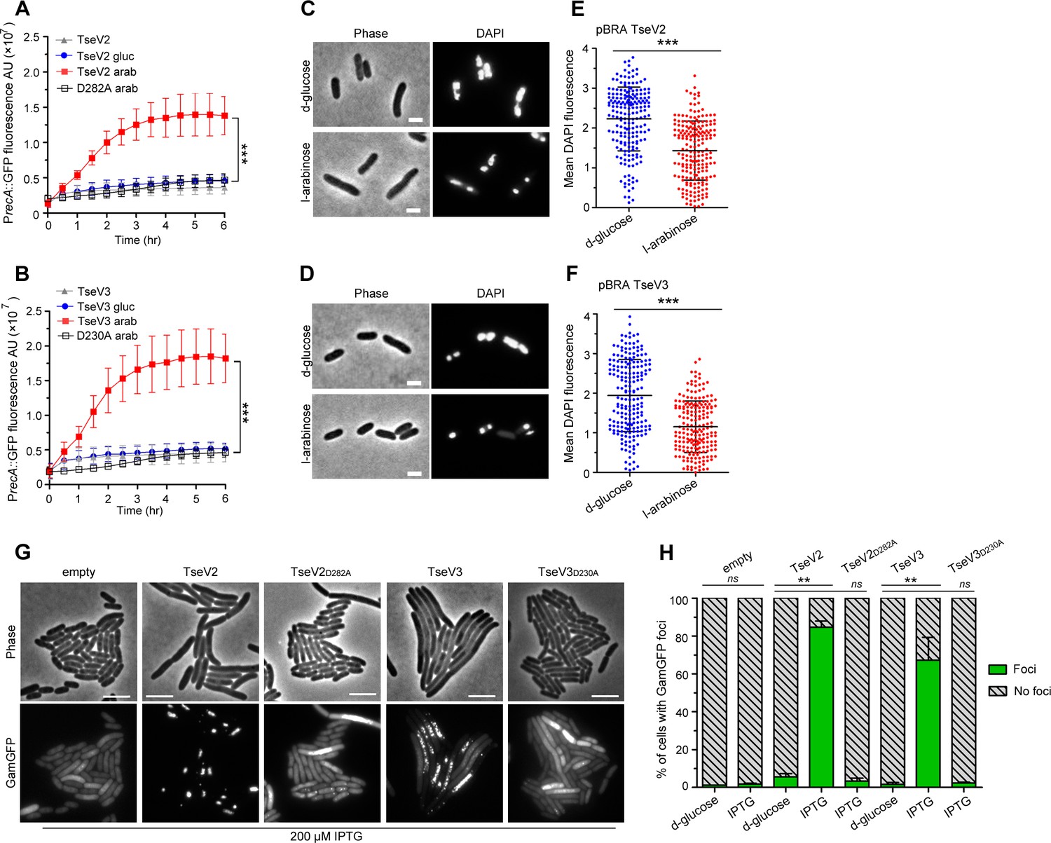
TseV2 and TseV3 induce DNA double-strand breaks.
Activation of the SOS response was analyzed using E. coli cells harboring the reporter plasmid pSC101-PrecA::GFP and pBRA TseV2 (A) or pBRA TseV3 (B), which were grown in AB defined media with d-glucose or l-arabinose. Data is the mean ± standard deviation (SD) of three independent experiments. ***p < 0.001 (Student’s t-test). Bright-field and DAPI images of E. coli cells carrying pBRA TseV2 (C) or pBRA TseV3 (D) grown in the presence of d-glucose (repressed) or l-arabinose (induced). Results are representative images of three independent experiments. (E, F) Quantification of the mean 4′,6-diamidino-2-phenylindole (DAPI) fluorescence per cell of 200 cells. Data correspond to the mean ± SD of a representative experiment. Scale bar 2 μm. ***p < 0.001 (Student’s t-test). (G) Representative bright-field and GFP images of E. coli coexpressing GamGFP and pEXT20 TseV2 or pEXT20 TseV3. Double-strand breaks appear as foci of GamGFP. Images are representatives of three independent experiments. Scale bar: 5 μm. (H) Quantification of the GamGFP foci shown in (G). Data are shown as the mean ± SD of the three independent experiments. **p < 0.01 (Student’s t-test).
-
Figure 4—source data 1
Values of GFP signal acquired for the SOS response experiment shown in Figure 4A, B.
- https://cdn.elifesciences.org/articles/82437/elife-82437-fig4-data1-v2.zip
-
Figure 4—source data 2
Values of 4′,6-diamidino-2-phenylindole (DAPI) fluorescence measured for each bacterium, and original images used for quantification shown in Figure 4C–F.
The files can be opened in ImageJ.
- https://cdn.elifesciences.org/articles/82437/elife-82437-fig4-data2-v2.zip
-
Figure 4—source data 3
Original images used to count GamGFP foci shown in Figure 4G, H and numbers of foci.
The image files can be visualized with ImageJ.
- https://cdn.elifesciences.org/articles/82437/elife-82437-fig4-data3-v2.zip
-
Figure 4—source data 4
Original images used to count GamGFP foci in bacteria carrying pEXT20 TseV2 or TseV2D282A in Figure 4G, H.
- https://cdn.elifesciences.org/articles/82437/elife-82437-fig4-data4-v2.zip
-
Figure 4—source data 5
Original images used to count GamGFP foci in bacteria carrying pEXT20 TseV3 or TseV3D230A in Figure 4G, H.
- https://cdn.elifesciences.org/articles/82437/elife-82437-fig4-data5-v2.zip
-
Figure 4—source data 6
Original images used to count GamGFP foci in bacteria carrying empty pEXT20 in Figure 4G, H.
- https://cdn.elifesciences.org/articles/82437/elife-82437-fig4-data6-v2.zip
Figure 4—figure supplement 1
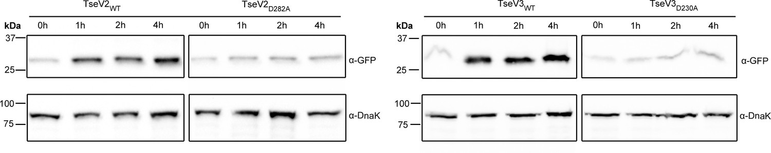
Western blot with anti-GFP antibody of protein extracts from E. coli carrying reporter plasmid pSC101 PrecA::GFP and pBRA with the indicated toxins after induction with 0.2% l-arabinose. anti-DnaK antibody was used as loading control.
-
Figure 4—figure supplement 1—source data 1
Original images of western blots.
- https://cdn.elifesciences.org/articles/82437/elife-82437-fig4-figsupp1-data1-v2.zip
Figure 5 with 1 supplement
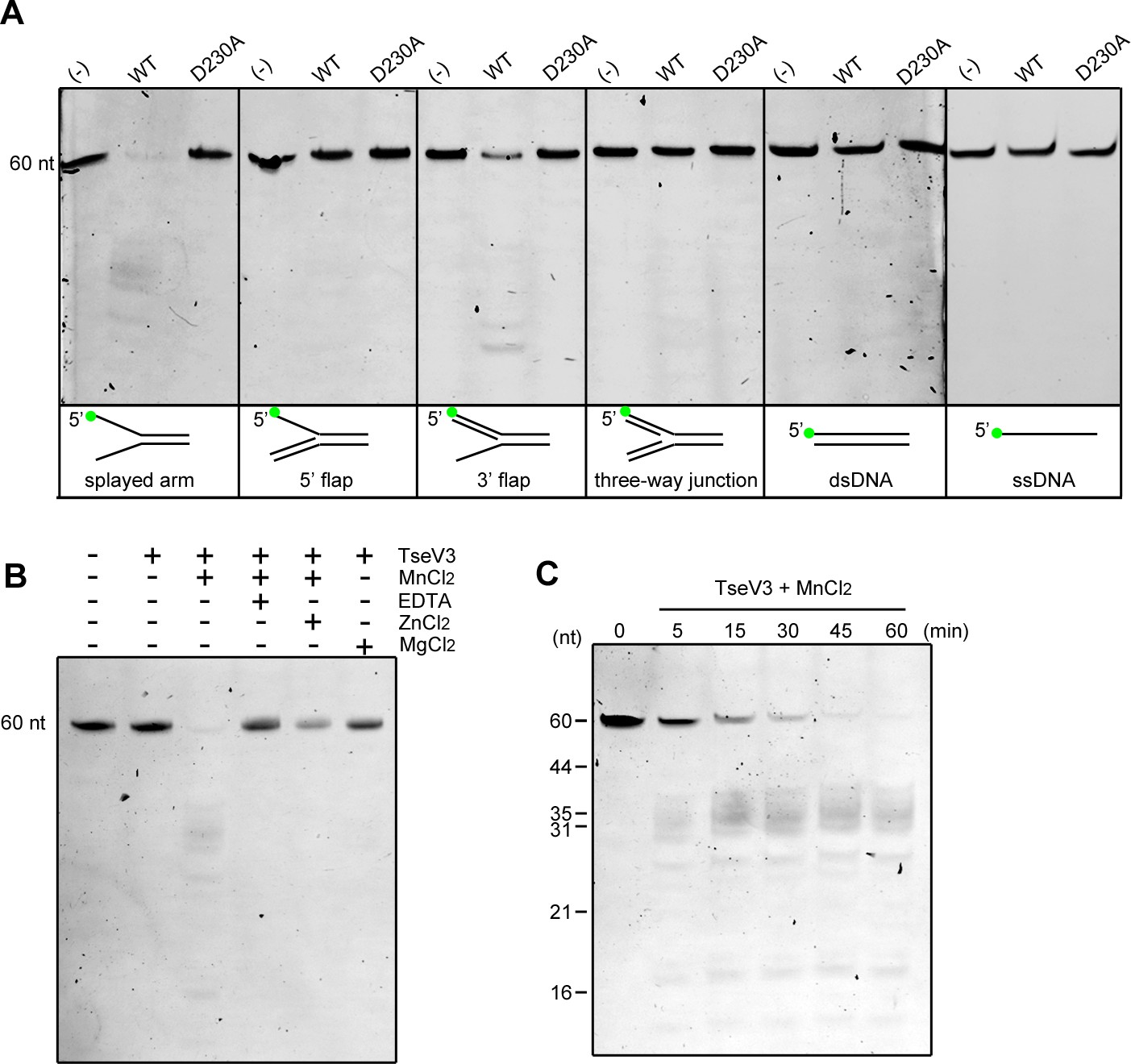
TseV3 is a Mn2+-dependent structure-specific nuclease.
(A) In vitro enzymatic assay with recombinant TseV3 or TseV3D230A coincubated with different DNA substrates at 37°C for 1 hr. Oligonucleotide F9 was labeled with FAM at the 5′ end (green circle). Image is representative of four independent experiments. (B) TseV3 was coincubated with splayed arm substrate at 37°C for 1 hr with 5 mM of the indicated cofactors. Image is representative of three independent experiments. (C) Time-course degradation of splayed arm by TseV3. Images are representative of three independent experiments.
-
Figure 5—source data 1
Original images of enzymatic assays.
- https://cdn.elifesciences.org/articles/82437/elife-82437-fig5-data1-v2.zip
-
Figure 5—source data 2
Original images of cofactors assays.
- https://cdn.elifesciences.org/articles/82437/elife-82437-fig5-data2-v2.zip
-
Figure 5—source data 3
Original images of time-course experiments.
- https://cdn.elifesciences.org/articles/82437/elife-82437-fig5-data3-v2.zip
Figure 5—figure supplement 1
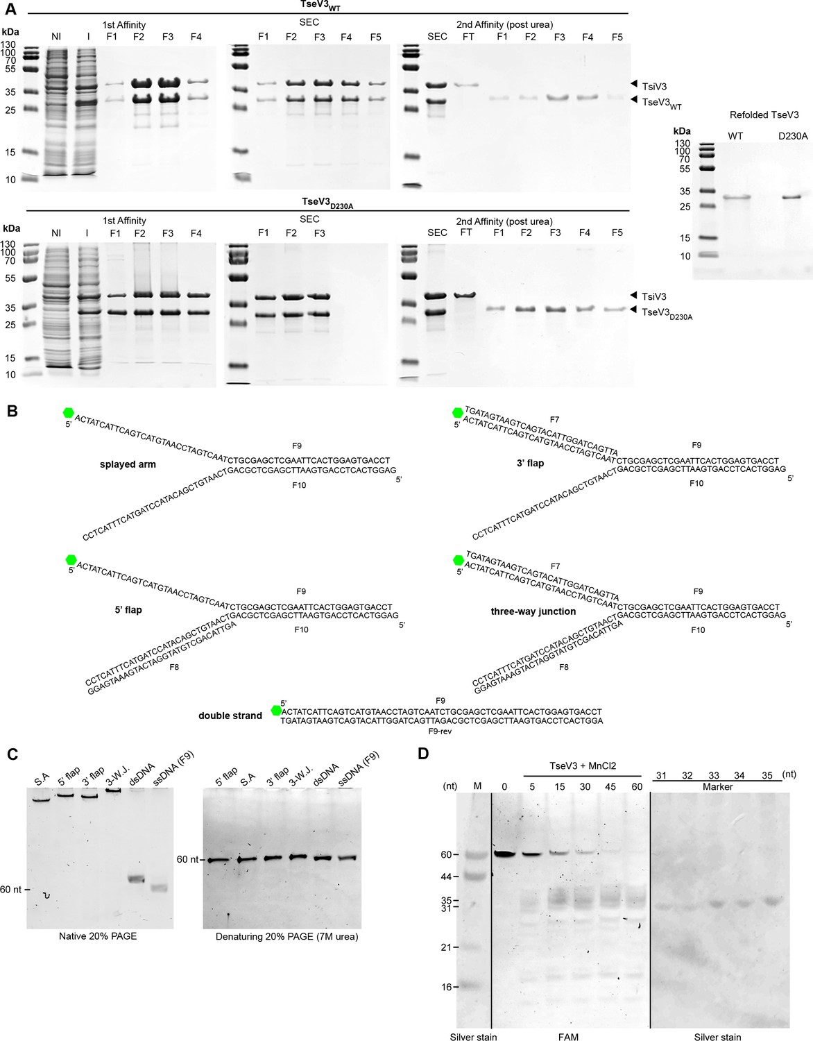
Recombinant protein purification and DNA substrates.
(A) Sodium dodecyl sulfate–polyacrylamide gel electrophoresis (SDS–PAGE) of recombinant proteins during purification steps to obtain purified protein for enzymatic assays. First affinity chromatography with noninduced culture extracts (NI), induced (I), and eluted fractions; size-exclusion chromatography (SEC) with eluted fractions; second affinity chromatography of samples after urea denaturation, flow through (FT), and eluted fraction; on the right, purified and refolded proteins TseV3WT and TseV3D230A used for enzymatic assays. (B) DNA substrate sequences with indicated oligonucleotides labeled with FAM at the 5′ end (green). (C) Annealed oligonucleotides visualized on native and denaturing 20% polyacrylamide gel. (D) Time-course degradation of splayed arm by TseV3 overlayed with silver-stained oligonucleotide markers.
-
Figure 5—figure supplement 1—source data 1
Raw images of sodium dodecyl sulfate–polyacrylamide gel electrophoresis (SDS–PAGE).
- https://cdn.elifesciences.org/articles/82437/elife-82437-fig5-figsupp1-data1-v2.zip
-
Figure 5—figure supplement 1—source data 2
Original images of 20% native and denaturing PAGE.
- https://cdn.elifesciences.org/articles/82437/elife-82437-fig5-figsupp1-data2-v2.zip
-
Figure 5—figure supplement 1—source data 3
Original images of time-course experiment and silver-stained gel.
- https://cdn.elifesciences.org/articles/82437/elife-82437-fig5-figsupp1-data3-v2.zip
Figure 6 with 1 supplement
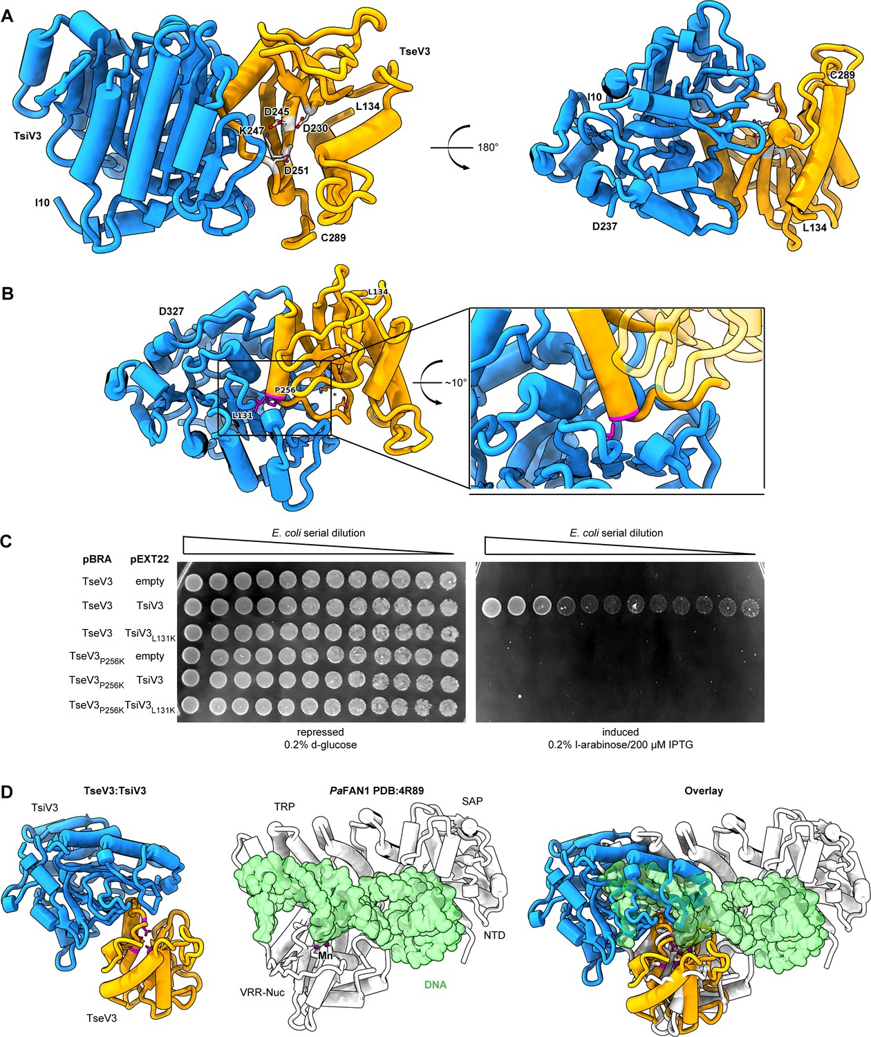
The effector–immunity complex reveals that TsiV3 blocks TseV3 substrate-binding site.
(A) Constrained model of the TseV3:TsiV3 heterodimer with two different views: TsiV3 in blue (I10–D327) nd TseV3 in orange (L134–C289). Models are labeled to assist interpretation. PD-(D/E)xK superfamily conserved residues of TseV3 (D230, D245, and K247) are shown in stick form and colored light gray, confirming that they converge to form a putative consensus active site. (B) Prediction of interface-compromising mutants in the TseV3:TsiV3 heterodimer. TsiV3 (blue) and TseV3 (orange) with putative active sites labeled with asterisk. Residues L131 of TsiV3 and P256 of TseV3 (both in stick form, magenta) form the closest point of contact in the heterodimer and are at the center of a hydrophobic-rich interface. (C) E. coli toxicity assay using cells carrying plasmids with wild-type or point mutations in TsiV3 (L131K) or TseV3 (P256K) as a potential means to destabilize the TseV3:TsiV3 complex interaction. (D) Superimposition of the TseV3:TsiV3 coordinates with those of the PaFAN1:DNA complex (PDB 4R89). PaFAN1 protein in white, DNA duplex in green, and catalytic Mn2+ are depicted as purple spheres. The overlay (right) is presented in the same orientation as the individual complexes: TseV3:TsiV3 (left, catalytic residues in magenta) and PaFAN1:DNA (middle).
-
Figure 6—source data 1
Original image of the E. coli plates shown in Figure 5D.
- https://cdn.elifesciences.org/articles/82437/elife-82437-fig6-data1-v2.zip
Figure 6—figure supplement 1
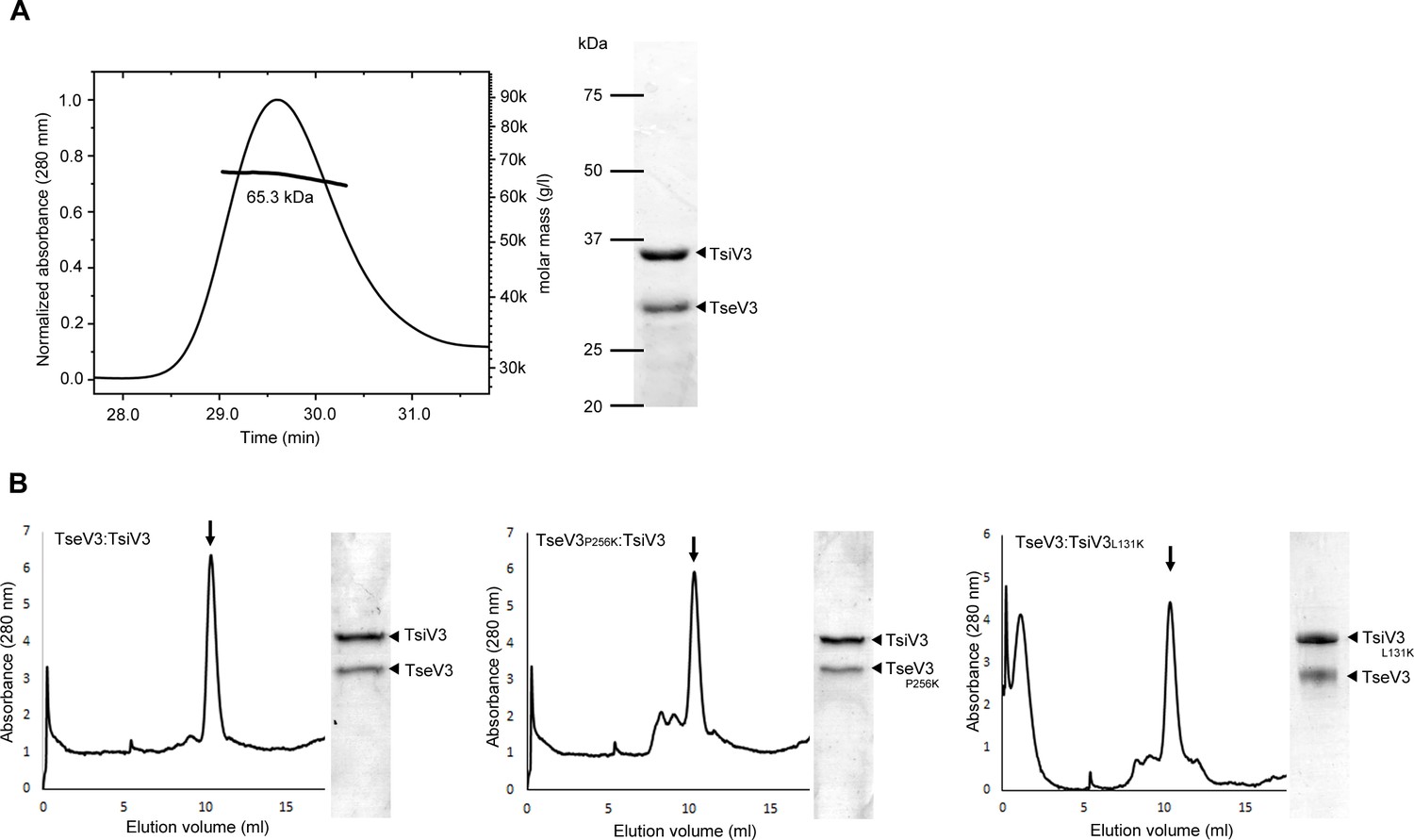
TseV3:TsiV3 SEC analysis.
(A) SEC-MALS analysis shows the formation of a stable complex between TseV3:TsiV3. The line corresponds to the calculated molecular mass. Right panel: Coomassie stained sodium dodecyl sulfate–polyacrylamide gel electrophoresis (SDS–PAGE) showing the apparent molecular mass of proteins eluted from SEC-MALS peak. (B) SEC showing the formation of a complex between TseV3:TsiV3, TseV3P256K:TsiV3 and TseV3:TsiV3L131K. Right: Coomassie stained SDS–PAGE showing proteins from the central peak.
-
Figure 6—figure supplement 1—source data 1
Raw image of the sodium dodecyl sulfate–polyacrylamide gel electrophoresis (SDS–PAGE) with labels and SEC-MALS data.
- https://cdn.elifesciences.org/articles/82437/elife-82437-fig6-figsupp1-data1-v2.zip
Additional files
-
Supplementary file 1
Amino acid sequence alignment of VgrGs.
(A) Amino acid sequence alignment of VgrG1, VgrG2, and VgrG3. (B) Amino acid sequence alignment of VgrG2 and VgrG3. Amino acids are color-coded according to their properties.
- https://cdn.elifesciences.org/articles/82437/elife-82437-supp1-v2.tif
-
Supplementary file 2
List of all homologs collected by JackHMMER searches and used to build the phylogenetic tree shown in Figure 3A.
- https://cdn.elifesciences.org/articles/82437/elife-82437-supp2-v2.xlsx
-
Supplementary file 3
Genomic context of members of each VRR-Nuc subfamily.
- https://cdn.elifesciences.org/articles/82437/elife-82437-supp3-v2.xlsx
-
Supplementary file 4
Plasmidial DNA extraction from E. coli expressing TseV2, TseV2D282A, TseV3, or TseV3D230A for 1–2 hr visualized on 1% agarose gel.
- https://cdn.elifesciences.org/articles/82437/elife-82437-supp4-v2.tif
-
Supplementary file 5
Amino acid sequence alignment of TseV1-4 and P. aeruginosa TseV (PA0822) based on secondary structures.
(A) Manual amino acid sequence alignment of TseV14 and P. aeruginosa TseV (PA0822) based on secondary structures. The secondary structures are indicated above the alignments with α-helixes represented by spirals and β-sheets by arrows. The conserved catalytic residues are highlighted in red with the logo underneath the alignments. TseV4 contains another start codon located upstream of the annotated one. (B) TseV1–4 structures predicted by the AlphaFold (Jumper et al., 2021). Underneath is the conserved PD-(D/E)xK enzymatic core with the absent structures marked in dashed red.
- https://cdn.elifesciences.org/articles/82437/elife-82437-supp5-v2.tif
-
Supplementary file 6
Crystallographic statistics of the TseV3:TsiV3 complex.
- https://cdn.elifesciences.org/articles/82437/elife-82437-supp6-v2.xlsx
-
Supplementary file 7
List of strains, plasmids and primers used in the study.
- https://cdn.elifesciences.org/articles/82437/elife-82437-supp7-v2.xlsx
-
MDAR checklist
- https://cdn.elifesciences.org/articles/82437/elife-82437-mdarchecklist1-v2.docx
Download links
A two-part list of links to download the article, or parts of the article, in various formats.
Downloads (link to download the article as PDF)
Open citations (links to open the citations from this article in various online reference manager services)
Cite this article (links to download the citations from this article in formats compatible with various reference manager tools)
Antibacterial T6SS effectors with a VRR-Nuc domain are structure-specific nucleases
eLife 11:e82437.
https://doi.org/10.7554/eLife.82437


