N-terminal domain on dystroglycan enables LARGE1 to extend matriglycan on α-dystroglycan and prevents muscular dystrophy
- Howard Hughes Medical Institute, Senator Paul D. Wellstone Muscular Dystrophy Specialized Research Center, Department of Molecular Physiology and Biophysics and Department of Neurology, Roy J. and Lucille A. Carver College of Medicine, The University of Iowa, United States
- Department Pharmaceutical Sciences, School of Pharmaceutical Sciences, University of Shizuoka, Japan
- Department of Neurology, School of Medicine, Teikyo University, Japan
Figures
Figure 1

Domain structure of dystroglycan (DG) and Δ-α-DGN.
Wild-type DG is a pre-proprotein with an N-terminal signal peptide (light green) that is translated in the rough endoplasmic reticulum. The globular N-terminal domain (α-DGN; orange) is present in wild-type DG but absent in the mutant (∆-α-DGN). The junction between α-DGN and the mucin-like domain (light teal) contains a furin convertase site. The globular extracellular C-terminal domain (CTD; pink) contains an SEA (sea urchin sperm protein, enterokinase and agrin) autoproteolysis site, which cleaves pro-DG into α-DG and β-DG (green). Glycosylation has been omitted for clarity.
Figure 2 with 2 supplements
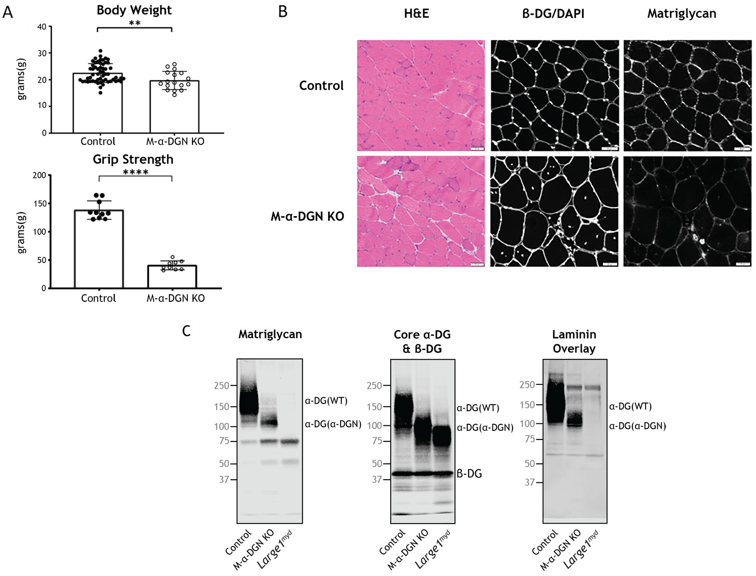
Characterization of mice with a muscle-specific loss of α-DG N-terminal (α-DGN).
(A) Body weight and grip strength of 12-week-old wild-type (WT) littermate (control) and muscle-specific α-DGN knockout (M-α-DGN KO) mice. Double and quadruple asterisks: statistical significance determined by Student’s unpaired t-test (**p-value = 0.005, ****p-value <0.0001). (B) Histological analyses of quadriceps muscles from 12-week-old control and M-α-DGN KO mice. Sections stained with H&E or used for immunofluorescence to detect β-DG (affinity purified rabbit anti-β-DG), DAPI, and matriglycan (IIH6). Scale bars = 50 μm. (C) Immunoblot analysis of skeletal muscle from control, M-α-DGN KO, and Large1myd mice. Glycoproteins were enriched using wheat-germ agglutinin (WGA)-agarose with 10 mM EDTA. Immunoblotting was performed to detect matriglycan (IIIH11), core α-DG, β-DG (AF6868), and laminin overlay. α-DG in WT control muscle (α-DG (WT)) and α-DG in α-DGN-deficient muscle (α-DG (Δα-DGN)) are indicated on the right. The number of KO mice was 17, and that of LC mice was 57. There were 6 male KO mice, 11 female KO mice, 23 male LC mice, and 34 female LC mice. Molecular weight standards in kilodaltons (kDa) are shown on the left.
-
Figure 2—source data 1
Full blots for Figure 2C.
- https://cdn.elifesciences.org/articles/82811/elife-82811-fig2-data1-v2.zip
Figure 2—figure supplement 1
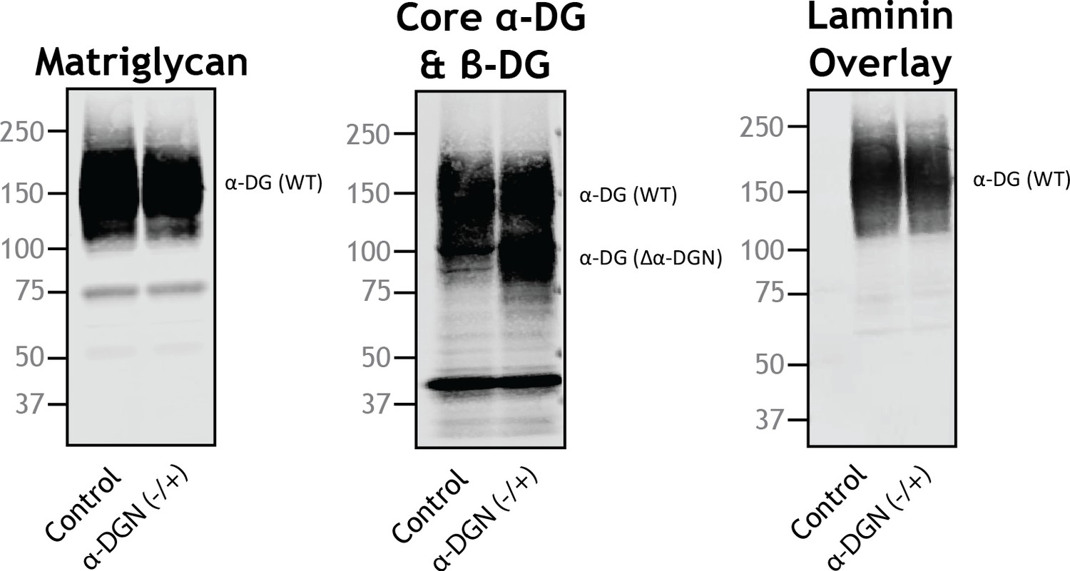
Mice heterozygous (+/-) for a constitutive deletion of α-DG N-terminal (α-DGN) have two different sizes of α-DG.
Immunoblot analysis of skeletal muscle from littermate controls or mice that are heterozygous for the α-DGN KO allele (α-DGN (-/+)). Glycoproteins were enriched from the quadriceps skeletal muscles of mice using wheat-germ agglutinin (WGA)-agarose with 10 mM EDTA. Immunoblotting was performed to detect matriglycan (IIIH11), core α-DG and β-DG (AF6868), and laminin overlay. α-DG in wild-type (WT) control muscle (α-DG(WT)) and α-DG in α-DGN-deficient muscle (α-DG(Δα-DGN)) are indicated on the right. Molecular weight standards in kilodaltons (kDa) are shown on the left.
-
Figure 2—figure supplement 1—source data 1
Full blots for Figure 2—figure supplement 1.
- https://cdn.elifesciences.org/articles/82811/elife-82811-fig2-figsupp1-data1-v2.zip
Figure 2—figure supplement 2
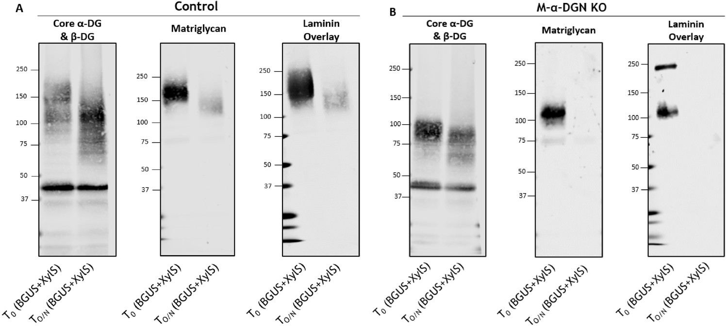
The short 100–120 kDa band in muscle-specific α-DGN knockout (M-α-DGN KO) muscle is matriglycan.
(A) Immunoblot analysis of total skeletal muscle from control mice after digestion with enzymes β-glucuronidase and α-xylosidase. Glycoproteins were enriched using wheat-germ agglutinin (WGA)-agarose with 10 mM EDTA and incubated overnight with β-glucuronidase (BGUS) and α-xylosidase (XyIS). Immunoblotting was performed to detect matriglycan (IIH6), core α-DG and β-DG (AF6868), and laminin overlay before (To) and after overnight digestion (TO/N). (B) Immunoblot analysis of M-α-DGN KO total skeletal muscle after digestion with enzymes BGUS and XyIS. Glycoproteins were enriched using WGA-agarose with 10 mM EDTA and incubated overnight with BGUS and XyIS. Immunoblotting was performed to detect matriglycan (IIH6), core α-DG and β-DG (AF6868), and laminin overlay before (To) and after digestion (TO/N). Molecular weight standards in kilodaltons (kDa) are shown on the left.
-
Figure 2—figure supplement 2—source data 1
Full blots for Figure 2—figure supplement 2.
- https://cdn.elifesciences.org/articles/82811/elife-82811-fig2-figsupp2-data1-v2.zip
Figure 3

α-DG N-terminal (α-DGN) deficiency results in post-synaptic defects.
Neuromuscular junctions (NMJs) from tibialis anterior (TA), extensor digitorum longus (EDL), and soleus (SOL) muscles obtained from 35- to 39-week-old control and muscle-specific α-DGN knockout (M-α-DGN KO) mice. (A) Representative images of post-synaptic terminals (α-BTX-488; green), motor axons (anti-neurofilament-H; red), and pre-synaptic terminals (anti-synaptophysin; red) from TA, EDL, and SOL muscles. Scale bars = 20 μm. (B) Scoring of post-synaptic defects by blinded observers (scoring criteria described in Materials and methods). Statistical significance determined by Student’s unpaired t-test; *p-value <0.05; **p-value <0.001; ***p-value <0.0001.
Figure 4 with 1 supplement
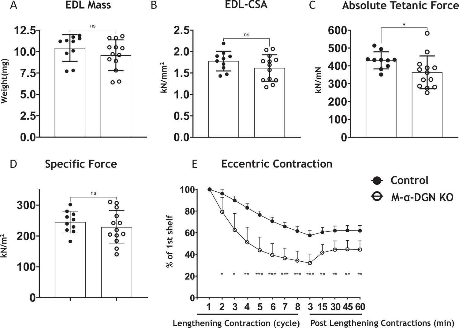
α-DG N-terminal (α-DGN)-deficient extensor digitorum longus (EDL) muscle demonstrates greater lengthening contraction-induced force decline.
(A) Weight (mg) of EDL muscles from wild-type (WT) littermates (controls) and muscle-specific α-DGN knockout (M-α-DGN KO) mice; p=0.2469, as determined by Student’s unpaired t-test. (B) Cross-sectional area of EDL muscles; p=0.1810, as determined by Student’s unpaired t-test. (C) Maximum absolute tetanic force production in EDL muscles. p=0.0488, as determined by Student’s unpaired t-test. (D) Specific force production in EDL muscles; p=0.4158, as determined by Student’s unpaired t-test. (E) Force deficit and force recovery after eccentric contractions in EDL muscles from 12- to 17-week-old male and female control (closed circles; n=7) and M-α-DGN KO (open circles; n=7) mice. *p<0.05; **p<0.01; ***p<0.001, as determined by Student’s unpaired t-test of at any given lengthening contractions cycle. Bars represent the mean ± the standard deviation.
Figure 4—figure supplement 1

α-DG N-terminal (α-DGN)-deficient muscle and protein O-mannose kinase (POMK)-deficient muscle with similar short forms of matriglycan exhibit similar lengthening contraction-induced force decline.
Force deficit and force recovery after eccentric contractions in extensor digitorum longus (EDL) muscles from 12- to 17-week-old male and female controls (closed circles; n=7), muscle-specific α-DGN knockout (M-α-DGN KO) (open circles; n=7), M-POMK littermate controls (closed triangles; n=3), and M-POMK KO (open triangles; n=4) mice. There is no significant difference in M-α-DGN KO vs. M-POMK KO EDL as determined by Student’s unpaired t-test at any given lengthening contractions cycle and post-lengthening contraction.
Figure 5 with 2 supplements
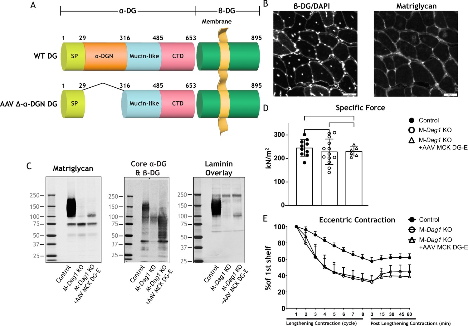
Exogenous α-DG N-terminal (α-DGN)-deficient dystroglycan (DG) also produces short matriglycan like M-Dag1 KO muscle.
(A) Schematic representation of a wild-type (WT) DG and an adeno-associated virus (AAV) carrying a mutant DG in which the N-terminal domain has been deleted (DG-E). α-DG is composed of a signal peptide (SP, amino acids 1–29), an N-terminal domain (amino acids 30–316), a mucin-like domain (amino acids 317–485), and a C-terminal domain (amino acids 486–653). The green box represents β-DG. (B) Immunofluorescence analyses of quadriceps muscles from 12-week-old M-Dag1 KO mice injected with AAV-MCK DG-E to detect β-DG, nuclei (DAPI), and matriglycan (IIH6). Scale bars = 50 μm. (C) Immunoblot analysis of skeletal muscle obtained from littermate controls (control), M-Dag1 KO mice, or M-Dag1 KO mice injected with AAV-MCK DG-E. Glycoproteins were enriched from skeletal muscles using wheat-germ agglutinin (WGA)-agarose. Immunoblotting was performed to detect matriglycan (IIIH11), core α-DG and β-DG (AF6868), and laminin (overlay). (D) Production of specific force in extensor digitorum longus ( EDL) muscles from 12- to 17-week-old male and female M-Dag1 KO mice (controls; closed circles, n=10); M-α-DGN KO mice (open circles, n=13); and M-Dag1 KO + AAV MCK DG-E mice (open triangles, n=6). p-Values determined by Student’s unpaired t-test; controls vs. M-Dag1 KO: p=0.4158; controls vs. M-Dag1 KO + AAV MCK DG-E: p=0.3632; M-Dag1 KO vs. M-Dag1 KO + AAV MCK DG-E: p=0.948. (E) Force deficits and recovery in EDL muscles from mice in D. There is no significant difference in M-Dag1 KO vs. M-Dag1 KO + AAV MCK DG-E as determined by Student’s unpaired t-test at any given lengthening contraction cycle or post-lengthening contraction.
-
Figure 5—source data 1
Full blots for Figure 5C.
- https://cdn.elifesciences.org/articles/82811/elife-82811-fig5-data1-v2.zip
Figure 5—figure supplement 1

Characteristics of M-Dag1 KO (Pax7cre; Dag1flox/flox) mice.
(A) Immunofluorescence analyses of quadriceps muscles from a 12-week-old wild-type (WT) littermate (control) or M-Dag1 KO mouse. Sections were stained to detect β-DG (AP83) and nuclei (DAPI). Scale bars = 50 μm. (B) Immunoblot analysis of skeletal muscle from control and M-Dag1 KO mice. Glycoproteins were enriched from skeletal muscles using wheat-germ agglutinin (WGA)-agarose with (+) and without (-) 10 mM EDTA. Immunoblotting was performed to detect matriglycan (IIIH11) and core α-DG and β-DG (AF6868). (C) Specific force in extensor digitorum longus (EDL) muscles of mice in indicated groups; p=0.0128, as determined by Student’s unpaired t-test. (D) Force deficit and force recovery after eccentric contractions in EDL muscles of 12- to 17-week-old male and female control (n=3) and M-Dag1 KO (n=6) mice.
-
Figure 5—figure supplement 1—source data 1
Full blots for Figure 5—figure supplement 1.
- https://cdn.elifesciences.org/articles/82811/elife-82811-fig5-figsupp1-data1-v2.zip
Figure 5—figure supplement 2
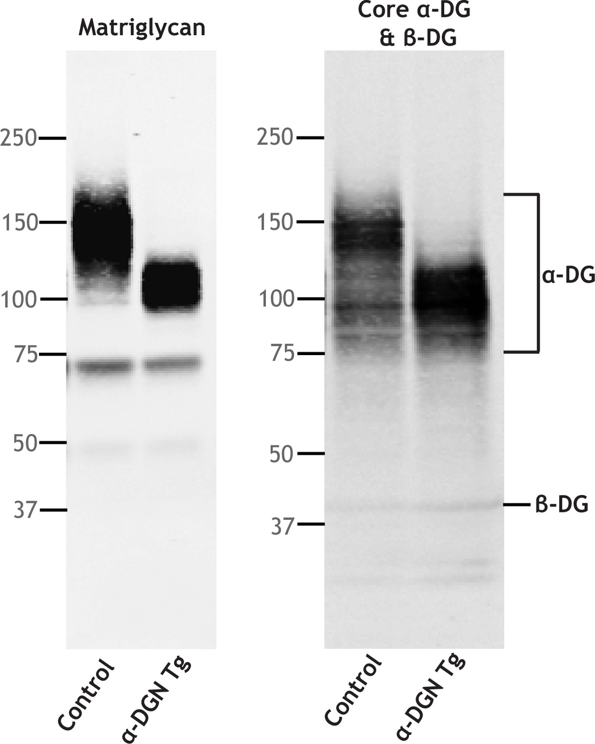
Excess free α-DG N-terminal (α-DGN) interferes with like-acetylglucosaminyltransferase-1 (LARGE1) elongation of matriglycan on α-DG.
Immunoblot analysis of skeletal muscle from control and α-DGN transgenic (α-DGN Tg) mice. Glycoproteins were enriched using wheat-germ agglutinin (WGA)-agarose. Immunoblotting was performed to detect matriglycan (IIH6), core α-DG, and β-DG (AF6868).
-
Figure 5—figure supplement 2—source data 1
Full blots for Figure 5—figure supplement 2.
- https://cdn.elifesciences.org/articles/82811/elife-82811-fig5-figsupp2-data1-v2.zip
Figure 6 with 1 supplement
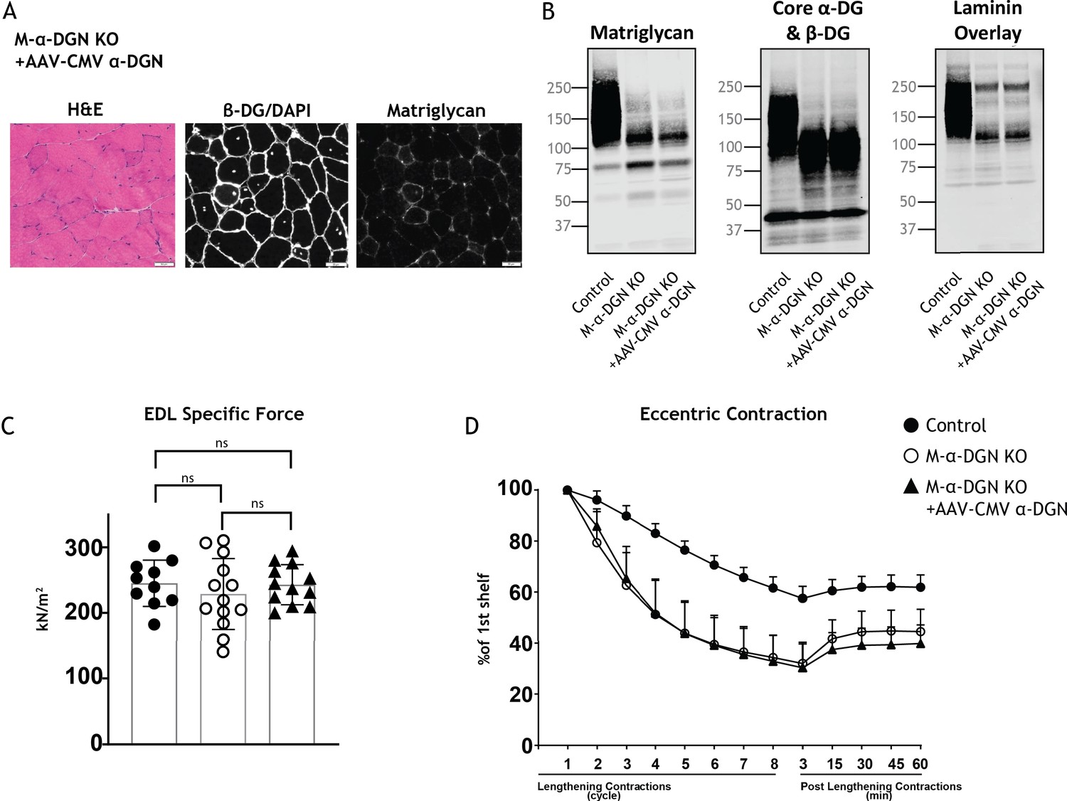
Expression of α-DG N-terminal (α-DGN) in muscle-specific α-DGN knockout (M-α-DGN KO) mice does not rescue matriglycan elongation.
(A) Representative sections of quadriceps muscles from 17-week-old M-α-DGN KO mice injected with AAV-CMV α-DGN. Sections were stained with H&E and immunofluorescence to detect matriglycan (IIH6) and β-DG (AP83). Scale bars = 50 μm.(B) Immunoblot analysis of skeletal muscle obtained from littermate controls or M-α-DGN KO mice and M-α-DGN KO mice injected with AAV-CMV α-DGN (M-α-DGN KO+AAV CMV α-DGN). Glycoproteins were enriched using wheat-germ agglutinin (WGA)-agarose with 10 mM EDTA. Immunoblotting was performed to detect matriglycan (IIIH11), core α-DG and β-DG (AF6868), and laminin overlay. (C) Production of specific force in extensor digitorum longus (EDL) muscles from 12- to 17-week-old male and female M-α-DGN wild-type (WT) littermates (controls; closed circles, n=10); M-α-DGN KO (open circles, n=13); and M-α-DGN KO+AAV CMV α-DGN (closed triangles, n=12). p-Values determined by Student’s unpaired t-test; controls vs. M-α-DGN KO+AAV CMV α-DGN: p=0.8759; controls vs. M-α-DGN KO: p=0.4333; M-α-DGN KO vs. M-α-DGN KO+AAV CMV α-DGN: p=0.4333. (D) Force deficit and force recovery after lengthening contractions in EDL muscles from 12- to 17-week-old male and female M-α-DGN KO WT littermates (controls, closed circles; n=6) and M-α-DGN KO (KO, open circles; n=7) mice, and in M-α-DGN KO mice injected with AAV-CMV α-DGN (KO+AAV CMV α-DGN, closed triangles; n=8). There is no significant difference in M-α-DGN KO vs. M-α-DGN KO+AAV CMV α-DGN as determined by Student’s unpaired t-test at any given lengthening contractions cycle or post-lengthening contractions.
-
Figure 6—source data 1
Full blots for Figure 6B.
- https://cdn.elifesciences.org/articles/82811/elife-82811-fig6-data1-v2.zip
Figure 6—figure supplement 1

Like-acetylglucosaminyltransferase-1 (LARGE1) overexpression does not extend matriglycan on dystroglycan (DG) lacking α-DG N-terminal (α-DGN).
AAV-MCK-Large1 was injected into the retro-orbital sinus 10- to-24-week-old muscle-specific α-DGN knockout (M-α-DGN KO) mice. Quadriceps skeletal muscle was dissected 10–22 weeks after injection from control, M-α-DGN KO, and M-α-DGN KO+AAV-MCK-mLarge1 and used for immunoblotting analysis. Glycoproteins were enriched using wheat-germ agglutinin (WGA)-agarose with 10 mM EDTA. Immunoblotting was performed to detect matriglycan (IIIH11), core α-DG and β-DG (AF6868), and laminin (overlay). Molecular weight standards in kilodaltons (kDa) are shown on the left.
-
Figure 6—figure supplement 1—source data 1
Full blots for Figure 6—figure supplement 1.
- https://cdn.elifesciences.org/articles/82811/elife-82811-fig6-figsupp1-data1-v2.zip
Figure 7
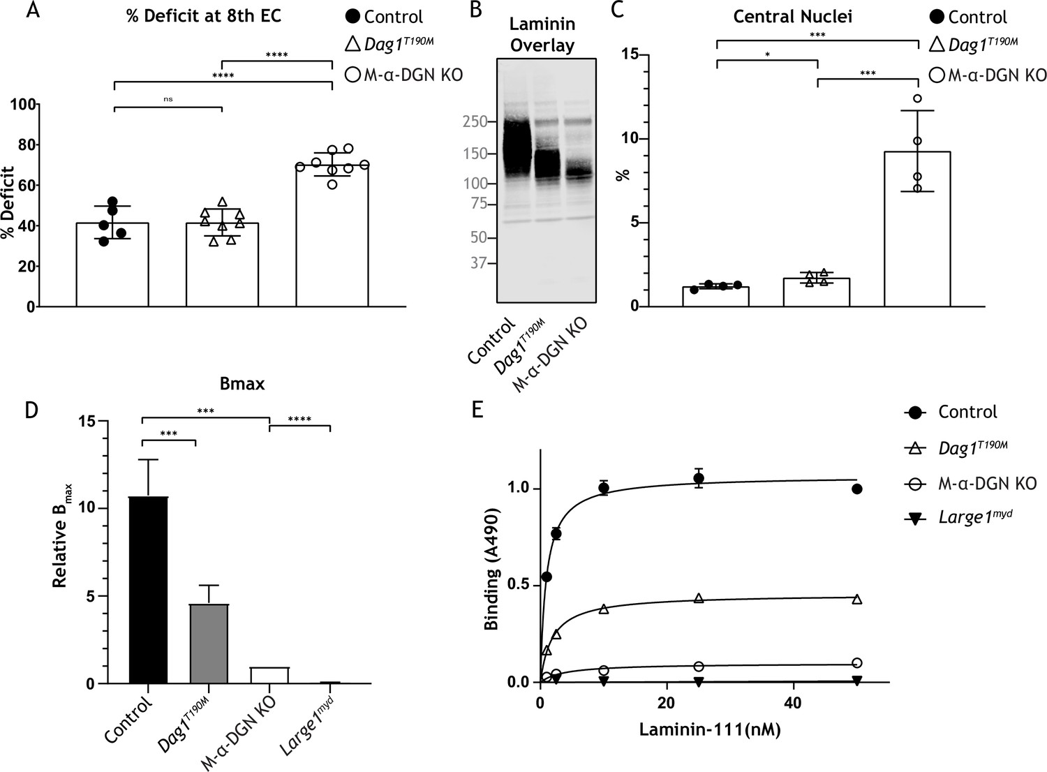
Relationship between matriglycan length and dystrophic phenotype.
(A) Percentage deficit of eight eccentric contraction (EC) in extensor digitorum longus (EDL) muscles from C57BL/6J wild-type (WT) (control), muscle-specific α-DGN knockout (M-α-DGN KO), and Dag1T190M mice. p-Values determined by Student’s unpaired t-test; control vs. Dag1T190M: p=0.0263; control and Dag1T190M vs. M-α-DGN KO: p<0.001. (B) Immunoblot analysis of quadriceps skeletal muscles from control, Dag1T190M and M-α-DGN KO mice. Glycoproteins were enriched using wheat-germ agglutinin (WGA)-agarose with 10 mM EDTA. Immunoblotting was performed with laminin (laminin overlay). (C) Percentage of muscle fibers with central nuclei in 12- to 19-week-old control, Dag1T190M and M-α-DGN KO mice; n=4 for all groups. p-Values determined by Student’s unpaired t-test; control and Dag1T190M vs. M-α-DGN KO: p<0.001; control vs. Dag1T190M: p=0.0263. (D) Comparison of average solid-phase determined relative Bmax values for laminin. Bmax values for M-α-DGN KO were set to 1 to allow for direct comparisons; error bars indicate s.e.m. p-Values determined using Student’s unpaired t-test; control vs. Dag1T190M and control vs. M-α-DGN KO: p<0.01, and M-α-DGN KO vs. Large1myd: p<0.001. (E) Solid-phase analysis of laminin-binding using laminin-111 in skeletal muscle from control, Dag1T190M, M-α-DGN KO, and Large1myd KO mice (three replicates for each group). Control Kd: 0.9664±0.06897 nM; Dag1T190M Kd: 1.902±0.1994 nM; and M-α-DGN KO Kd: 2.322±0.6114 nM.
-
Figure 7—source data 1
Full blot for Figure 7B.
- https://cdn.elifesciences.org/articles/82811/elife-82811-fig7-data1-v2.zip
Tables
Appendix 1—key resources table
| Reagent type (species) or resource | Designation | Source or reference | Identifiers | Additional information |
|---|---|---|---|---|
| Genetic reagent (Mus musculus) | Pax7Cre C57BL/6J | The Jackson Laboratory, Bar Harbor, ME, USA | JAX:010530, RRID:IMSR_JAX:010530 | Pax7tm1(cre)Mrc |
| Genetic reagent (Mus musculus) | Largemyd | The Jackson Laboratory, Bar Harbor, ME, USA | JAX:000300, RRID:IMSR_JAX:000300 | MYD/Le-Os+/+Largemydmyd/J |
| Genetic reagent (Mus musculus) | Largemyd | Campbell Lab | Described in Materials and Methods: Animals (92.5% C57BL/6J) | |
| Genetic reagent (Mus musculus) | Dag1ΔH30-A316 | PMID:31097590 DOI:10.1073/pnas.1904493116 | Dag1Δα-DGN | |
| Genetic reagent (Mus musculus) | Dag1flox | PMID:12230980 DOI: 10.1016/s0092-8674(02)00907–8 | JAX:006834, RRID:IMSR_JAX:006834 | B6.129-Dag1tm2Kcam/J |
| Genetic reagent (Mus musculus) | Dag1T190M | PMID: 21388311 DOI: 10.1056/NEJMoa1006939 | ||
| Genetic reagent (Mus musculus) | α-DGN Tg | This paper | Described in Materials and methods: Animals | |
| Genetic reagent (Mus musculus) | Pomkflox | PMID:32975514 DOI:10.7554/eLife.61388 | ||
| Antibody | Anti-DG; sheep polyclonal | R&D Systems | Cat# AF6868, RRID:AB_10891298 | WB (1:500) |
| Antibody | Anti-α-DG (IIH6C4); mouse monoclonal | Development Studies Hybridoma Bank/Campbell Lab | Cat# IIH6 C4, RRID:AB_2617216 | Described in Materials and Methods: Animals IF (1:10-1:100) |
| Antibody | Anti-α-DG (IIH6C4); mouse monoclonal | MilliporeSigma Campbell Lab | Cat# 05–593, RRID:AB_309828 | Described in Materials and Methods: Animals WB (1:1000–1:2000) |
| Antibody | Anti-Laminin; rabbit polyclonal | MilliporeSigma | Cat# L9393, RRID:AB_477163 | WB (1:1000), Solid Phase Assay (1:5000) |
| Antibody | Anti-β-DG; rabbit polyclonal | Campbell Lab PMID: 1741056 DOI: 10.1038/355696a0 | AP83 | Described in Materials and Methods: Animals IF (1:50) |
| Antibody | Anti-β-DG mouse IgM; mouse monoclonal | Leica Biosystems | Cat# NCL-b-DG, RRID:AB_442043 | IF (1:50 to 1:200) |
| Antibody | Anti-sheep IgG; donkey polyclonal | Rockland | Cat# 613-731-168, RRID:AB_220181 | WB (1:2000) |
| Antibody | Anti-mouse IgG (H+L); donkey polyclonal | LI-COR Biosciences | Cat# 926–32212, RRID:AB_621847 | WB (1:15,000), IF (1:800) |
| Antibody | Anti-rabbit IgG (H+L); donkey polyclonal | LI-COR Biosciences | Cat# 926–32213, RRID:AB_621848 | WB (1:15,000), IF (1:800) |
| Antibody | Anti-mouse IgM; goat polyclonal | LI-COR Biosciences | Cat# 926–32280, RRID:AB_2814919 | WB (1:2500) |
| Antibody | Anti-mouse IgG1; goat polyclonal | LI-COR Biosciences | Cat# 926–32350, RRID:AB_2782997 | WB (1:2000, 1:10,000) |
| Antibody | Anti-rabbit IgG (H+L); goat polyclonal | Thermo Fisher Scientific | Cat# A-11034, RRID:AB_2576217 | IF (1:1000 to 1:2000) |
| Antibody | Anti-mouse IgM; goat polyclonal | Thermo Fisher Scientific | Cat# A-21042, RRID:AB_2535711 | IF (1:1000 to 1:2000) |
| Antibody | Anti-human Synaptophysin (SP11); rabbit monoclonal | Thermo Fisher Scientific | Cat# MA5-14532, RRID:AB_10983675 | IF (1:100) |
| Antibody | Neurofilament NF-H; chicken polyclonal | EnCor Biotechnology | Cat# CPCA-NF-H, RRID:AB_2149761 | IF (1:1000) |
| Antibody | Anti-chicken IgY (H+L); goat polyclonal | Thermo Fisher Scientific | Cat# A32759, RRID:AB_2762829 | IF (1:1000) |
| Antibody | Anti-α-DG (IIIH11); mouse monoclonal | Campbell Lab | Described in Materials and Methods: Animals WB (1:100–1:1000) | |
| Chemical compound, drug | Pepstatin A | MilliporeSigma | Cat# 516481 | |
| Chemical compound, drug | Calpain Inhibitor I | MilliporeSigma | Cat# A6185 | |
| Chemical compound, drug | Aprotinin from bovine lung | MilliporeSigma | Cat# A1153 | |
| Chemical compound, drug | Leupeptin | MilliporeSigma | Cat# 108975 | |
| Chemical compound, drug | PMSF | MilliporeSigma | Cat# P7626 | |
| Chemical compound, drug | Immobilon-FL PVDF | MilliporeSigma | Cat# IPFL00010 | |
| Chemical compound, drug | Calpeptin | Thermo Fisher Scientific | Cat# 03-340-05125M | |
| Chemical compound, drug | Bis-acrylamide solution-30% (37.5:1) | Hoefer, Inc | Cat# GR337-500 | |
| Chemical compound, drug | Benzamidine Hydrochloride Hydrate | MP Biomedicals | Cat# 195068 | |
| Chemical compound, drug | WGA agarose bound | Vector Labs | Cat# AL-1023, RRID:AB_2336862 | |
| Chemical compound, drug | Precision Plus Protein All Blue Standards-500 µL | Bio-Rad | Cat# 161–0373 | |
| Chemical compound, drug | Ethylenediamine Tetraacetic acid, disodium salt dihydrate, EDTA | Thermo Fisher Scientific | Cat# S311-500 | |
| Peptide, recombinant protein | Enzymes, β-glucuronidase α-xylosidase | This paper and PMID: 27526028 DOI: 10.1038/nchembio.2146 | Described in Materials and methods: Digestion of α-DG with exoglycosidases | |
| Software, algorithm | SigmaPlot | SigmaPlot | RRID:SCR_003210 | |
| Software, algorithm | Excel | Microsoft | RRID:SCR_016137 | |
| Software, algorithm | GraphPad Prism | GraphPad | RRID:SCR_002798 | Version 8.3 |
| Software, algorithm | FlowJo | Becton, Dickinson & Company (BD) | RRID:SCR_008520 | Version 7.6.5 |
| Software, algorithm | Image Studio Acquisition Software | LI-COR Biosciences | RRID:SCR_015795 | |
| Software, algorithm | Fiji | National Institutes of Health | RRID:SCR_002285 | |
| Software | Adobe Illustrator | Adobe | RRID:SCR_010279 | Version 27.1.1 |
| Software, algorithm | UniProt Proteomes | RRID:SCR_018666 | ||
| Software, algorithm | IUPRED | RRID:SCR_014632 | ||
| Other | Streptavidin, Alexa Fluor 594 conjugate | Thermo Fisher Scientific | Cat# S11227 | IF (1:1000 to 1:2000) |
| Other | Western Blot Imager | LI-COR Biosciences | Odyssey CLx RRID:SCR_014579 | |
| Other | Isolated Mouse Muscle System | Aurora Scientific | 1200A | |
| Other | Mouse Grip Strength Meter | Columbus Instruments | 1027 Mouse | |
| Other | Tabletop ultracentrifuge | Beckman Coulter | Optima MAX, 130K | |
| Other | Ultracentrifuge | Beckman Coulter | Optima-L-100 XP | |
| Other | Centrifuge | Beckman Coulter | Avanti J-E HPC | |
| Other | Slide Scanner Microscope | Olympus | VS120-S5-FL RRID:SCR_018411 | |
| Other | Confocal Microscope | Olympus | FLUOVIEW FV3000 RRID:SCR_017015 | |
| Other | Cryostat | Leica Biosystems | CM3050S RRID:SCR_016844 | |
| Other | Imaging System | LI-COR Biosciences | Odyssey CLx Infrared RRID:SCR_014579 | |
| Other | 96-Well Plates | Corning Inc | Cat# 3590 | Described in Materials and methods |
| Other | Hematoxylin (Certified Biological Stain) | Fisher Scientific | Cat# H345-100 | Described in Materials and methods |
| Other | Eosin 515 LT | Leica Biosystems | Cat# 3801619 | Described in Materials and methods |
| Other | AAV-MCK DG (ΔH30-A316) | This paper | Described in Materials and methods: AAV vector production and AAV injection | |
| Other | AAV-CMV-α-DGN | This paper | Described in Materials and methods: AAV vector production and AAV injection | |
| Other | AAV-MCK-mLarge1 | This paper | Described in Materials and methods: AAV vector production and AAV injection | |
| Other | α BTX-488 (α-Bungarotoxin) | Thermo Fisher Scientific | Cat# B13423 | IF (1:500) |
| Other | Laminin (Natural, mouse) Lam-111 | Gibco | Cat# 23017–015 | |
| Other | Enzymes, β-glucuronidase α-xylosidase | This paper and PMID: 27526028 DOI: 10.1038/nchembio.2146 | Described in Materials and methods: Digestion of α-DG with exoglycosidases |
Additional files
Download links
A two-part list of links to download the article, or parts of the article, in various formats.
Downloads (link to download the article as PDF)
Open citations (links to open the citations from this article in various online reference manager services)
Cite this article (links to download the citations from this article in formats compatible with various reference manager tools)
N-terminal domain on dystroglycan enables LARGE1 to extend matriglycan on α-dystroglycan and prevents muscular dystrophy
eLife 12:e82811.
https://doi.org/10.7554/eLife.82811


