Mitochondrial MICOS complex genes, implicated in hypoplastic left heart syndrome, maintain cardiac contractility and actomyosin integrity
- Development, Aging and Regeneration Program, Center for Genetic Disorders & Aging Research, Sanford Burnham Prebys Medical Discovery Institute, United States
- Department of Bioengineering, Sanford Consortium for Regenerative Medicine, UCSD, School of Medicine, United States
- Cardiovascular Genetics Research Laboratory, Mayo Clinic, United States
- Division of Computational Biology, Department of Quantitative Health Sciences, Mayo Clinic, United States
- Department of Pediatrics, UCSD School of Medicine, La Jolla, Rady’s Hospital MC 5004, United States
- Center for Regenerative Medicine, Division of Pediatric Cardiology, Department of Pediatric and Adolescent Medicine, Division of General Internal Medicine, Department of Molecular and Pharmacology and Experimental Therapeutics, Mayo Clinic, United States
- Department of Cardiovascular Medicine, Division of Pediatric Cardiology, Department of Pediatric & Adolescent Medicine, Cardiovascular Genetics Research Laboratory, Mayo Clinic, United States
Figures
Figure 1 with 1 supplement
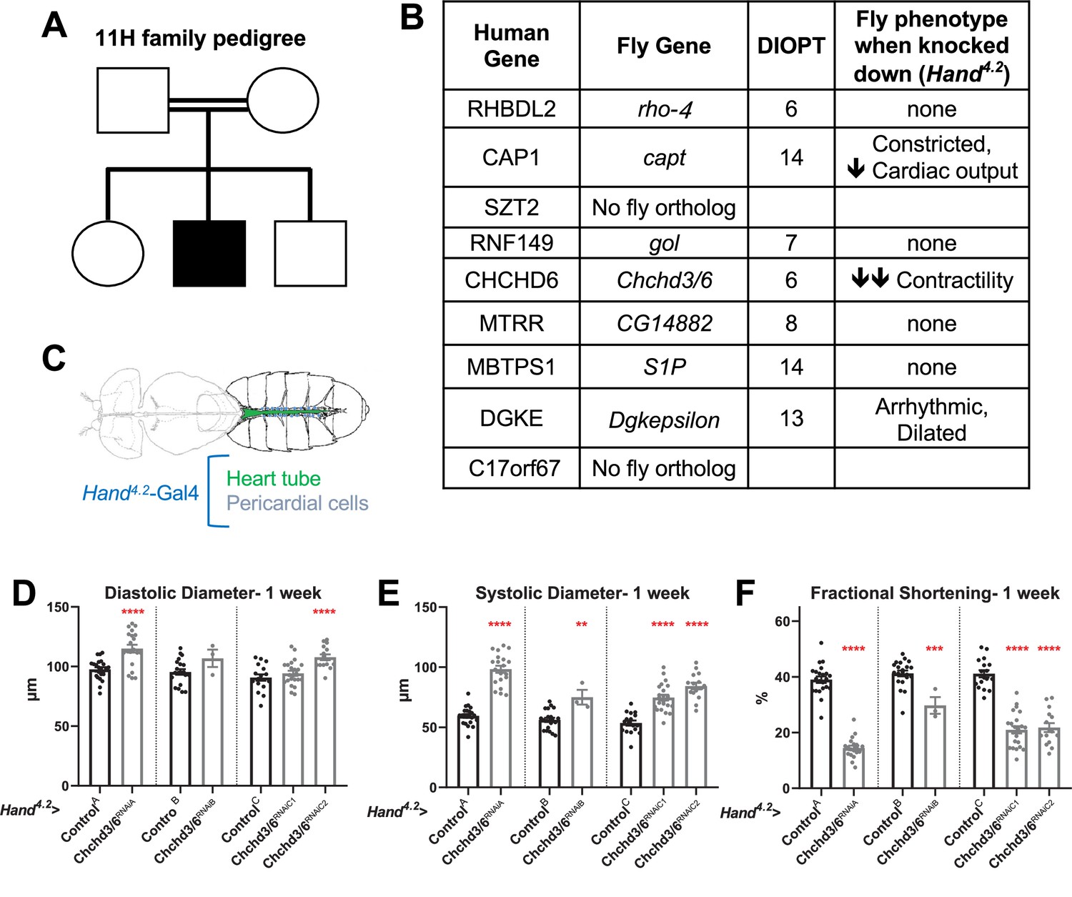
Prioritization of CHCHD6 in HLHS proband and its Drosophila ortholog Chchd3/6.
(A) Pedigree of index family 11 H. The family includes consanguineous parents (denoted by double horizontal lines) without cardiac defects, one son with HLHS (proband), and two siblings without cardiac defects. (B) List of 9 candidate genes derived from proband 11 H with corresponding Drosophila orthologs. Orthology based on DIOPT score. Conserved Drosophila candidate HLHS genes were knocked down individually in the Drosophila heart using the Hand4.2-Gal4 driver. The functional phenotypes listed were significantly different relative to ControlA or ControlB and were measured in 1-week-old female Drosophila hearts. (C) Schematic of Drosophila highlighting the abdominal region which includes the heart tube and flanking pericardial cells, where the Hand4.2-Gal4 driver is expressed. Image adapted from Figure 1A of Xie et al., 2013. (D) End-Diastolic diameter (EDD), (E) End-systolic diameter (ESD), and (F) fractional shortening (FS) from 1-week-old female Hand4.2-Gal4>Chchd3/6 flies.
-
Figure 1—source data 1
List of 9 candidate genes derived from proband 11 H with corresponding Drosophila orthologs.
Orthology based on DIOPT score. Conserved Drosophila candidate HLHS genes were knocked down individually in the Drosophila heart using the Hand4.2-Gal4 driver. The functional phenotypes listed were significantly different relative to ControlA or ControlB and were measured in 1-week-old female Drosophila hearts.
- https://cdn.elifesciences.org/articles/83385/elife-83385-fig1-data1-v2.xlsx
Figure 1—figure supplement 1
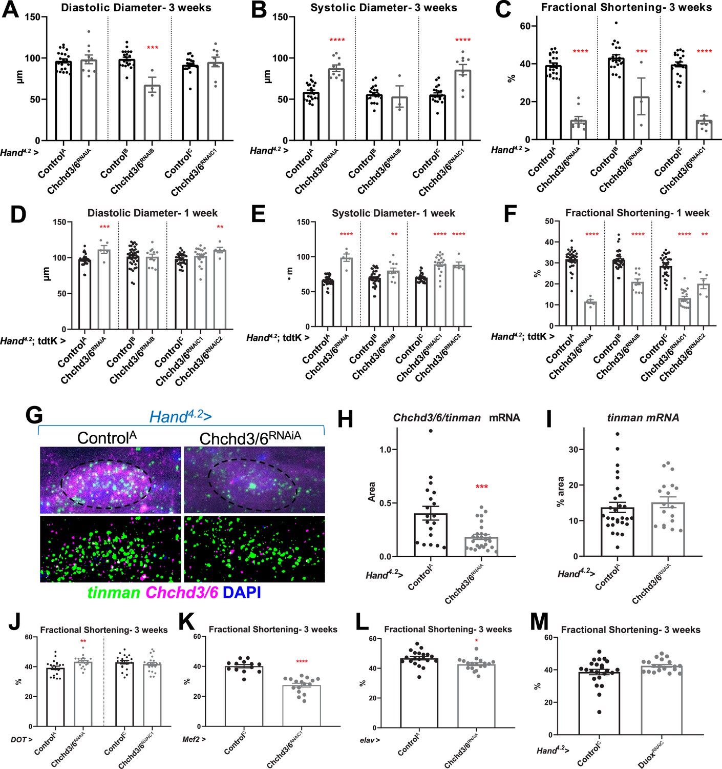
Chchd3/6 KD in the Drosophila heart causes reduced fractional shortening due to systolic dysfunction.
(A) EDD, (B) ESD and (C) FS from 3 week old Hand4.2>Chchd3/6 female flies. Very low number of Hand4.2>Chchd3/6RNAiB flies reaching 3 weeks-of-age indicates lethality possibly due to stronger KD of Chchd3/6. (D–F) 1-week-old Hand4.2-Gal4, tdtK >Chchd3/6 female flies measured for (D) EDD, (E) ESD and (F) FS. (G) Confocal images of Chchd3/6 mRNA and tinman mRNA inside adult cardiomyocytes (top; nuclei are outlined) and processed images (bottom). (H–I) Chchd3/6 and tinman mRNA in 1-week-old female Drosophila hearts. (H) Chchd3/6 mRNA relative to tinman mRNA is reduced in Hand4.2>Chchd3/6RNAiA heart tissue. (I) tinman mRNA % area, used as marker to normalize expression to. Fractional shortening in control and Chchd3/6 KD female flies at 3 weeks of age with a (J) pericardial cell-specific driver (DOT-Gal4), (K) all muscle cell specific driver (Mef2-Gal4), and (L) neuronal driver (elav-Gal4). Note that Mef2 >Chchd3/6RNAiA was lethal at pupal stages. (M) Fractional shortening measurements from Hand4.2>DuoxRNAiC 3-week-old flies. Unpaired two-tailed t-test, *p≤0.05, **p≤0.01, ***p≤0.001, ****p≤0.0001; error bars represent SEM.
Figure 2
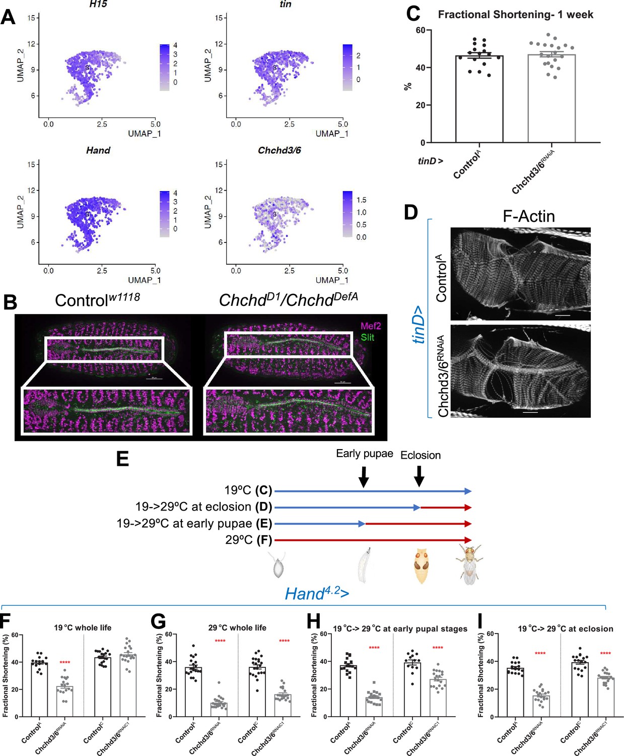
Chchd3/6 expression is important for adult cardiac function around larval stages and early adult stages.
(A) UMAP (uniform manifold approximation and projection) plot from CB-specific single-cell transcriptomics (Vogler, 2021a) showing expression of Chchd3/6 in CBs, as identified by cardiac TFs tin, H15, and Hand. (B) Stage 16–17 embryos (late stage cardiogenesis) were collected from a Chchd3/6 loss of function line (ChchdD1) line crossed to a Chchd3/6 deficiency line (ChchdDefA) and stained for Mef2 (all muscle transcription factor, magenta) and Slit (secreted protein of the lumen, green). 50 µm scale. (C) tinD >ControlA or>Chchd3/6RNAiA were reared at 29 °C and females were filmed and imaged at 1 week of age. (A) tinD >Chchd3/6RNAiA did not have a significant reduction in fractional shortening compared to tinD >ControlA flies. (D) F-actin was unchanged between tinD >ControlA and tinD >Chchd3/6RNAiA flies at 1 week of age; 20 µm scale. (E) Schematic overview of temperature shift experiments. (F–I) Fractional shortening measurements from 1-week-old female flies reared at (F) 19 °C for whole life, (G) at 29 °C for whole life, (H) 19 °C, and moved to 29 °C at early pupal stages, or (I) 19 °C, and moved to 29 °C once eclosed (virgin flies), Unpaired two-tailed t-test, ****p≤0.0001, error bars represent SEM.
Figure 3 with 1 supplement
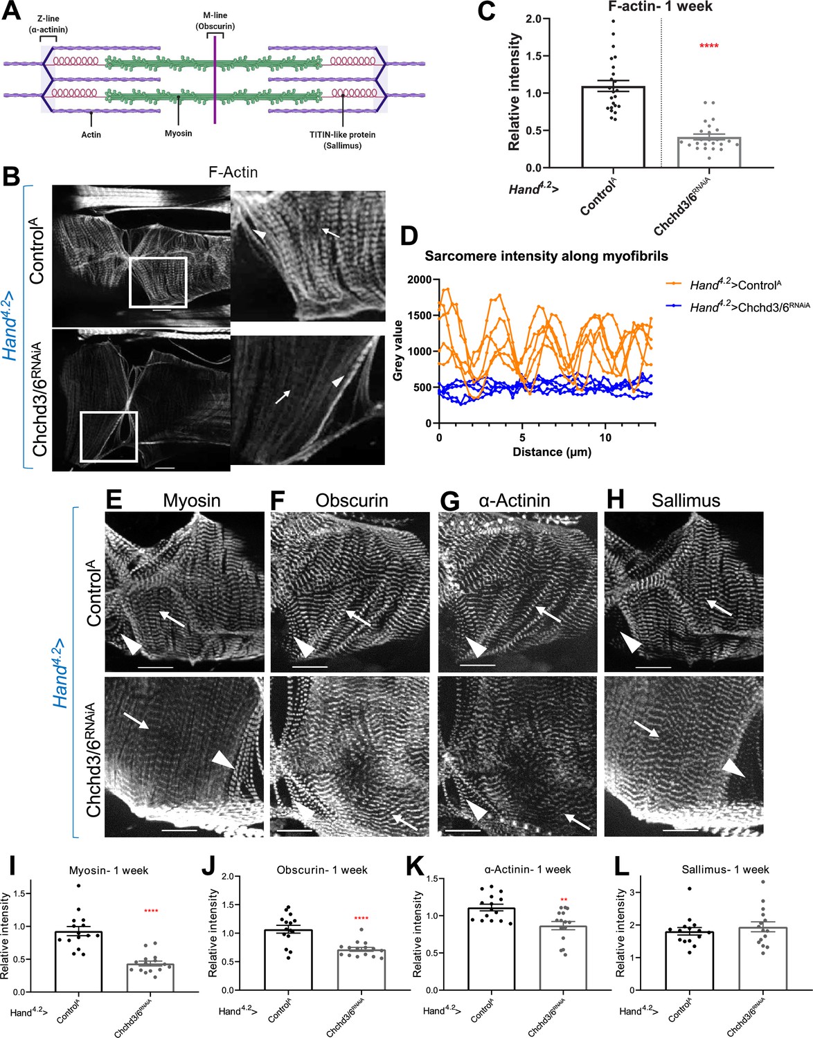
Cardiac tissue from heart-specific Chchd3/6 KD flies exhibit reduced and altered sarcomeric proteins in the myocardial tissue.
(A) Schematic of sarcomeric protein distribution inside myofibrils (image created with BioRender.com). (B) F-actin staining in 1-week-old female Drosophila hearts with Hand4.2-Gal4 KD of Chchd3/6. Arrowheads indicate ostial myofibrils and arrows point to myocardial myofibrils (non-ostial). (C) F-actin intensity measured as mean gray value (gray value/# of pixels) along myocardial myofibrils relative to mean gray value of ostial myofibrils. (D) Mean intensity of F-actin along individual myofibrils. One-week-old Drosophila hearts with Hand4.2-Gal4 driven KD of control or Chchd3/6 stained for antibodies against (E) Myosin, (F) Obscurin, (G) α-Actinin, or (H) Sallimus. Arrowheads indicate ostial myofibrils and arrows point to working cardiomyocyte tissue (non-ostial). (I–L) Mean fluorescence intensity along myocardial myofibrils relative to ostia myofibrils in 1-week-old Hand4.2-Gal4>CHCHD3/6RNAiA adults stained for sarcomeric proteins (I) Myosin, (J) Obscurin, (K) α-Actinin, or (L) Sallimus. Unpaired two-tailed t-test, **p≤0.01, ****p≤0.0001; error bars represent SEM. 20 µm scale.
Figure 3—figure supplement 1
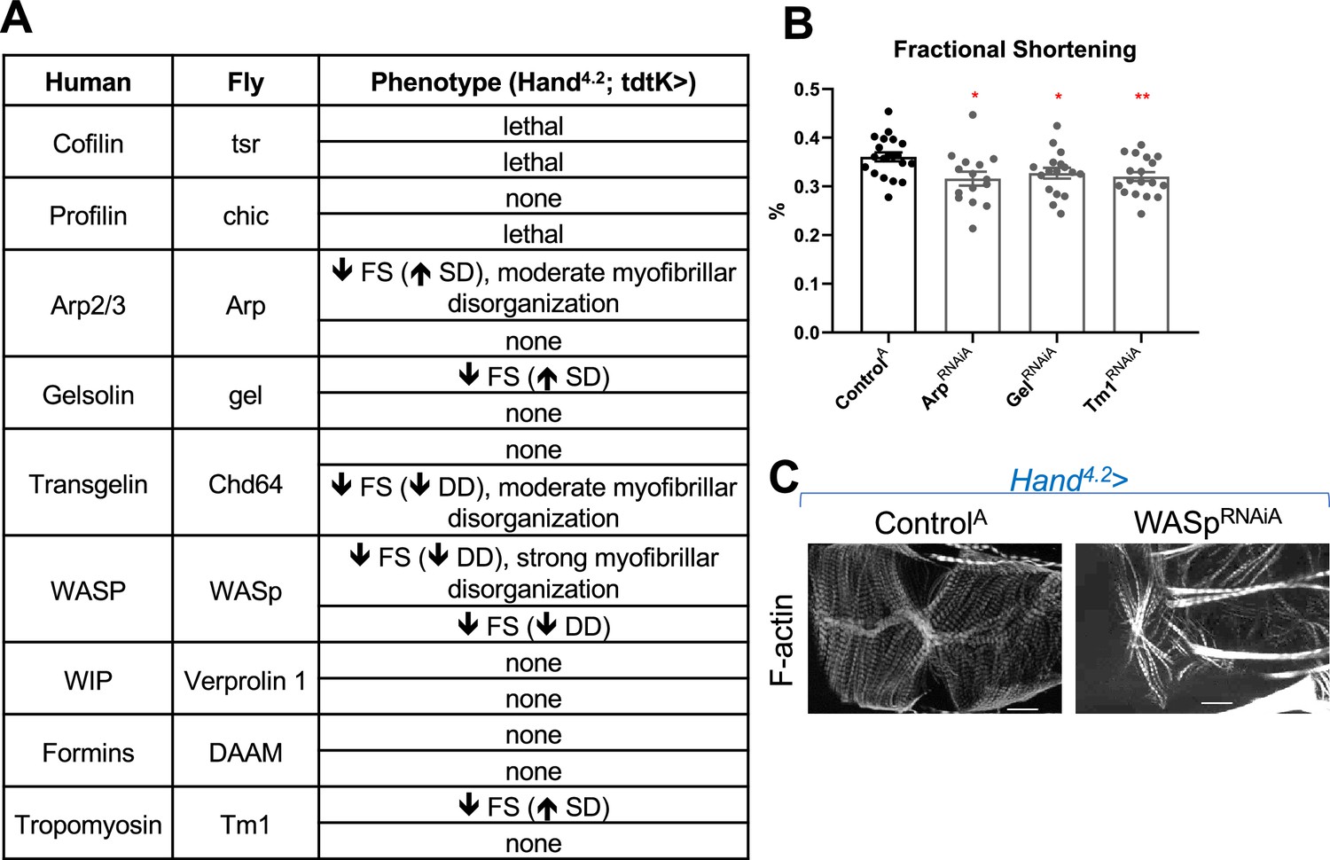
KD of actin polymerizing and depolymerizing genes did not recapitulate the phenotype of Chchd3/6 KD.
(A-C) Candidate genes involved in polymerization/de-polymerization of F-actin phenotypes upon KD using a Hand4.2-Gal4;tdtKattP2 driver, measured at 1 week of age. (B) Fractional shortening from RNAiA lines. (C) F-actin phenotype of hits. 20 µm scale. Unpaired two-tailed t-test, *p≤0.05, **p≤0.01; error bars represent SEM. FS = fractional shortening, DD = End diastolic diameter, SD = Systolic diameter.
-
Figure 3—figure supplement 1—source data 1
Candidate genes involved in polymerization/de-polymerization of F-actin phenotypes upon KD using a Hand4.2-Gal4;tdtKattP2 driver, measured at 1 week of age.
- https://cdn.elifesciences.org/articles/83385/elife-83385-fig3-figsupp1-data1-v2.xlsx
Figure 4 with 2 supplements
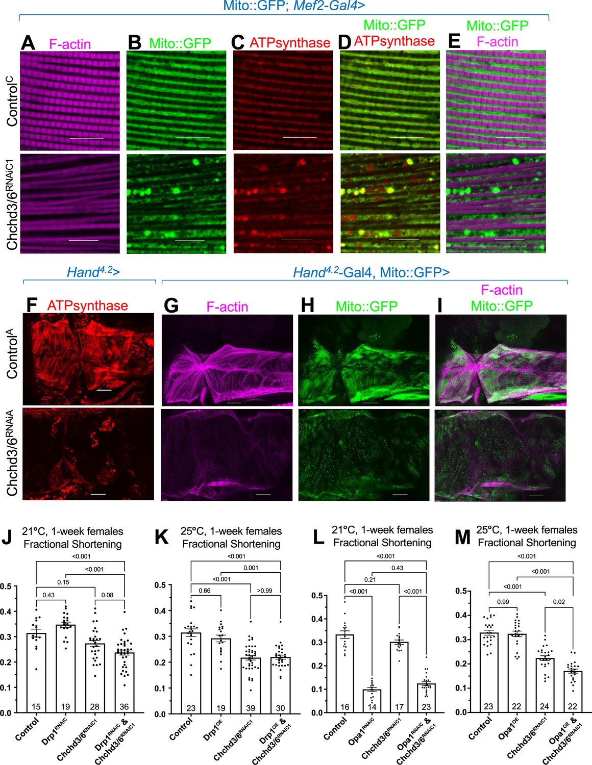
Mitochondrial fission-fusion defects were observed in cardiac Chchd3/6 KD.
(A–E) Visualization of F-actin and mitochondria in Drosophila indirect flight muscles (IFMs). 1–2 day-old male Drosophila IFMs with Mito::GFP; Mef2-Gal4 stained for (A) F-actin, (B) GFP (Mito::GFP (GFP tagged COX8A)), (C) ATP synthase, (D) merged image of B+C, and (E) merged image of A+B,10µm scale. (F) Hand4.2>Chchd3/6RNAiA heart tissue at 1 week of age. (G–I) F-actin and Mito::GFP staining in 1-week-old female hearts using the Hand4.2-Gal4; Mito::GFP driver, 20 µm scale. (J–M) Fractional shortening results are displayed from 1-week-old females with manipulation of mitochondrial fission-fusion genes and Chchd3/6RNAiC1. (J) Chchd3/6 and Drp1 KD at 21 °C had little effect on their own, but in combination caused a reduction in fractional shortening, displaying a significant genetic interaction (Figure 3—figure supplement 1C). (K) At 25 °C Chchd3/6 KD reduced fractional shortening substantially, whereas Drp1 OE by itself or in combination with Chchd3/6RNAiC1 had no effect, thus no genetic interaction was observed (Figure 3—figure supplement 1F). (L) Even at 21 °C, Opa1 KD drastically reduced contractility, which in combination with Chchd3/6 KD slightly improved, which resulted in a significant genetic interaction (Figure 3—figure supplement 1I). (M) At 25 °C, Opa1 OE (M) had no effect, but in combination with Chchd3/6 KD contractility was further reduced significantly, although the interaction p-value did not reach significance (Figure 3—figure supplement 1L). One-way ANOVA with multiple comparisons shows mean with SEM and associated p-values. Sample size is shown at bottom of each bar.
Figure 4—figure supplement 1
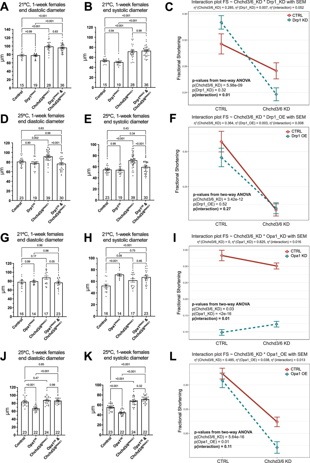
Genetic interactions between mitochondrial fission-fusion genes and Chchd3/6 KD.
Diastolic and systolic diameters and Two-way ANOVA interaction plots of fractional shortening. (A–C) KD of Drp1 and Chchd3/6, (D–F) Drp1 OE and Chchd3/6 KD, (G–I) Opa1 KD and Chchd3/6 KD, and (J–L) Opa1 OE and Chchd3/6 KD. One-way ANOVA with multiple comparisons shows mean with SEM and detailed p-values. Sample size is shown at bottom of each bar. The two-way ANOVA analyses and interaction plots in (C, F, I and L) were obtained in R. η2 is the effect size used in ANOVA, showing the extent that each factor – here including Chchd3/6, mitochondrial fission-fusion genes, their interaction and the error - accounts for the variance in the results. Except Opa1 KD, in all the other interaction experiments, Chchd3/6 was the main effect as shown by larger effect size η2 than the other factors.
Figure 4—figure supplement 2
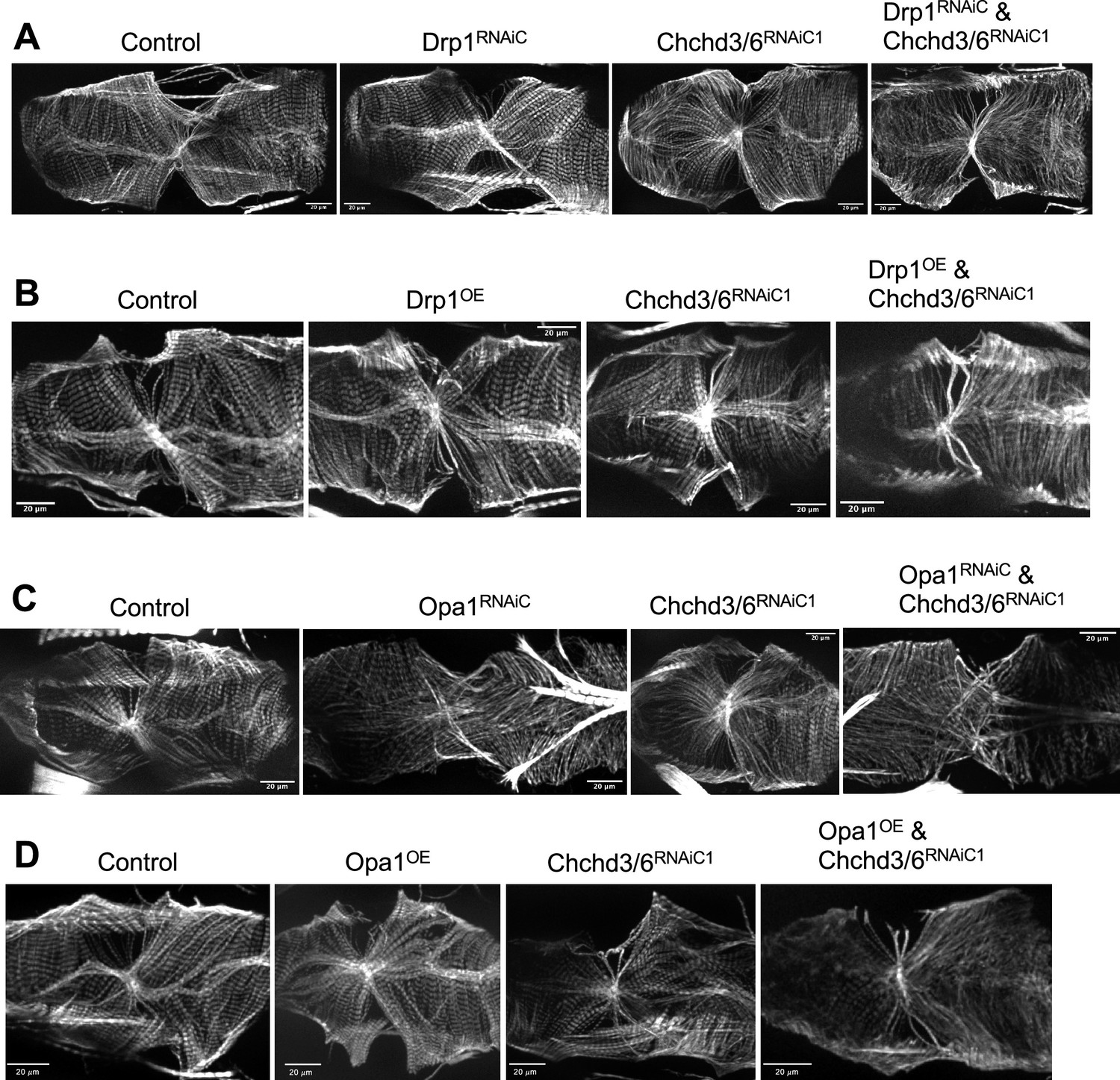
F-actin staining of the fly hearts in the interaction experiments involving mitochondrial fission-fusion genes and Chchd3/6 KD.
(A) Single and double KD of Drp1 and Chchd3/6 at 21 °C. (B) Single and double Drp1 OE and Chchd3/6 KD at 25 °C. (C) Single and double KD of Opa1 and Chchd3/6 KD at 21 °C. (D) Single and double Opa1 OE and Chchd3/6 KD at 25 °C.
Figure 5 with 1 supplement
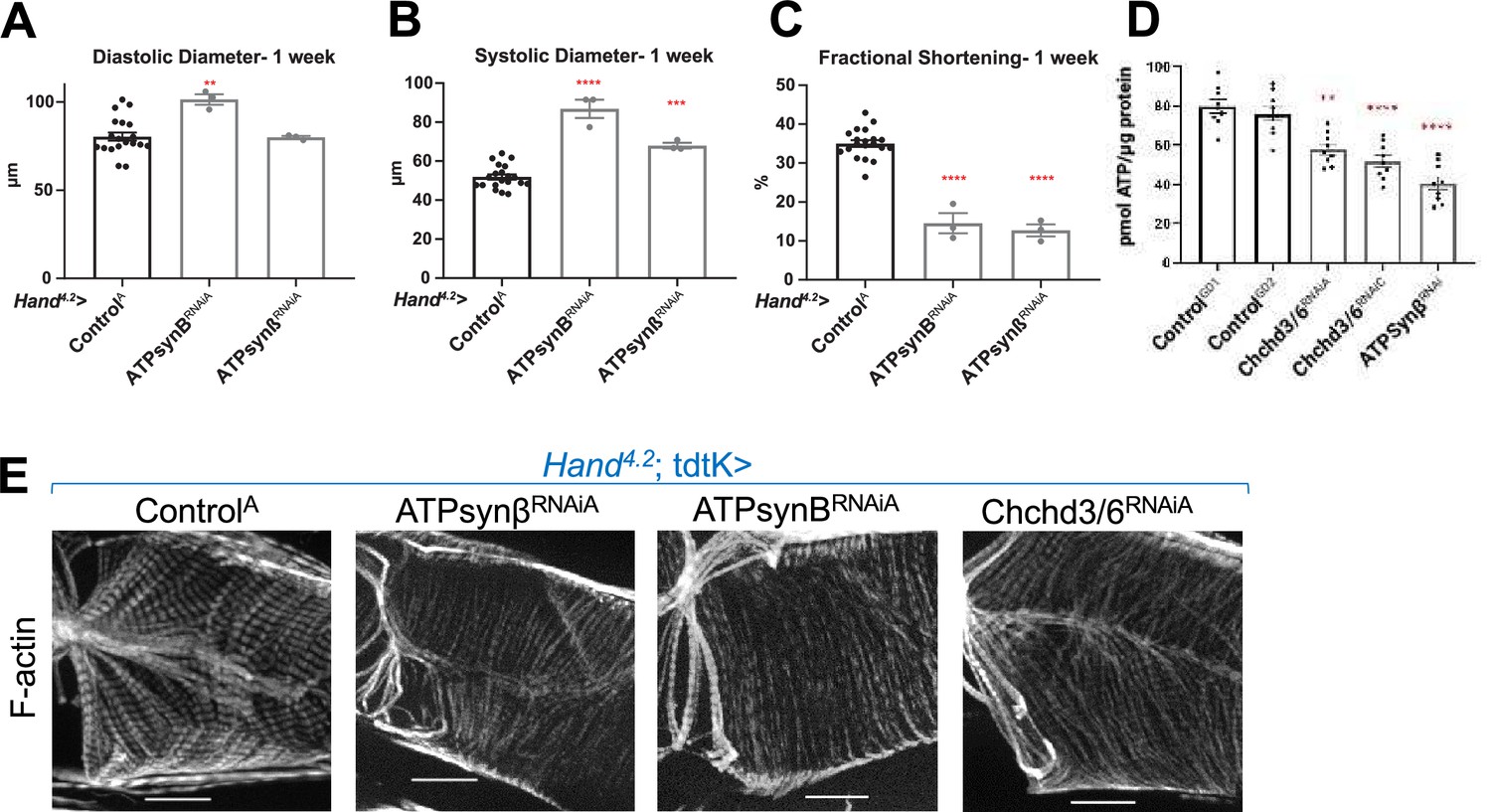
KD of ATP synthase subunit B and beta reduced both fractional shortening and F-actin staining.
(A–C)Hand4.2-Gal4; tdtK driven KD of ATP synthase subunits at 1 week of age measuring (A) diastolic diameter, (B) systolic diameter, and (C) fractional shortening. Data is plotted as ± SEM and significance indicated relative to ControlGD2. ****p ≤ 0.0001, **p ≤ 0.01. (D) Quantification of ATP levels from hearts of 1-week-old flies (10–12 hearts per sample). ATP measurements were plotted relative to protein content. (E) One-week-old Hand4.2-Gal4; tdtK driven KD of ATP synthase subunits with altered F-actin (Chchd3/6 KD is depicted to contrast the structural phenotypes). Statistical differences were calculated by one-way ANOVA followed by Tukey’s post hoc test for multiple comparisons.
Figure 5—figure supplement 1
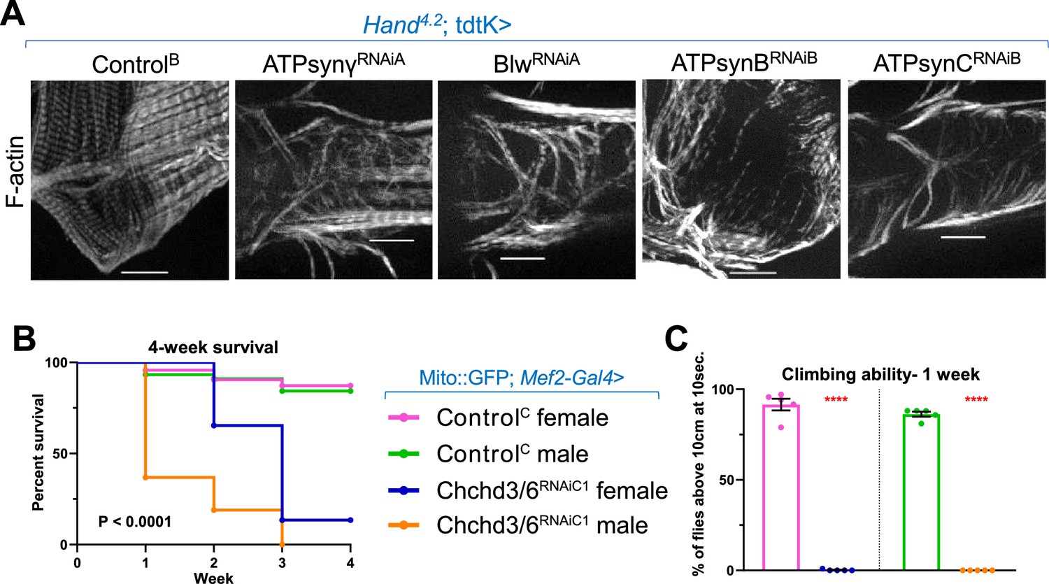
Cardiac KD of ATP synthase components disrupted or diminished F-actin staining.
Chchd3/6 KD in all muscle cells reduced the viability and climbing ability of the fly. (A) KD of mitochondrial ATP synthase subunits using Hand4.2-Gal4; tdtK produced strong F-actin phenotypes. 20 µm scale. (B & C) Chchd3/6 was knocked down using a Mito::GFP; Mef2-Gal4 line; note that Mito::GFP; Mef2-Gal4>Chchd3/6RNAiA is pupal lethal. (B) Viability was significantly reduced in male and female Chchd3/6RNAiC1 flies over a 4-week time course. Mantel-Cox test. (C) Negative Geotaxis assay (average number of flies above a 10 cm mark after being tapped down in a long vial) measured in 1-week-old male and female flies. Unpaired two-tailed t-test, ****p≤0.0001; error bars represent SEM.
Figure 6 with 2 supplements
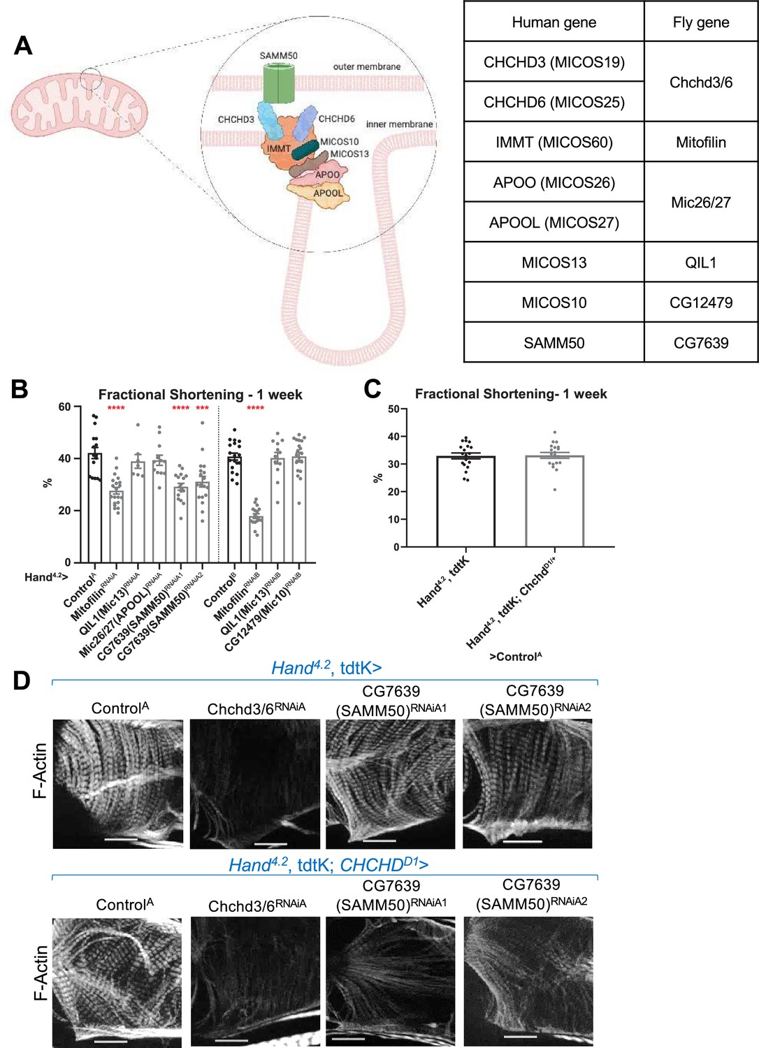
Assessment of other MICOS subunits in the Drosophila heart.
(A) Schematic of the MICOS complex and SAM50. Human MICOS subunits and their respective Drosophila homologs are listed (image created with BioRender.com). (B) Fractional shortening measured from 1-week-old female flies with KD of individual MICOS subunits and Sam50 using a Hand4.2-Gal4 driver. Unpaired two-tailed t-test, ***p≤0.001, ****p≤0.0001; error bars represent SEM. (C) Hand4.2-Gal4, tdtK; ChchdD1/+ line was crossed out with ControlA. Unpaired two-tailed t-test, error bars represent SEM. (D) 1 week old F-actin-stained Drosophila hearts with or without heterozygous loss-of-function ChchdD1/+ in the background, 20 µm scale.
-
Figure 6—source data 1
Human MICOS subunits and their respective Drosophila homologs.
- https://cdn.elifesciences.org/articles/83385/elife-83385-fig6-data1-v2.xlsx
Figure 6—figure supplement 1
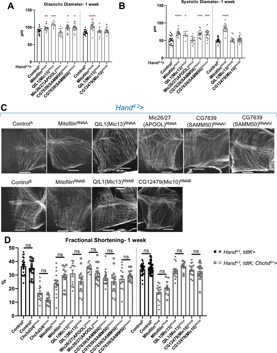
Heart function and F-actin staining in flies with KD of other components of MICOS and SAMM50.
(A) End-diastolic diameter and (B) End-systolic diameter measured from 1-week-old female flies with KD of individual MICOS subunits and CG7639 (SAMM50) using a Hand4.2-Gal4 driver. (C) F-actin cardiac sarcomere staining in 1-week-old female flies with Hand4.2-Gal4 KD. (D) All MICOS subunits and Sam50 were knocked down with Hand4.2-Gal4, tdtK (filled squares) or using the sensitizer line Hand4.2-Gal4, tdtK; ChchdD1/+ (empty squares) at 25 °C. Two-way ANOVA with Tukey’s multiple comparisons test, only statistical comparisons between same RNAi lines are shown; error bars represent SEM, 20 µm scale.
Figure 6—figure supplement 2
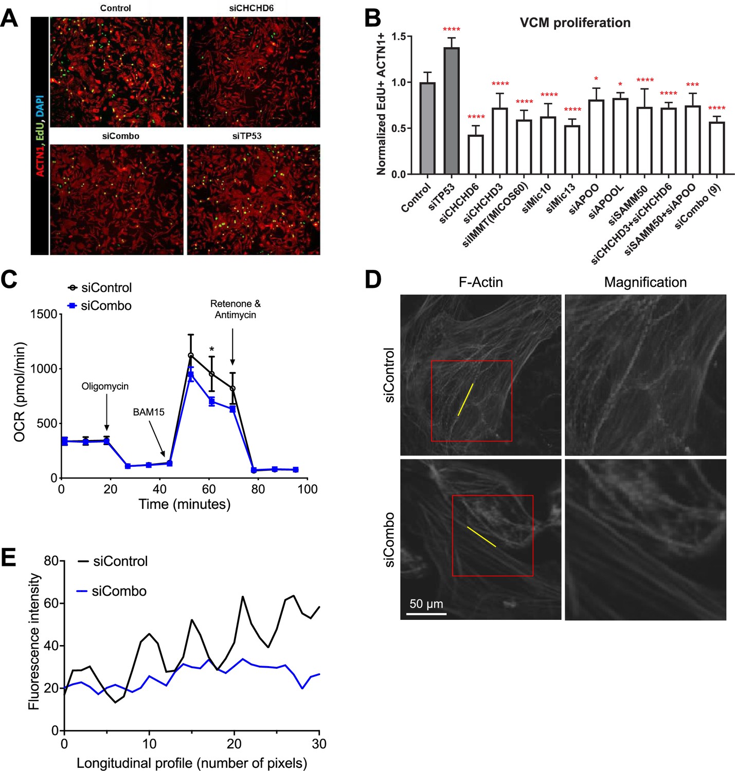
Cardiac cell proliferation and contractility assay following MICOS complex subunit KD.
(A) VCMs showed reduced proliferation with siCHCHD6. (B) Quantification of proliferation between siRNA mediated KD of individual MICOS subunits and MICOS subunit combination KDs in VCMs (siTP53 is a positive control). One-way ANOVA with Dunnett’s multiple comparisons test, *p≤0.05, ***p≤0.001, ****p≤0.0001; error bars represent standard deviation. (C) OCR in hiPSC-vCMs after combo KD of CHCHD3 and CHCHD6 compared to control. (D&E) F-actin staining and fluorescence intensity along the F-actin fiber in combo KD of CHCHD3 and CHCHD6 compared to control.
Figure 7
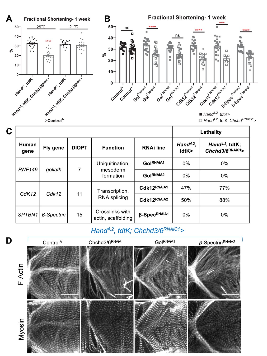
HLHS CHCHD3 and CHCHD6 family-based gene interaction screen reveals three hits.
(A) A Hand4.2-Gal4, tdtK; Chchd3/6RNAiC1 sensitizer line show reduced fractional shortening at 25ºC, which is no longer significant at 21ºC. Unpaired two-tailed t-test, ****p≤ 0.0001; error bars represent SEM. (B) Genetic interaction of Chchd3/6 and prioritized HLHS candidates. Two-way ANOVA with Tukey’s multiple comparisons test, only statistical comparisons between the same RNAi lines are shown; ***p≤ 0.001, ****p≤ 0.0001; error bars represent SEM. (C) Functional overview of human and Drosophila orthologs. KD of Ckd12 with Chchd3/6 KD led to increased lethality of eclosed flies by 1 week-of-age.
-
Figure 7—source data 1
Functional overview of human and Drosophila orthologs.
KD of Ckd12 with Chchd3/6 KD led to increased lethality of eclosed flies by 1 week-of-age.
- https://cdn.elifesciences.org/articles/83385/elife-83385-fig7-data1-v2.xlsx
Additional files
-
MDAR checklist
- https://cdn.elifesciences.org/articles/83385/elife-83385-mdarchecklist1-v2.docx
-
Supplementary file 1
Prioritized candidate genes from HLHS proband.
Nine candidate genes harbored rare homozygous variants predicted to impact protein structure or gene regulation. Hom = homozygous, Het = heterozygous, WT = wildtype, gnomAD = Genome Aggregation Database, MAF = minor allele frequency. rsID: reference single nucleotide polymorphism ID. RegulomeDB variants range from 1 (high functional evidence) to 6 (least functional evidence). CADD score (Combined Annotation-Dependent Depletion), higher percentile predicts higher possibility of functionality or pathogenicity.
- https://cdn.elifesciences.org/articles/83385/elife-83385-supp1-v2.xlsx
-
Supplementary file 2
Mitochondrial gene screen in the Drosophila heart.
RNAi lines of different mitochondrial functional groups were selected using the FlyBase.org gene ontology (GO) term mitochondrion (GO:0005739). The RNAi lines were crossed to Hand4.2-Gal4, tdtK and their progeny were assessed for contractility defects at 1-week of adult age using Hand4.2-Gal4; tdtK. Note that structural phenotype refers to any visible F-actin phenotype, not specifically to Chchd3/6 KD F-actin phenotype. FS = fractional shortening, DD = End diastolic diameter, SD = systolic diameter.
- https://cdn.elifesciences.org/articles/83385/elife-83385-supp2-v2.xlsx
-
Supplementary file 3
CHCHD3 and CHCHD6 MICOS variants in HLHS probands.
Rare, predicted-damaging variants in CHCHD3 and CHCHD6 were identified in 6 of 183 HLHS probands in the Mayo Clinic cohort and 1 in the Pediatric Cardiac Genomics Consortium (PCGC). Transcript and relevant protein variants are listed. rsID: reference single nucleotide polymorphism ID. RegulomeDB variants range from 1 (high functional evidence) to 6 (least functional evidence). CADD score (Combined Annotation-Dependent Depletion), higher percentile predicts higher possibility of functionality or pathogenicity. Corresponding Drosophila orthologs and orthology scores (DIOPT). *Annotation of variant for a non-canonical transcript (ENST00000448878).
- https://cdn.elifesciences.org/articles/83385/elife-83385-supp3-v2.xlsx
-
Supplementary file 4
Candidate genes from HLHS probands harboring CHCHD3 or CHCHD6 variants.
The majority of HLHS candidate genes has a Drosophila ortholog (genes without ortholog highlighted in green).
- https://cdn.elifesciences.org/articles/83385/elife-83385-supp4-v2.xlsx
Download links
A two-part list of links to download the article, or parts of the article, in various formats.
Downloads (link to download the article as PDF)
Open citations (links to open the citations from this article in various online reference manager services)
Cite this article (links to download the citations from this article in formats compatible with various reference manager tools)
Mitochondrial MICOS complex genes, implicated in hypoplastic left heart syndrome, maintain cardiac contractility and actomyosin integrity
eLife 12:e83385.
https://doi.org/10.7554/eLife.83385


