The effect of weight loss following 18 months of lifestyle intervention on brain age assessed with resting-state functional connectivity
- Department of Brain and Cognitive Sciences, Ben-Gurion University of the Negev, Israel
- The Health & Nutrition Innovative International Research Center, Faculty of Health Sciences, Ben-Gurion University of the Negev, Israel
- Department of Internal Medicine D, Chaim Sheba Medical Center, Israel
- Department of Medicine, University of Leipzig, Germany
- Department of Diagnostic Imaging, Soroka Medical Center, Israel
- Department of Psychology, Ben-Gurion University of the Negev, Israel
- Department of Nutrition, Harvard T.H. Chan School of Public Health, United States
Figures
Figure 1
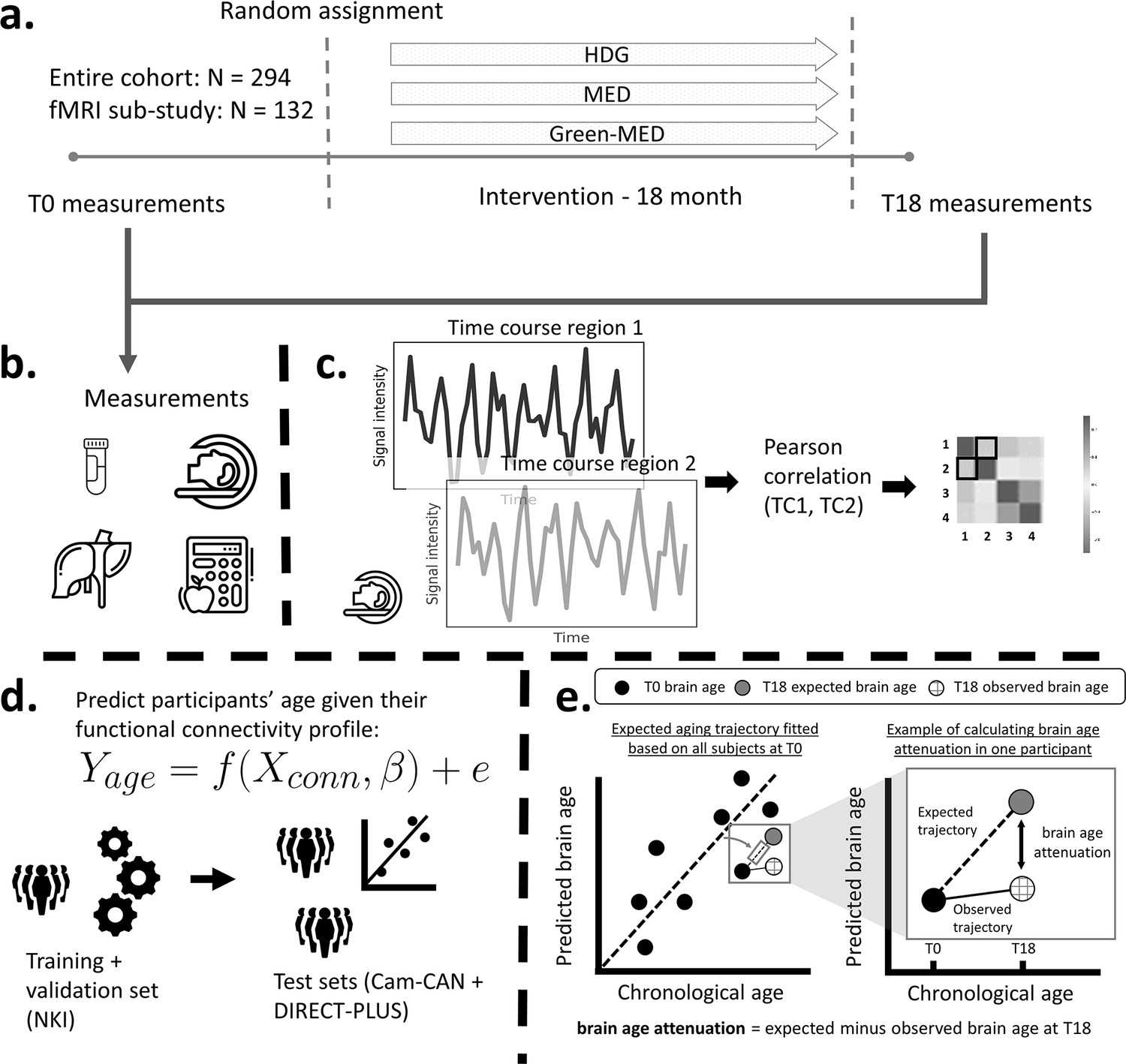
Study design and workflow.
The Dietary Intervention Randomized Controlled Trial Polyphenols Unprocessed Study (DIRECT-PLUS) trial examined the effect of successful weight loss following 18-month lifestyle intervention on adiposity, cardiometabolic, and brain health across intervention groups. (a) Participants in the functional connectivity sub-study (N=132) completed the baseline measurements at T0. They were randomly assigned to three intervention groups: healthy dietary guidelines (HDG), an active control group, Mediterranean diet (MED), and green-MED. All groups were combined with physical activity (PA). Eighteen months following intervention onset, all measurements were retaken (T18). (b) Measurements included anthropometric measurements, blood biomarkers, fat deposition, and structural and functional brain imaging. (c) Functional brain imaging was conducted while subjects were at rest and was used to estimate resting-state functional connectivity (RSFC). RSFC measures the correlation between the time series of pairs of brain regions. (d) We fitted a linear support vector regression to predict chronological age from all pairwise correlations. We fitted the model on the Nathan Kline Institute (NKI) dataset, then tested and applied it to the Cambridge Centre for Ageing and Neuroscience (Cam-CAN) and the DIRECT-PLUS data. (e, left scatter plot) Based on the T0 data, we first computed the expected aging trajectory as the linear relation between the chronological and predicted age of all subjects. The fitted line represents the increase in the predicted age in relation to chronological age in the absence of an intervention. (e, right scatter plot) The fitted line was used to estimate the expected brain age at T18, given each participant’s T0 brain age and the time passed between the T0 and T18 magnetic resonance imaging (MRI) scans. We computed the observed brain age by applying the brain age model to the T18 scans. Brain age attenuation was calculated as the expected brain age minus the observed at T18.
Figure 2 with 1 supplement
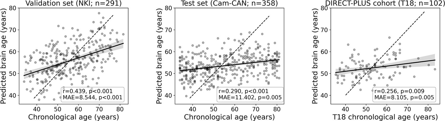
Prediction accuracy within the validation and test cohorts.
The scatter plots depict the data points and regression line between the predicted (y-axis) and observed (x-axis) age. The predicted-observed correlation is presented for the validation data (left), the Cambridge Centre for Ageing and Neuroscience (Cam-CAN) test data (middle), and the Dietary Intervention Randomized Controlled Trial Polyphenols Unprocessed Study (DIRECT-PLUS) data at baseline. The shaded area around the regression lines represents a 95% confidence interval estimated using bootstrapping. Pearson’s correlation, MAE (mean absolute error), and corresponding p-values are shown at the bottom of each plot. The dotted lines represent a perfect correlation for reference (predicted = observed).
Figure 2—figure supplement 1
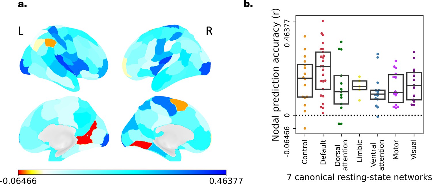
Brain age prediction accuracy of individual nodes.
We reiterated the model training procedure for each of the nodes separately. We extracted the row of each node in the resting-state functional connectivity (RSFC) matrix, which represents all the nodes’ connections with the rest of the brain, or its ‘connectivity fingerprint’. As in the original model, we used a linear support vector regression fitted on the Nathan Kline Institute (NKI) dataset, then tested it on the Cambridge Centre for Ageing and Neuroscience (Cam-CAN) dataset. Prediction accuracy was quantified as the correlation between chronological age and predicted age. (a) Prediction accuracy was depicted on the brain surface on a lateral (top) and medial (bottom) view. (b) Box plot showing the prediction accuracy (y-axis) in each of the seven canonical resting-state networks Zelicha et al., 2019. Each dot represents a single node.
Figure 3 with 1 supplement
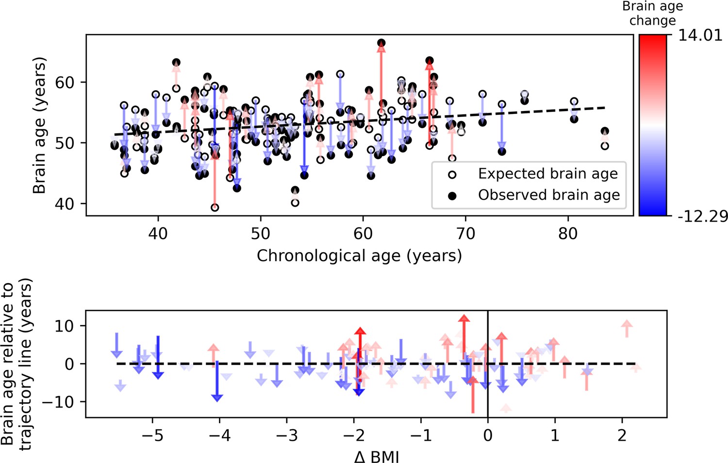
Observed compared to expected brain age at T18.
The upper panel depicts the chronological age (x-axis) and the observed (empty circles) and expected (full circles) brain age (y-axis) of each subject. The dashed line represents the expected brain age trajectory fitted based on the T0 data (see regression line in Figure 1e, left). Arrows point from the expected to the observed age of a single individual, corresponding to brain age attenuation. Arrows’ colors correspond to the extent of brain age attenuation (blue shades indicate attenuation, red shades indicate an acceleration in brain age). The observed age was lower than expected in 56.8% of the subjects, while the opposite was found in 43.1% (X2=1.922, p=0.166). In the lower panel, arrows were reordered by subjects’ body mass index (BMI) change over the 18 months of intervention. A significant correlation was found between the BMI and brain age change (r=0.319, p<0.001). This is evident in the graph, such that most of the blue arrows are located on the left side of the x-axis (negative values), and most of the red arrows appear on the right side (positive values).
-
Figure 3—source data 1
Participants’ demographics predicted and observed age and weight values.
A processed version of the data that includes participants’ demographics, predicted, and observed age and weight values for T0 and T18.
- https://cdn.elifesciences.org/articles/83604/elife-83604-fig3-data1-v2.csv
Figure 3—figure supplement 1
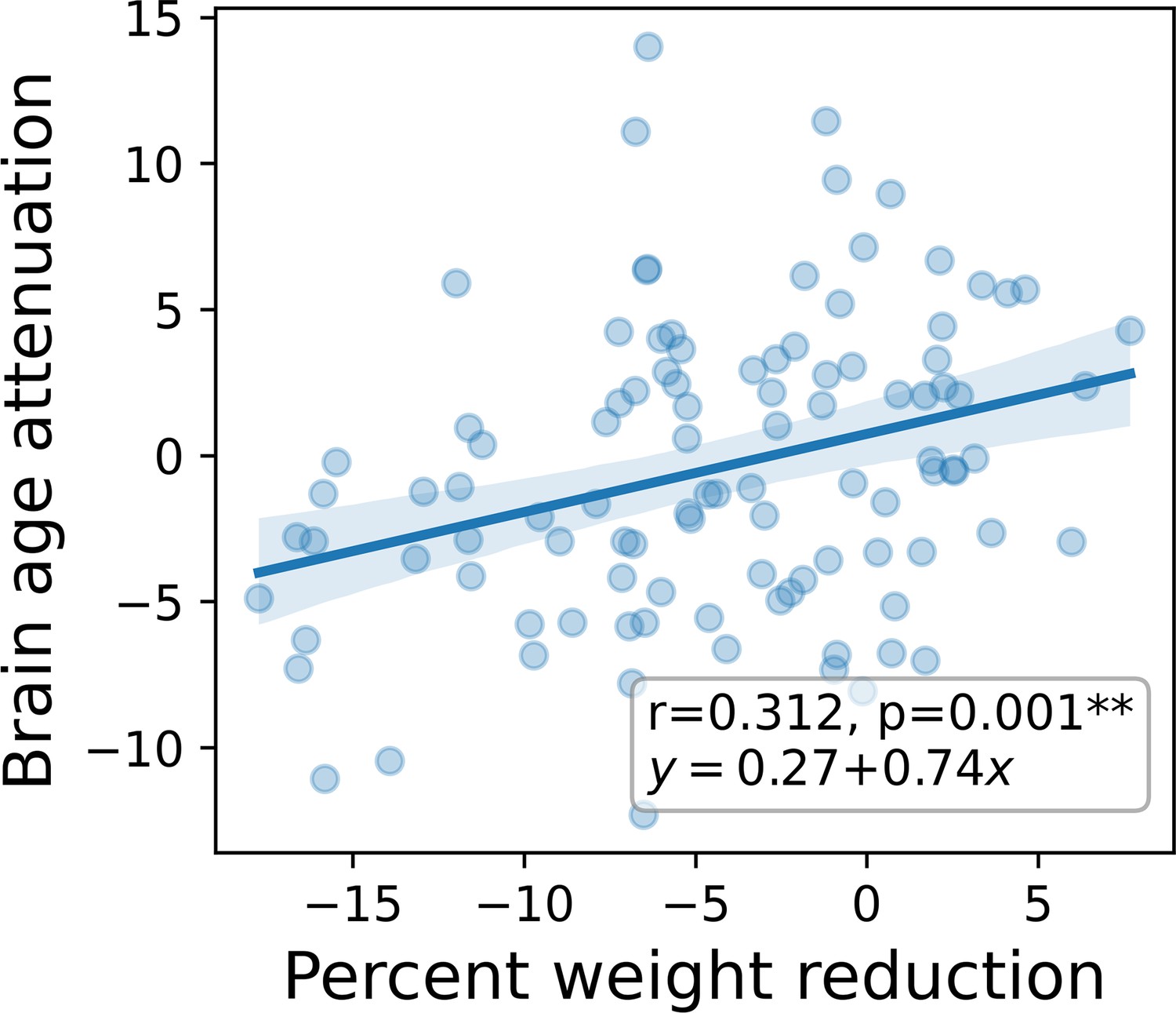
Brain age attenuation compared to percent weight reduction from baseline.
A scatter plot depicting the linear relationship between percent weight reduction from baseline (x-axis) and years of brain age attenuation (y-axis). Also shown on the graph are the correlation coefficient and the parameters of the linear relation between the two variables. One percent of body weight loss corresponded to an 8.9 months’ attenuation of brain age. The shaded area around the regression line represent a 95% confidence interval estimated using bootstrap.
Figure 4
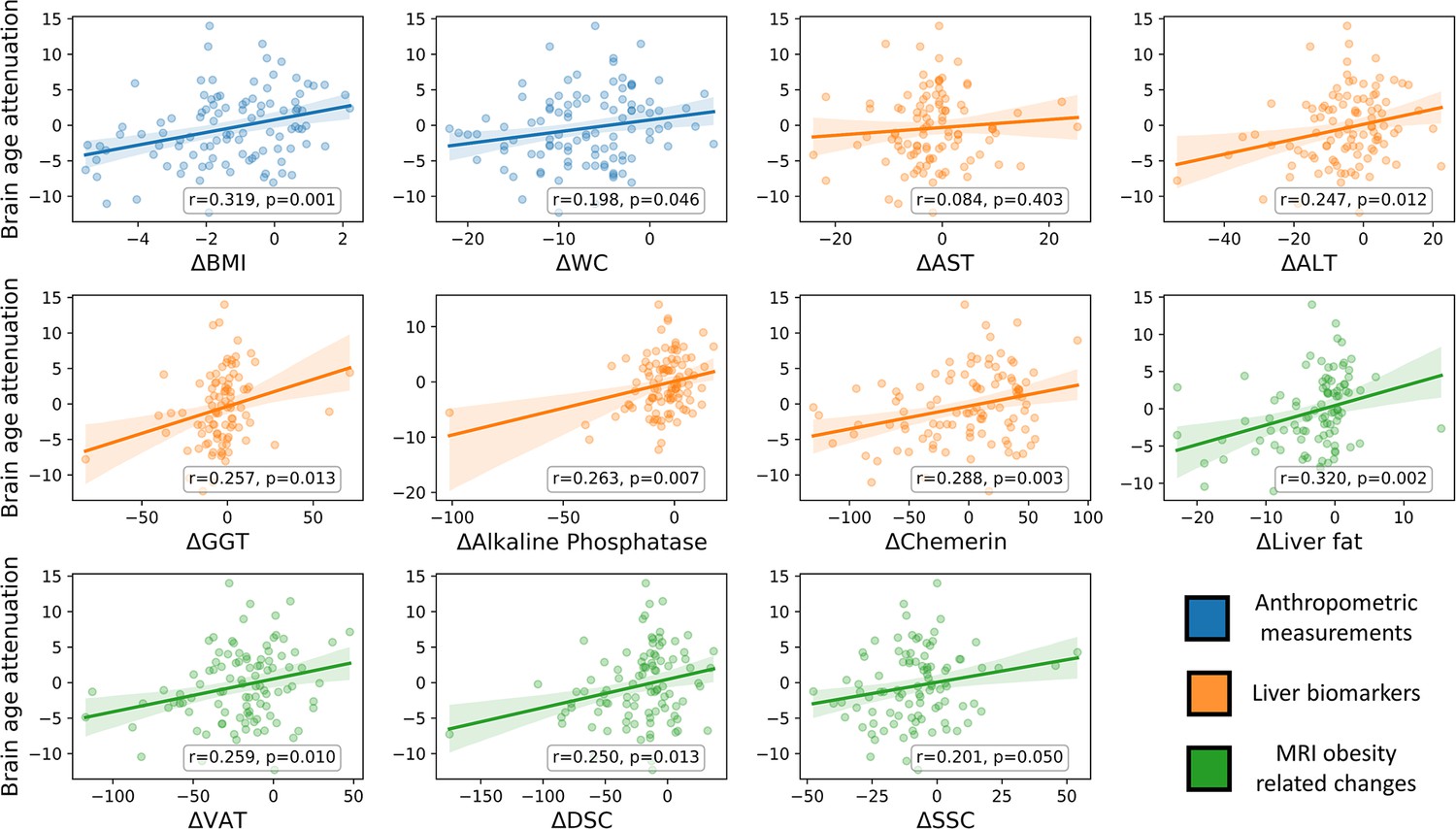
Brain age attenuation association with clinical measurements.
The scatter plots depict the data points and regression line between brain age attenuation (y-axis) and each clinical measurement (x-axis). Clinical measurements include anthropometry (blue), liver markers (orange), glycemic markers (brown), lipid profile (red), and fat deposition measured using magnetic resonance imaging (MRI) (green). The shaded area around the regression line represents a 95% confidence interval estimated using bootstrapping. Pearson’s correlation and the corresponding p-value are shown at the bottom of each plot. Significant associations following false discovery rate (FDR) correction are marked in bold (*=p < 0.05, **=p < 0.01).
Tables
Table 1
Association between baseline characteristics and age, predicted age, and brain age gap.
| Age | Brain age | T0 brain age gap(bias corrected) | |||||
|---|---|---|---|---|---|---|---|
| r | p-Value | r | p-Value | r | p-Value | ||
| BMI (kg/m²) | –0.110 | 0.272 | 0.067 | 0.504 | 0.097 | 0.334 | |
| Chemerin (ng/mL) | 0.153 | 0.124 | 0.247 | 0.012 | 0.216 | 0.029 | |
| HOMA IR | 0.177 | 0.079 | 0.179 | 0.074 | 0.141 | 0.163 | |
| HbA1c (%) | 0.315 | 0.001 | 0.117 | 0.240 | 0.042 | 0.677 | |
| HDL-C (mg/dL) | 0.158 | 0.113 | –0.095 | 0.343 | –0.138 | 0.168 | |
| LDL-C (mg/dL) | –0.152 | 0.129 | –0.024 | 0.811 | 0.013 | 0.893 | |
| Triglycerides (mg/dL) | –0.023 | 0.815 | 0.145 | 0.147 | 0.155 | 0.120 | |
| Liver fat (cm²) | –0.039 | 0.711 | 0.156 | 0.132 | 0.170 | 0.101 | |
| VAT (cm²) | 0.495 | 0.000 | 0.329 | 0.001 | 0.225 | 0.025 | |
Additional files
-
Supplementary file 1
Intervention outline by group.
A table describing the details of the intervention for each of the three groups.
- https://cdn.elifesciences.org/articles/83604/elife-83604-supp1-v2.xlsx
-
Supplementary file 2
Association between baseline characteristics and age, predicted age, and brain age gap.
A table describing the Pearson’s correlation between baseline characteristics and age, predicted age, and brain age gap.
- https://cdn.elifesciences.org/articles/83604/elife-83604-supp2-v2.xlsx
-
Supplementary file 3
Correlation and partial correlation of brain age attenuation and the measured biomarkers.
A table describing the Pearson’s correlation and partial correlation of brain age attenuation and the change in the measured biomarkers.
- https://cdn.elifesciences.org/articles/83604/elife-83604-supp3-v2.xlsx
-
Supplementary file 4
Relation between brain age attenuation and food consumption.
A table describing the Kendall’s tau correlation coefficient of brain age attenuation and food consumption, as reported using a food frequency questionnaire (FFQ).
- https://cdn.elifesciences.org/articles/83604/elife-83604-supp4-v2.xlsx
-
MDAR checklist
- https://cdn.elifesciences.org/articles/83604/elife-83604-mdarchecklist1-v2.docx
Download links
A two-part list of links to download the article, or parts of the article, in various formats.
Downloads (link to download the article as PDF)
Open citations (links to open the citations from this article in various online reference manager services)
Cite this article (links to download the citations from this article in formats compatible with various reference manager tools)
The effect of weight loss following 18 months of lifestyle intervention on brain age assessed with resting-state functional connectivity
eLife 12:e83604.
https://doi.org/10.7554/eLife.83604


