Structures of human dynein in complex with the lissencephaly 1 protein, LIS1
- Department of Cellular and Molecular Medicine, University of California, San Diego, United States
- Department of Molecular, Cellular and Developmental Biology, University of Michigan, United States
- Department of Cell and Developmental Biology, University of California, San Diego, United States
- Howard Hughes Medical Institute, United States
- Department of Molecular Biology, University of California, San Diego, United States
Figures
Figure 1 with 2 supplements
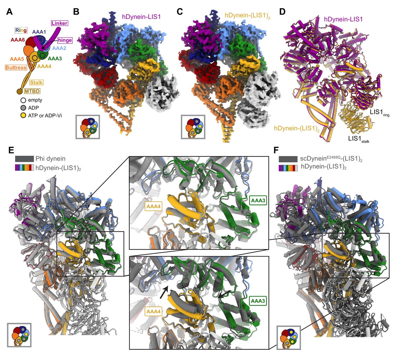
Structures of human dynein bound to LIS1.
(A) Cartoon schematic of dynein showing domain organization. The names of the major structural elements in dynein are indicated inside boxes. MTBD: MicroTubule Binding Domain. The four AAA+ domains that can bind nucleotide are indicated with the black circles. The color coding used throughout the figures to indicate their nucleotide state is shown below dynein’s cartoon. (B, C) Cryo-EM maps of the motor domain of human dynein bound to (B) one (dynein-LIS1) or (C) two (dynein-(LIS1)2) LIS1 β-propellers. (D) An overlay of the two human dynein-LIS1 structures solved here. (E) An overlay of the human Phi dynein (PDB 5NVU) and the human dynein-(LIS1)2 structures, aligned on AAA4. The inset shows that the conformation of AAA3 and AAA4 are the same. (F) An overlay of the yeast dynein-(Pac1)2 (carrying a point mutation at E2488Q; PDB 7MGM) and the human dynein-(LIS1)2 structures, aligned on AAA4. The inset shows there is a slight difference in AAA4 relative to AAA3 between the two structures.
Figure 1—figure supplement 1
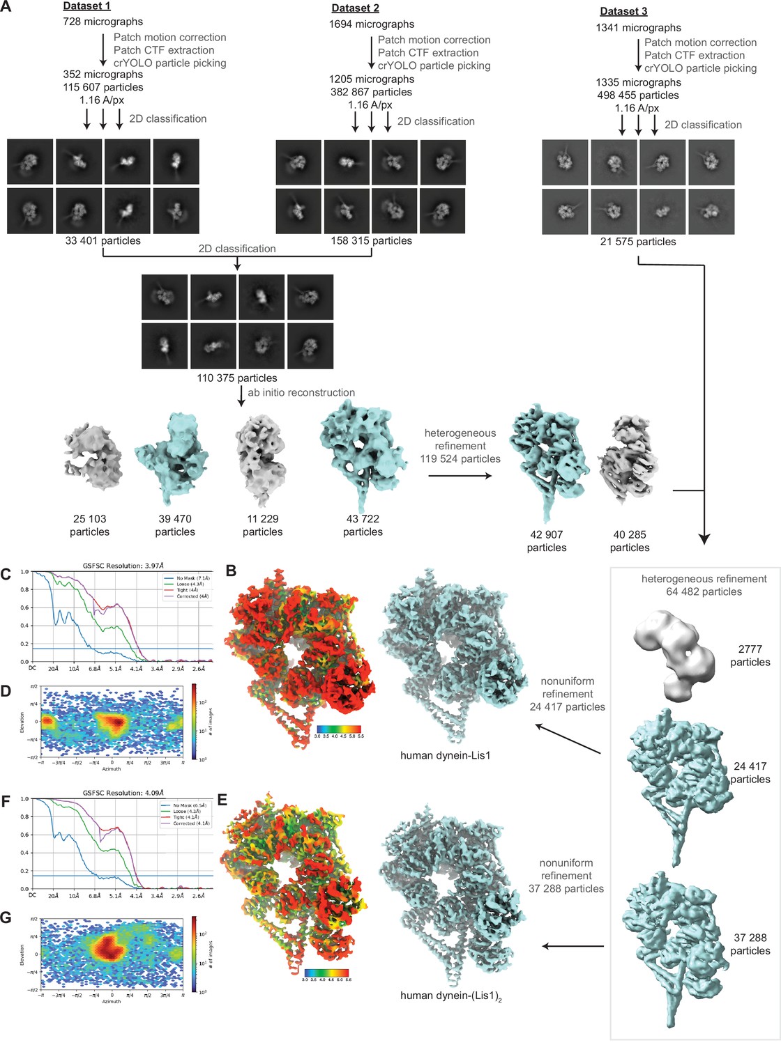
Cryo-EM data processing workflow.
(A) CryoEM data processing pipeline for human dynein bound to LIS1. Particle extraction was carried out in Relion 3.0 and all other jobs were performed in cryoSPARC. Particles belonging to volumes colored in teal were included in subsequent processing jobs. (B, C, D) Local resolution map (B), and FSC (C) and angular distribution (D) plots for human dynein-LIS1 (one β-propeller). (E, F, G) Local resolution map (E), and FSC (F) and angular distribution (G) plots for human dynein-(LIS1)2 (two β-propellers).
Figure 1—figure supplement 2
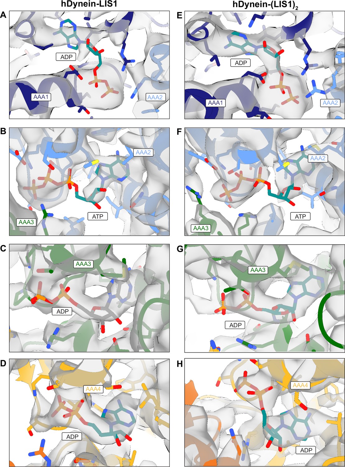
Nucleotides bound to dynein.
(A–D) Nucleotides bound to dynein in the dynein-LIS1 (one β-propeller) structure at AAA1 (A), AAA2 (B), AAA3 (C) and AAA4 (D). (E–H) Nucleotides bound to dynein in the dynein-(LIS1)2 (twp β-propellers) structure at AAA1 (E), AAA2 (F), AAA3 (G) and AAA4 (H).
Figure 2

Structure of LIS1 binding to dynein at sitering.
(A) LIS1 at sitering interacts with dynein via the AAA3-AAA4 bridging loop, a AAA4 helix and a AAA5 loop. (B) An overlay of the human and yeast dynein structures bound to LIS1 /Pac1, aligned on AAA4 (human, light grey; yeast, dark grey). (C) LIS1 (light grey) and Pac1 (dark grey) from panel (B) are viewed facing the β propeller, with dynein removed for clarity. This panel shows the rotation, highlighted by the purple markers and arrows of LIS1 relative to Pac1 at sitering. (D) The AAA3-AAA4 bridging loop contacts LIS1 and preserves a hydrophobic pocket in AAA4.
Figure 3
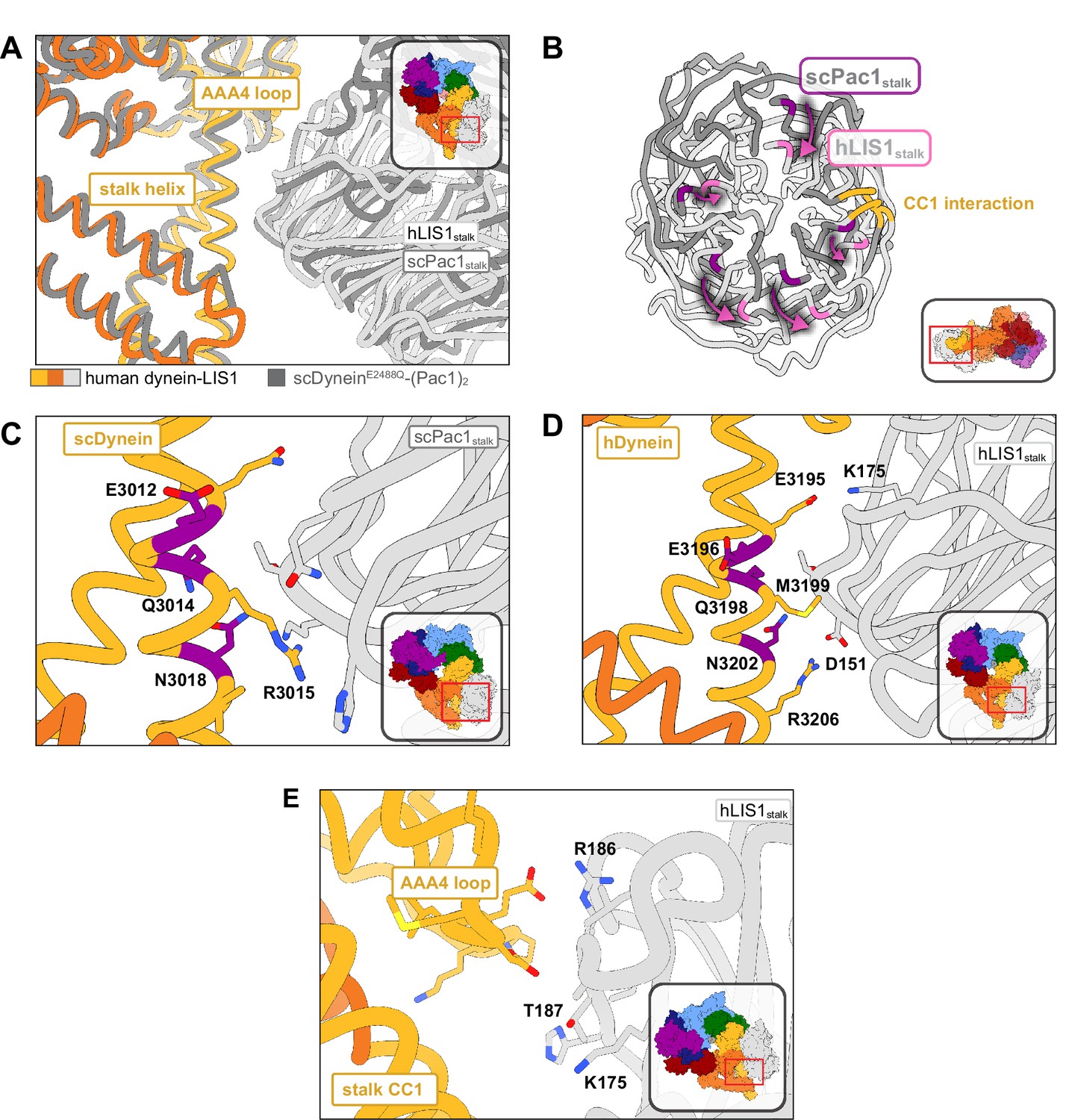
Structure of LIS1 binding to dynein at sitestalk.
(A) An overlay of human and yeast dynein bound to LIS1/Pac1, aligned on AAA4. (B) LIS1 (light grey) and Pac1 (dark grey) from panel (B) are viewed facing the β-propeller, with dynein removed for clarity. This panel shows the rotation, highlighted by the purple markers and arrows of LIS1 relative to Pac1 at sitestalk. The area where LIS1/Pac1 interacts with dynein’s CC1 stalk helix is shown in yellow. (C) The yeast dynein-Pac1stalk interaction. (D) The human dynein-LIS1stalk interaction. (E) The AAA4 loop–LIS1stalk interaction.
Figure 4
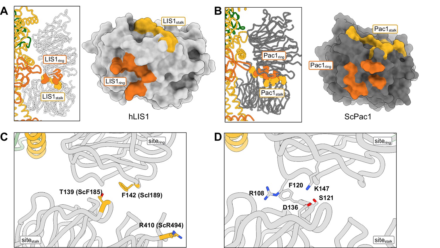
Structure of the LIS1-LIS1 interface.
(A, B) Residues involved in the LIS1-LIS1 interaction are shown in the context of the dynein-(LIS1/Pac1)2 structures and mapped onto a surface representation of LIS1 (A) or Pac1 (B). Residues involved in the interaction with sitering (LIS1ring) are shown in orange and those involved in the interaction with sitestalk (LIS1stalk) are shown in yellow. (C) The human LIS1-LIS1 interaction does not use residues equivalent to those in the yeast Pac1-Pac1 interaction. (D) The human LIS1-LIS1 interface.
Figure 5
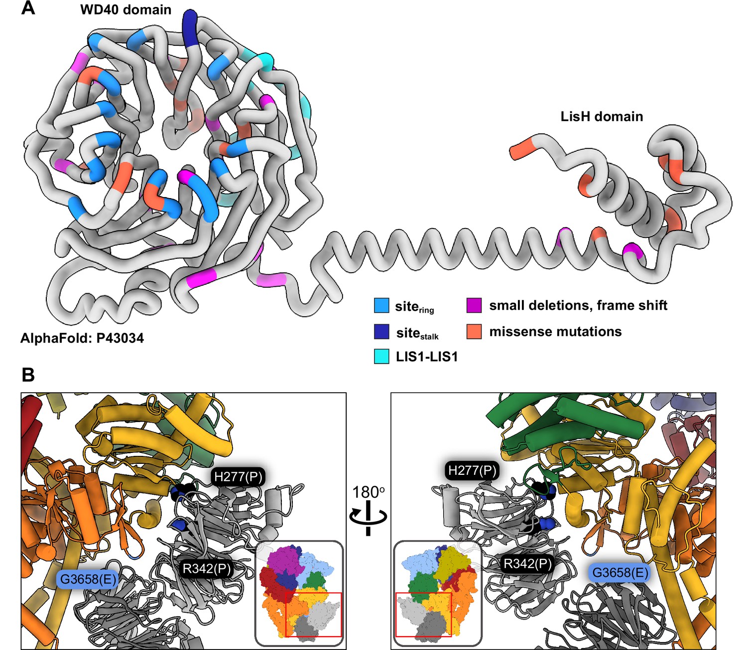
Lissencephaly disease causing mutations.
(A) AlphaFold (Jumper et al., 2021; Senior et al., 2020) model of full length human LIS1 with residues colored by either interface or lissencephaly mutation. Sitering, medium blue; sitestalk, dark blue; LIS1-LIS1, turquoise; lissencephaly small deletions leading to a frame shift, purple; missense mutations, salmon. (B) Two views are shown of disease-linked mutations in dynein located near sites of LIS1 binding. H277P, a lissencephaly mutation, and R342P, a double cortex syndrome mutation, are close to sitering. G3658E, associated with intellectual disability, is located at the tip of the AAA5 beta hairpin loop that is part of sitering.
Videos
Video 1
Comparison of the human dynein-(LIS1)2 and yeast dynein-(Pac1)2 structures.
The video compares the human (dynein-(LIS1)2) and yeast (dynein-(Pac1)2; PDB 7MGM) structures, highlighting some of the major interactions, and the differences in the positions adopted by LIS1/Pac1 at sitering and sitestalk in the two systems.
Video 2
Disease mutations in dynein and LIS1.
This video shows the location of amino acids in LIS1 mutated in type-1 lissencephaly, and residues in dynein that are mutated in several neurodevelopmental or nondegenerative disorders (Charcot-Marie-Tooth, Spinal Muscular Atrophy, Autism Spectrum Disorders, and Malformations of cortical development/ Intellectual disability). We only show residues where we determined that the reported mutation(s) do not have an obvious destabilizing effect based on an inspection of the structure.
Additional files
-
Supplementary file 1
Cryo-EM data information and model validation.
CryoEM data collection parameters, reconstruction information and model refinement statistics for the structures of human dynein bound to one and two LIS1 β-propellers.
- https://cdn.elifesciences.org/articles/84302/elife-84302-supp1-v2.docx
-
Supplementary file 2
Disease mutations in dynein and LIS1 shown in Video 2.
This table lists the mutations in dynein and LIS1 that are associated with different disease and are shown in Video 2, along with the references reporting them. Video 2 shows (and this Table lists) only residues where we determined that the reported mutation(s) do not have an obvious destabilizing effect based on an inspection of the structure. The color coding in the table corresponds to that used in Video 2.
- https://cdn.elifesciences.org/articles/84302/elife-84302-supp2-v2.docx
-
MDAR checklist
- https://cdn.elifesciences.org/articles/84302/elife-84302-mdarchecklist1-v2.pdf
Download links
A two-part list of links to download the article, or parts of the article, in various formats.
Downloads (link to download the article as PDF)
Open citations (links to open the citations from this article in various online reference manager services)
Cite this article (links to download the citations from this article in formats compatible with various reference manager tools)
Structures of human dynein in complex with the lissencephaly 1 protein, LIS1
eLife 12:e84302.
https://doi.org/10.7554/eLife.84302


