Distinct roles of forward and backward alpha-band waves in spatial visual attention
- Cerco, CNRS Université de Toulouse, France
- Artificial and Natural Intelligence Toulouse Institute, France
Figures
Figure 1
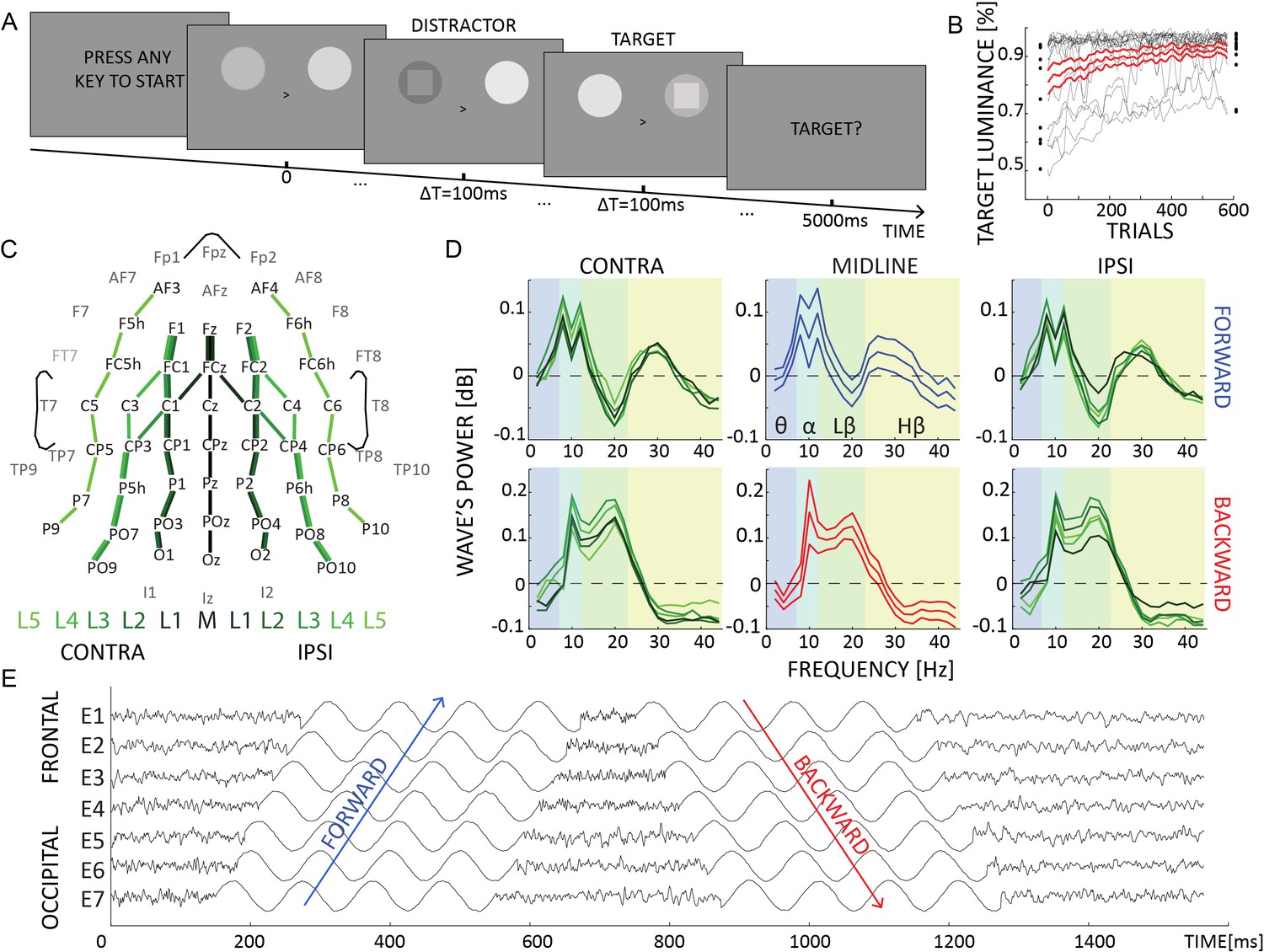
Experimental design and waves’ spectral profile.
(A) Each trial lasted 5 s, in which two flickering stimuli were presented to both hemifield. Participants were instructed to attend either the left or the right hemifield, as indicated by a central cue. In some trials, a target or a distractor appeared for 100 ms as a square either in the attended or unattended location. (B) The target and distractor luminance changed over trials due to the QUEST algorithm, which kept participants’ performance around 80%. (C) We quantified traveling waves along 11 electrodes lines, running along the anterior–posterior axis. These lines were located in the contralateral or the ipsilateral hemisphere to the attended location. (D) The amount of waves in dB computed for forward (in blue) and backward (in red) waves in the midline (central subplots, thinner lines represent standard errors of the mean) and in the ipsi- and contralateral hemisphere (left and right panels, respectively). These waves were computed on trials without target or distractors. Positive (negative) values reflect more (less) waves than the chance level (as quantified by the surrogate distribution), whereas values around 0 indicate no difference between the real and the null distribution. (E) Simulated data providing a schematic representation of forward and backward waves in the time domain in a given line of electrodes (from more frontal E1 to more occipital E7). A positive and a negative phase shift characterized forward and backward waves, respectively.
Figure 2
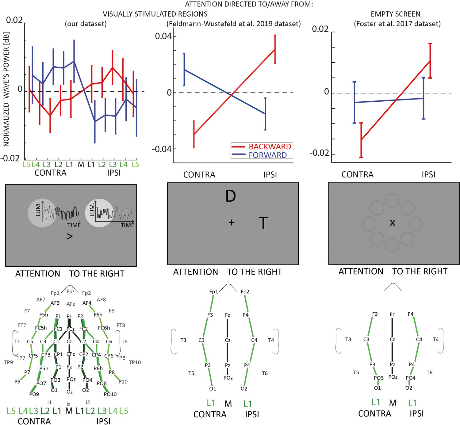
Traveling waves block analysis.
Each column in the figure represents a different EEG dataset involving experiments with visual stimulation (left and middle columns) and without visual stimulation (right column). In the upper panels, the net amount of forward (blue) and backward (red) waves is represented along different lines of electrodes, normalized to the midline. The left and central panels reveal an increase (decrease) of forward (backward) waves contralateral to the attended location when participants attended to visual stimulation. The right column shows that when participants attended an empty screen (data from Foster et al., 2017), only backward waves were modulated by visual attention, and no effect was observed in the forward waves without visual stimulation. Error bars represent standard errors of the mean. The middle row shows schematic representations of the screen during the tasks: the central panel illustrates the task from Feldmann-Wüstefeld and Vogel, 2019, where D and T stand for Distractor and Target, respectively. In the task from Foster et al., 2017, the screen was empty, as the eight circles were not displayed during the task but here illustrate the stimulus positions (Foster et al., 2017). The lower panels represent the lines of electrodes in all datasets.
Figure 3
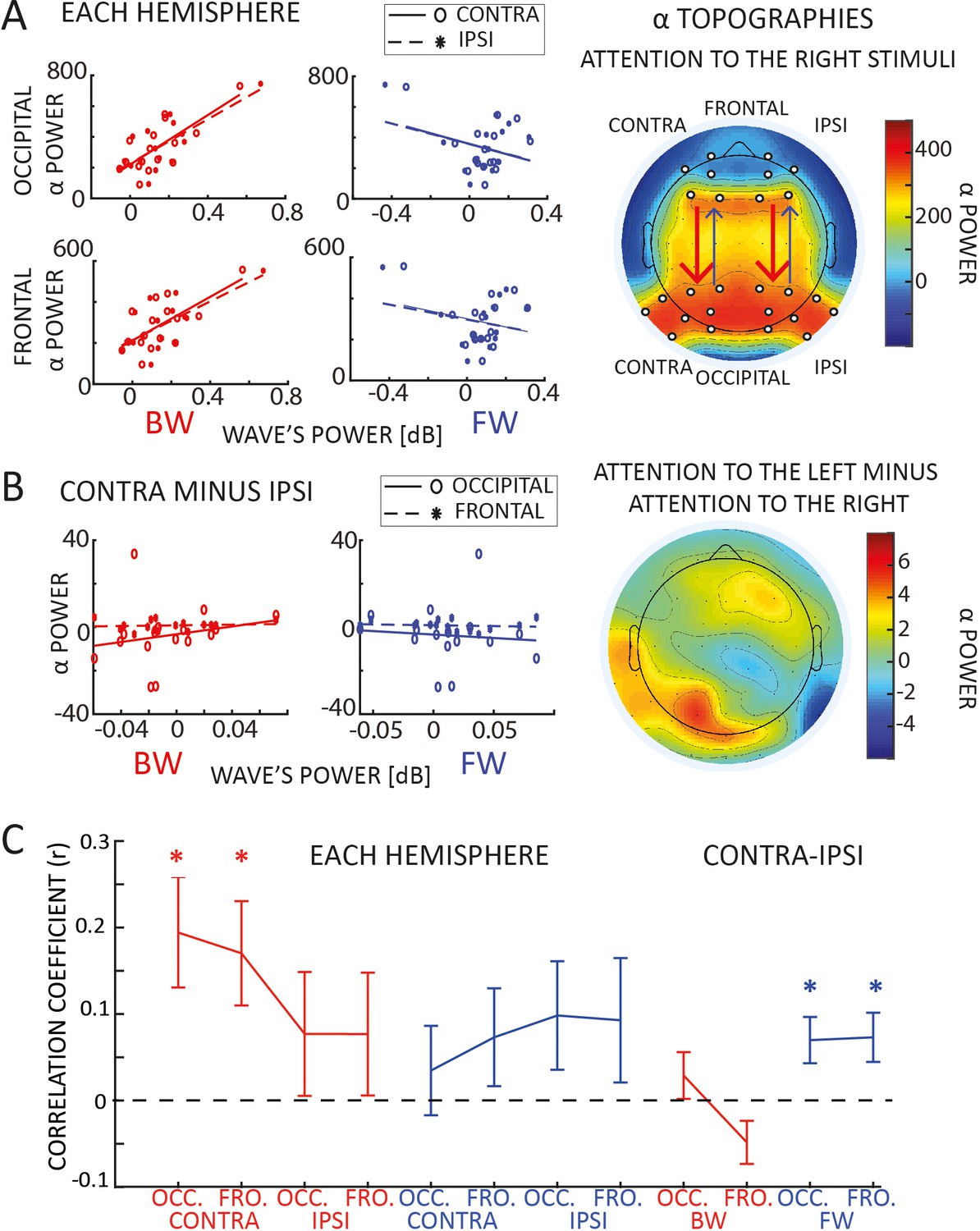
Correlation with alpha-band power.
(A) Panel A reveals a correlation between backward waves and alpha power (static, standing power, i.e., measured via wavelets transform), in both frontal and occipital areas, in both hemispheres. We did not observe such correlation with forward waves. The plot to the right reveals the topographic distribution of alpha power when participants attended to the right hemifield (we included the ‘left’ condition by flipping the electrodes symmetrically to the midline). The white dots indicate the electrodes used for the correlation. (B) The plots to the left show the correlation between the laterality effect in the alpha power and in the waves (laterality measured as the mean difference between contra- and ipsilateral hemispheres for both alpha power and the waves – for the waves we computed the difference using lines of electrodes symmetrical to the midline). We did not observe any correlation in neither forward nor backward waves, with neither frontal nor occipital alpha power. The topography to the right reveals a lateralization effect in the alpha power (attention to the left minus attention to the right), confirming the presence of alpha power lateralization, in line with previous studies (Sauseng et al., 2005; Thut et al., 2006; Händel et al., 2011). (C) Panel C shows the trial-by-trial correlation coefficients averaged across participants for different conditions (as indicated in the x-axis). Confirming the results in panel A, we found a positive correlation across participants between backward waves and alpha power, specifically in the contralateral hemisphere. We also observed a positive global effect of the laterality condition across participants in the forward waves, even though the combined p values for the trial-by-trial correlation did not reach the significant threshold. Error bars represent standard errors of the mean.
Figure 4
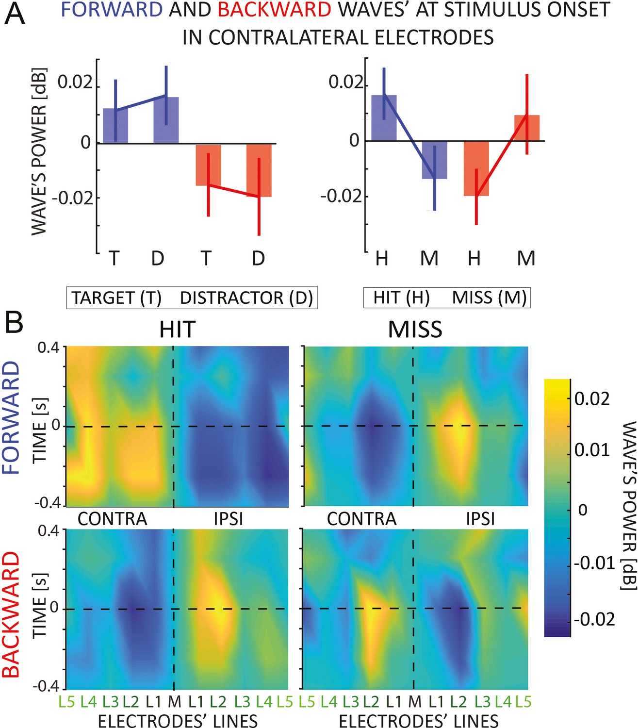
Event analysis.
(A) The figure shows the amount of forward (in blue) and backward (in red) contralateral waves around the onset of the target/distractor (left) or hit and missed targets (right panel). Error bars are standard error of the mean. We found an interaction effect when we analyzed the hit versus missed target. (B) The 2D maps represent the amount of waves in the 11 lines of electrodes (x-axis) and around the onset time (y-axis) for forward and backward waves, and for hits and missed targets separately. The opposite pattern for hits versus misses, already visible before the target onset, suggests that missed targets are due to a failure of attentional allocation rather than sensory processing; and consequently, that proper attentional allocation is characterized by contralateral forward waves and ipsilateral backward waves.
Figure 5
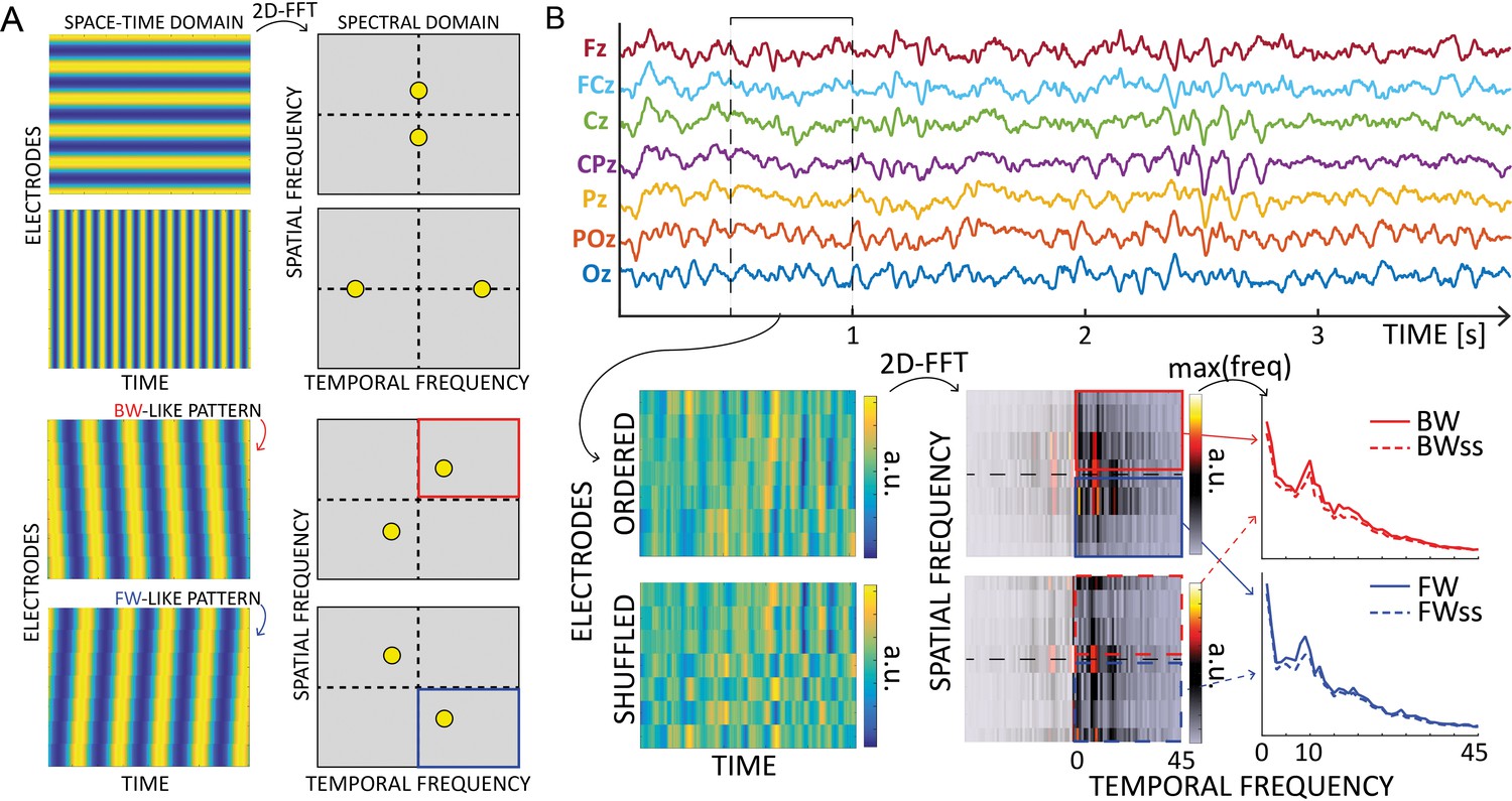
Waves analysis.
(A) The 2D-Fast Fourier Trasform (2D-FFT) decomposes an image (e.g., a space-time representation of an EEG signal) into its spectral components. The upper part shows the decomposition of a 2D sinusoid propagating along the vertical or horizontal axis of the image. The corresponding peaks are found on the axis in the spectral domain, and their position depends on the frequency of the oscillations. The lower part of the figure shows how the spectra change when the oscillations propagate with a backward- or forward- like pattern. Importantly, the spectral peaks rotate in two of the four quadrants depending on the direction, providing a reliable measure of forward or backward waves in the image. (B) Schematic of the waves’ quantification method. After defining time windows over each electrode line, we computed 2D Fourier transformation to quantify the amount of forward (in blue) and backward (in red) waves. From the upper and lower quadrants of the 2D-FFT spectra, we consider the maximum value over spatial frequencies, providing a 1D spectrum of forward and backward waves in the temporal frequency domain. The same procedure after shuffling the electrodes’ order provides a surrogate measure, used as a baseline. Notably, such surrogate distribution captures the 1/f trend and the alpha-band peak, accounting for these factors in the final waves’ quantification.
Author response image 1

Laterality index for occipital electrodes, quantifying α-band lateralization during attention allocation.
All electrodes go in the expected direction, revealing an increase of α-band power in the ipsilateral occipital hemisphere.
Author response image 2

Amount of forward and backward waves in the hemisphere contralateral to the attended location as a function of time in trials with no targets nor distractors.
Data are the average over the 16 subjects, ± standard errors of the mean.
Tables
Table 1
Correlation with alpha-band power.
The table reports the Pearson’s correlation coefficient and the Bayes Factor (BF10 supporting the alternative hypothesis, that is the presence of a correlation) between frontal and occipital electrodes and forward (FW) and backward (BW) waves, in both contra- and ipsilateral hemispheres. Values in bold reflect Bayes Factors providing strong evidence in favor of the alternative hypothesis. All correlations were computed on trials when neither a target nor a distractor was displayed.
| Pearsonr (BF₁₀) | FW | BW | |||
|---|---|---|---|---|---|
| CONTRA | IPSI | CONTRA | IPSI | ||
| OCC. | CONTRA | −0.297 (0.549) | −0.350 (0.697) | 0.720 (28.519) | 0.698 (19.503) |
| IPSI | −0.305 (0.566) | −0.342 (0.669) | 0.786 (116.990) | 0.746 (47.512) | |
| FRONT. | CONTRA | −0.222 (0.422) | −0.252 (0.465) | 0.772 (84.225) | 0.712 (24.645) |
| IPSI | −0.327 (0.625) | −0.354 (0.710) | 0.747 (48.448) | 0.705 (21.841) | |
Additional files
Download links
A two-part list of links to download the article, or parts of the article, in various formats.
Downloads (link to download the article as PDF)
Open citations (links to open the citations from this article in various online reference manager services)
Cite this article (links to download the citations from this article in formats compatible with various reference manager tools)
Distinct roles of forward and backward alpha-band waves in spatial visual attention
eLife 12:e85035.
https://doi.org/10.7554/eLife.85035


