β-Carotene accelerates the resolution of atherosclerosis in mice
- Division of Nutritional Sciences, University of Illinois Urbana Champaign, United States
- Department of Food Science and Human Nutrition, University of Illinois Urbana Champaign, United States
- The University of Jordan, School of Agriculture, Department of Nutrition and Food Technology, Jordan
- The Leon H. Charney Division of Cardiology, Department of Medicine, The Marc and Ruti Bell Program in Vascular Biology, New York University Grossman School of Medicine, NYU Langone Medical Center, United States
Figures
Figure 1 with 1 supplement
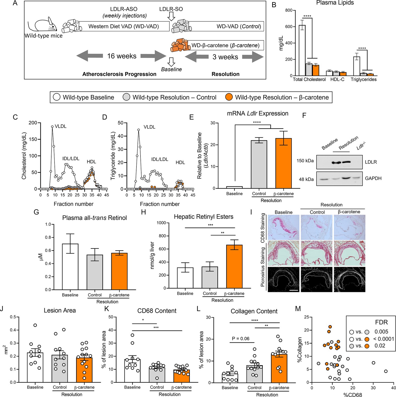
β-Carotene accelerates atherosclerosis resolution in wild-type mice infused with antisense oligonucleotide targeting the low-density lipoprotein receptor (ASO-LDLR).
(A) Four-week-old male and female wild-type mice were fed a purified Western diet deficient in vitamin A (WD-VAD) and injected with ASO-LDLR once a week for 16 wk to induce atherosclerosis. After 16 wk, a group of mice was harvested (baseline) and the rest of the mice were injected once with sense oligonucleotide (SO-LDLR) to inactivate ASO-LDLR and promote atherosclerosis resolution. Mice undergoing resolution were either kept on the same diet (resolution–control) or switched to a Western diet supplemented with 50 mg/kg of β-carotene (resolution–β-carotene) for three more weeks. (B) Plasma lipid levels at the moment of the sacrifice. (C) Cholesterol and (D) triglyceride distribution in fast performance liquid chromatography (FPLC)-fractioned plasma (data pooled from five mice/group). (E) Relative low-density lipoprotein receptor (LDLR) mRNA, and (F) protein expressions in the liver. (G) Circulating vitamin A (all-trans retinol), and (H) hepatic retinyl ester stores determined by HPLC. (I) Representative images for macrophage (CD68+, top panels) and picrosirius staining to identify collagen using the bright-field (middle panels) or polarized light (bottom panels). (J) Plaque size, (K) relative CD68 content, and (L) collagen content in the lesion. (M) Descriptive discriminant analysis employing the relative CD68 and collagen contents in the lesion as variables highlighting the false discovery rate (FDR) for each comparison. Each dot in the plot represents an individual mouse (n = 10–12/group). Values are represented as means ± SEM. Statistical differences were evaluated using one-way ANOVA with Tukey’s multiple comparisons test. Differences between groups were considered significant with a p-value<0.05. *p<0.05; **p<0.01; ***p<0.005; ****p<0.001. Size bar = 200 µm.
Figure 1—figure supplement 1
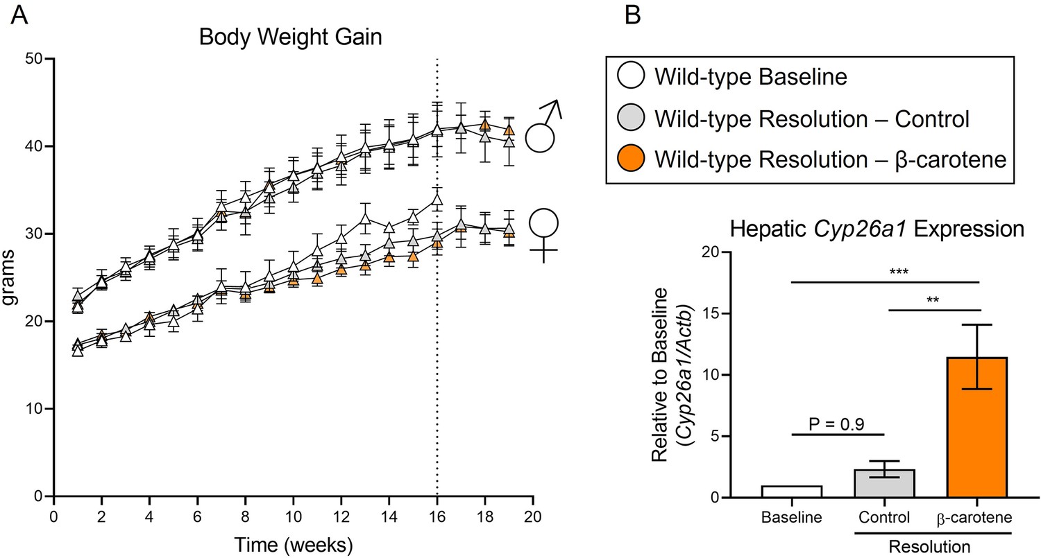
Four-week-old male and female wild-type mice were fed a purified Western diet deficient in vitamin A (WD-VAD) and injected with antisense oligonucleotide targeting the low-density lipoprotein receptor (ASO-LDLR) once a week for 16 wk to induce atherosclerosis.
After 16 wk (dotted line), a group of mice was harvested (baseline) and the rest of the mice were injected once with sense oligonucleotide (SO-LDLR) to inactivate ASO-LDLR and promote atherosclerosis resolution. Mice undergoing resolution were either kept on the same diet (resolution–control) or switched to a Western diet supplemented with 50 mg/kg of β-carotene (resolution–β-carotene) for three more weeks. (A) Body weight progression and (B) hepatic expression mRNA expression for Cyp26a1 referred to Actb as a housekeeping control. N = 5–10 mice/group, Values are represented as means ± SEM. Statistical differences were evaluated using one-way ANOVA with Tukey’s multiple comparisons test. Differences between groups were considered significant with a p-value<0.05. **p<0.01; ***p<0.005.
Figure 2 with 1 supplement
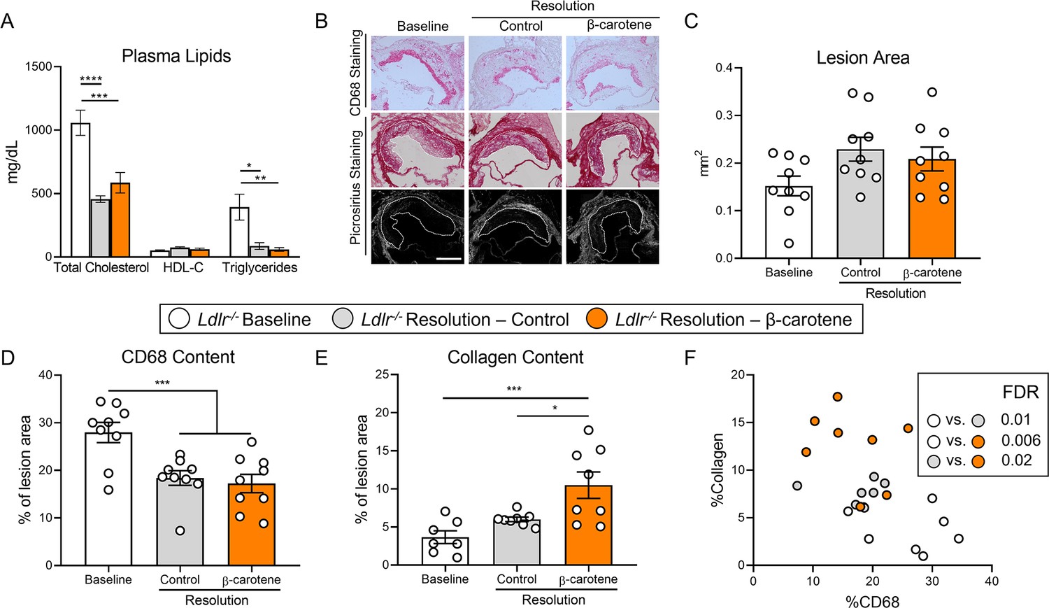
β-Carotene accelerates atherosclerosis resolution in low-density lipoprotein-deficient (Ldlr-/-) mice subjected to dietary switch.
Four-week-old male and female Ldlr-/- mice were fed a purified Western diet deficient in vitamin A (WD-VAD) for 12 wk to induce atherosclerosis. After 12 wk, a group of mice was harvested (baseline) and the rest of the mice were switched to a standard diet (resolution–control) or the same diet supplemented with 50 mg/kg of β-carotene (resolution–β-carotene) for four more weeks. (A) Total cholesterol plasma levels at the moment of the sacrifice. (B) Representative images for macrophage (CD68+, top panels) and picrosirius staining to identify collagen using the bright-field (middle panels) or polarized light (bottom panels). (C) Plaque size, (D) relative CD68 content, and (E) collagen content in the lesion. (F) Descriptive discriminant analysis employing the relative CD68 and collagen contents in the lesion as variables highlighting the false discovery rate (FDR) for each comparison. Each dot in the plot represents an individual mouse (n = 9–12/group). (A–E) Values are represented as means ± SEM. Statistical differences were evaluated using one-way ANOVA with Tukey’s multiple comparisons test. Differences between groups were considered significant with a p-value<0.05. *p<0.05; ***p<0.005; ****p<0.001. Size bar = 200 µm.
Figure 2—figure supplement 1
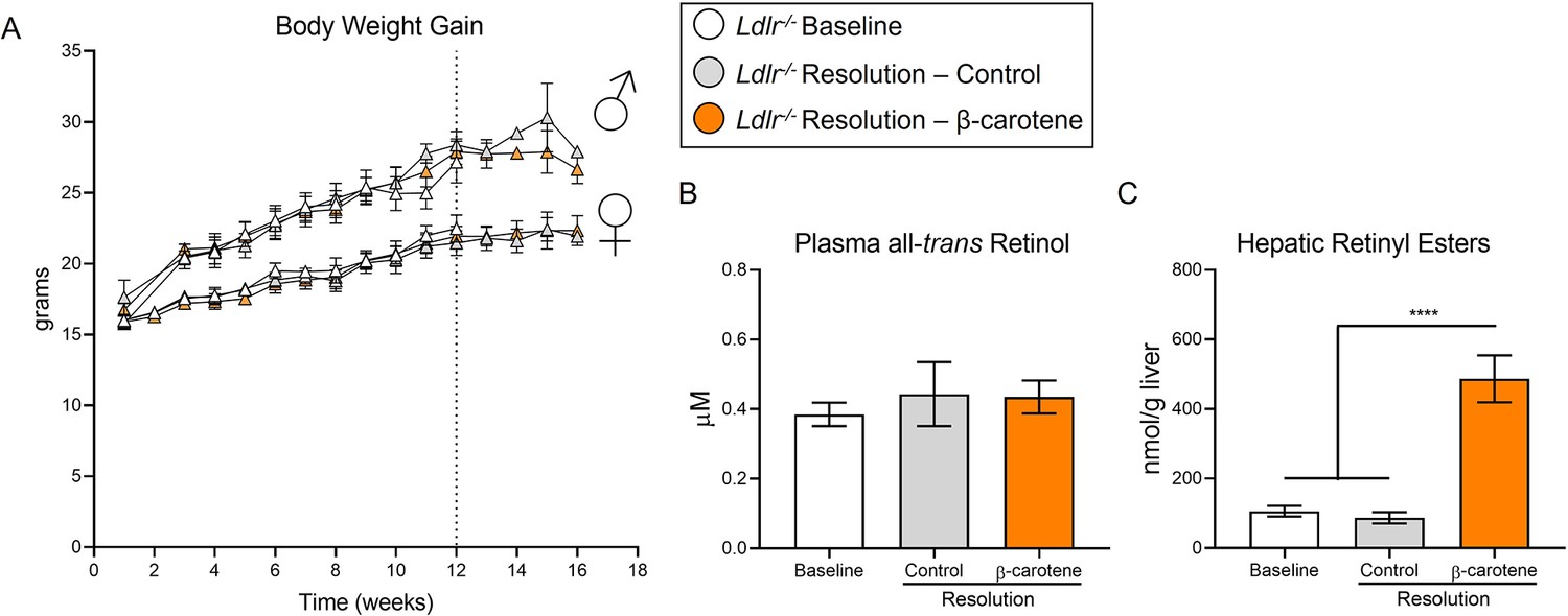
Four-week-old male and female Ldlr-/- mice were fed a purified Western diet deficient in vitamin A (WD-VAD) for 12 wk to induce atherosclerosis.
After 12 wk (dotted line), a group of mice was harvested (baseline) and the rest of the mice were switched to a standard diet (resolution–control) or the same diet supplemented with 50 mg/kg of β-carotene (resolution–β-carotene) for four more weeks. (A) Body weight progression. (B) Circulating vitamin A (all-trans retinol), and (C) hepatic retinyl ester stores. N = 5–10 mice/group, Values are represented as means ± SEM. Statistical differences were evaluated using one-way ANOVA with Tukey’s multiple comparisons test. Differences between groups were considered significant with a p-value<0.05. ****p<0.001.
Figure 3 with 1 supplement
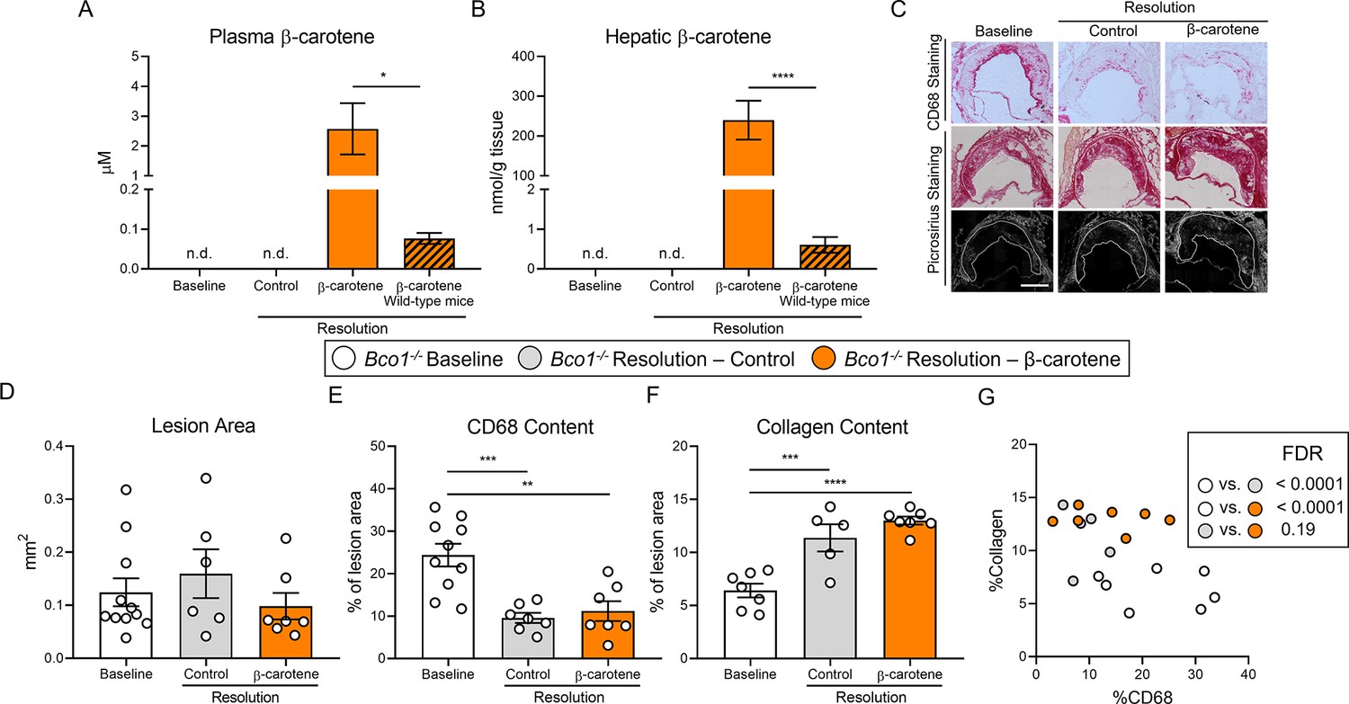
β-Carotene supplementation does alter atherosclerosis resolution in β-carotene oxygenase 1-deficient (Bco1-/-) mice infused with antisense oligonucleotide targeting the low-density lipoprotein receptor (ASO-LDLR).
Four-week-old male and female Bco1-/- mice were fed a purified Western diet deficient in vitamin A (WD-VAD) and injected with ASO-LDLR once a week for 16 wk to induce the development of atherosclerosis. After 16 wk, a group of mice was harvested (baseline) and the rest of the mice were injected once with sense oligonucleotide (SO-LDLR) to block ASO-LDLR (resolution). Mice undergoing resolution were either kept on the same diet (resolution–control) or switched to a Western diet supplemented with 50 mg/kg of β-carotene (resolution–β-carotene) for three more weeks. (A) β-Carotene levels in plasma and (B) liver at the sacrifice determined by HPLC. (C) Representative images for macrophage (CD68+, top panels) and picrosirius staining to identify collagen using the bright-field (middle panels) or polarized light (bottom panels). (D) Plaque size, (E) relative CD68 content, and (F) collagen content in the lesion. (G) Descriptive discriminant analysis employing the relative CD68 and collagen contents in the lesion as variables highlighting the false discovery rate (FDR) for each comparison. Each dot in the plot represents an individual mouse (n = 5–11/group). (A–F) Values are represented as means ± SEM. Statistical differences were evaluated using one-way ANOVA with Tukey’s multiple comparisons test. Differences between groups were considered significant with a p-value<0.05. *p<0.05; **p<0.01; ***p<0.005; ****p<0.001. Size bar = 200 µm.
Figure 3—figure supplement 1
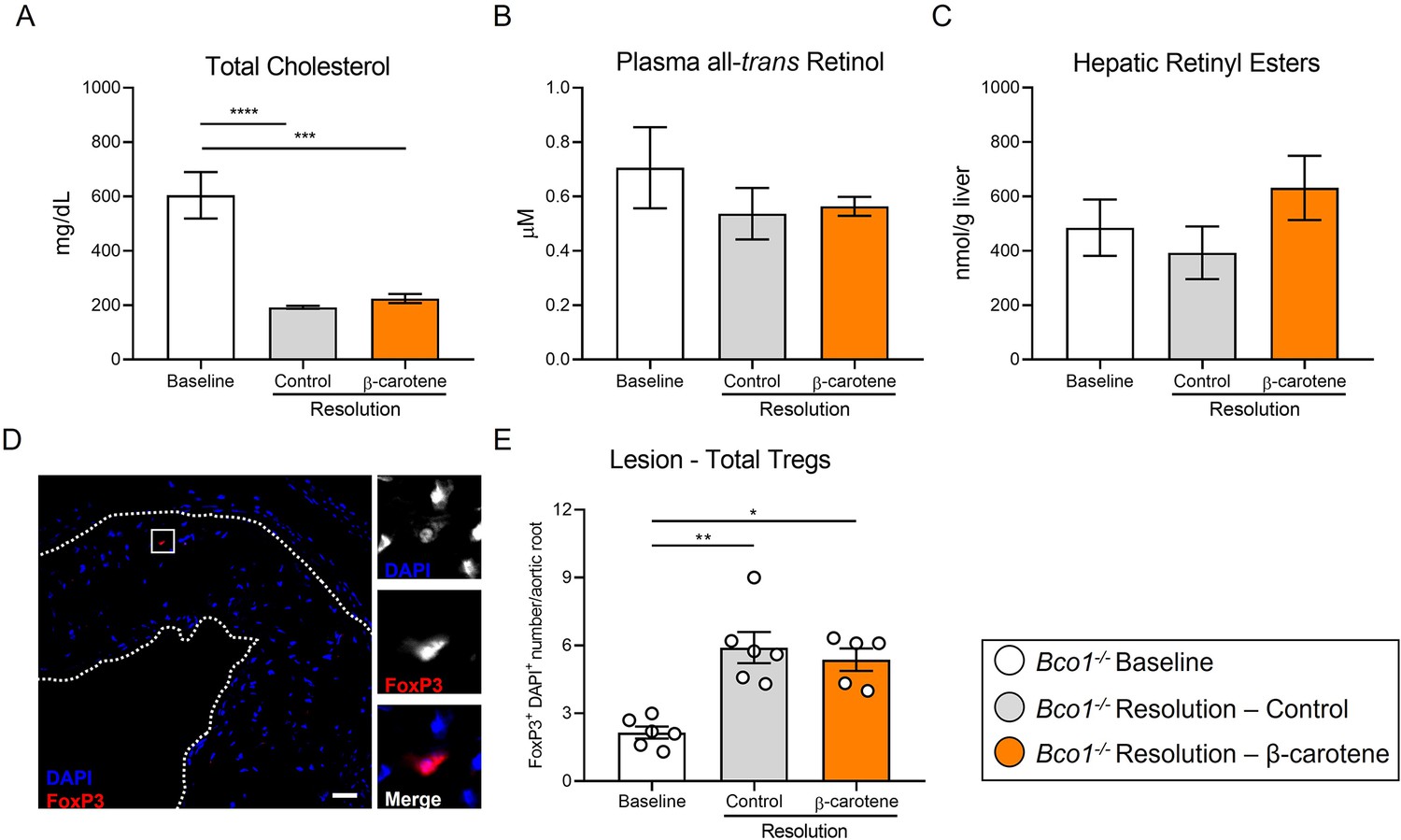
Four-week-old male and female Bco1-/- mice were fed a purified Western diet deficient in vitamin A (WD-VAD) and injected with antisense oligonucleotide targeting the low-density lipoprotein receptor (ASO-LDLR) once a week for 16 wk to induce the development of atherosclerosis.
After 16 wk, a group of mice was harvested (baseline) and the rest of the mice were injected once with sense oligonucleotide (SO-LDLR) to block ASO-LDLR (resolution). Mice undergoing resolution were either kept on the same diet (resolution–control) or switched to a Western diet supplemented with 50 mg/kg of β-carotene (resolution–β-carotene) for three more weeks. (A) Total plasma cholesterol, and (B) vitamin A (all-trans retinol). (C) Hepatic vitamin A (retinyl ester) stores. (D) Representative confocal image and (E) quantification of total Tregs in the lesion. Size bar 50 µm. N = 5–10 mice/group, Values are represented as means ± SEM. Statistical differences were evaluated using one-way ANOVA with Tukey’s multiple comparisons test. Differences between groups were considered significant with a p-value<0.05. ***p<0.005, ****p<0.001.
Figure 4 with 1 supplement
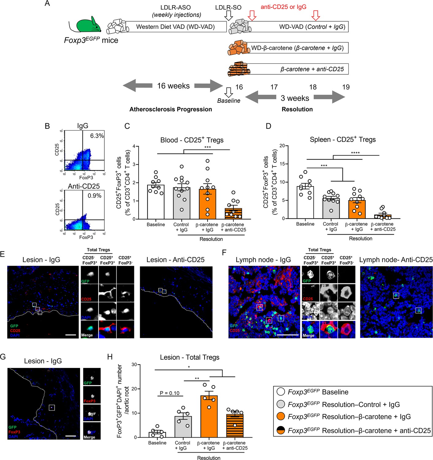
Effect of anti-CD25 treatment on Treg number.
(A) Four-week-old male and female mice expressing enhanced green fluorescence protein (EGFP) under the control of the forkhead box P3 (Foxp3) promoter (Foxp3EGFP mice) were fed a purified Western diet deficient in vitamin A (WD-VAD) and injected with antisense oligonucleotide targeting the low-density lipoprotein receptor (ASO-LDLR) once a week for 16 wk to induce the development of atherosclerosis. After 16 wk, a group of mice was harvested (baseline) and the rest of the mice were injected once with sense oligonucleotide (SO-LDLR) to block ASO-LDLR (resolution). Mice undergoing resolution were either kept on the same diet (resolution–control) or switched to a Western diet supplemented with 50 mg/kg of β-carotene (resolution–β-carotene) for three more weeks. An additional group of mice fed with β-carotene was injected twice before sacrifice with anti-CD25 monoclonal antibody to deplete Treg (resolution–β-carotene+anti-CD25). The rest of the resolution groups were injected with IgG isotype control antibody. (B) Representative flow cytometry panels showing splenic CD25+FoxP3+ (CD25+ Treg) cells in mice injected with IgG or anti-CD25. (C) Quantification of the splenic and (D) circulating blood levels of CD25+ Treg cells determined by flow cytometry. (E) Representative confocal images show the presence of total Tregs (CD25-FoxP3+ + CD25+ Tregs) and CD25+FoxP3- T cells in the lesion of mice injected with IgG (left panel) or anti-CD25 (right panel). (F) Representative confocal images show the presence of total Tregs and CD25+FoxP3- T cells in lymph nodes of mice injected with IgG (left panel) or anti-CD25 (right panel). quantification of CD25+ Tregs in the lesions. (G) Representative confocal image and (H) quantification of total Tregs in the lesion. Each dot in the plot represents an individual mouse (n = 5–11 mice/group). Values are represented as means ± SEM. Statistical differences were evaluated using one-way ANOVA with Tukey’s multiple comparisons test. Differences between groups were considered significant with a p-value<0.05. *p<0.05; **p<0.01; ***p<0.005; ****p<0.001.
Figure 4—figure supplement 1
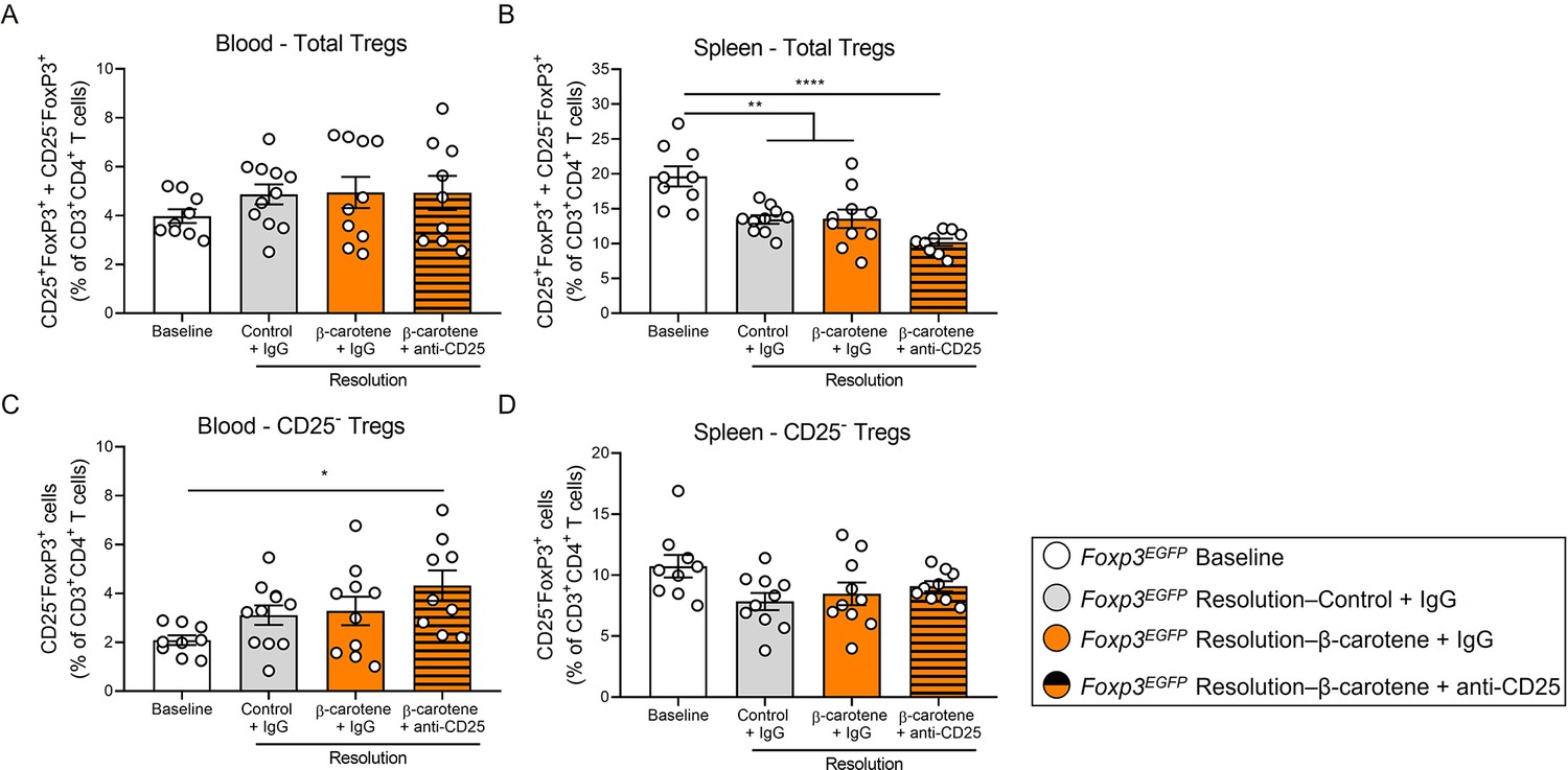
Four-week-old male and female mice expressing enhanced green fluorescence protein (EGFP) under the control of the forkhead box P3 (Foxp3) promoter (Foxp3EGFP mice) were fed a purified Western diet deficient in vitamin A (WD-VAD) and injected with antisense oligonucleotide targeting the low-density lipoprotein receptor (ASO-LDLR) once a week for 16 wk to induce the development of atherosclerosis.
After 16 wk, a group of mice was harvested (baseline) and the rest of the mice were injected once with sense oligonucleotide (SO-LDLR) to block ASO-LDLR (resolution). Mice undergoing resolution were either kept on the same diet (resolution–control) or switched to a Western diet supplemented with 50 mg/kg of β-carotene (resolution–β-carotene) for three more weeks. An additional group of mice fed with β-carotene was injected twice before sacrifice with anti-CD25 monoclonal antibody to deplete Treg (resolution–β-carotene+anti-CD25). The rest of the resolution groups were injected with IgG isotype control antibody. (A) Quantification of the circulating and (B) splenic CD25+FoxP3+ and CD25-FoxP3+ (total Tregs) measured by flow cytometry. (C) Circulating and (D) splenic CD25-FoxP3+ Tregs. Size bars = 50 μm. N = 4–10 mice/group. Values are represented as means ± SEM. Statistical differences were evaluated using one-way ANOVA with Tukey’s multiple comparisons test. Differences between groups were considered significant with a p-value<0.05. *p<0.05.
Figure 5 with 1 supplement
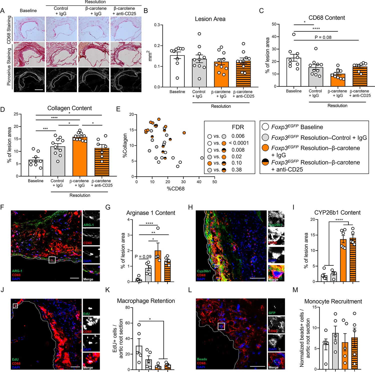
Effect of anti-CD25 treatment on lesion composition and monocyte/macrophage trafficking.
Four-week-old male and female expressing enhanced green fluorescence protein (EGFP) under the control of the forkhead box P3 (Foxp3) promoter (Foxp3EGFP mice) were fed a purified Western diet deficient in vitamin A (WD-VAD) and injected with antisense oligonucleotide targeting the low-density lipoprotein receptor (ASO-LDLR) once a week for 16 wk to induce the development of atherosclerosis. After 16 wk, a group of mice was harvested (baseline) and the rest of the mice were injected once with sense oligonucleotide (SO-LDLR) to block ASO-LDLR (resolution). Mice undergoing resolution were either kept on the same diet (resolution–control) or switched to a Western diet supplemented with 50 mg/kg of β-carotene (resolution–β-carotene) for three more weeks. An additional group of mice fed with β-carotene was injected twice before sacrifice with anti-CD25 monoclonal antibody to deplete Treg (resolution–β-carotene+anti-CD25). The rest of the resolution groups were injected with IgG isotype control antibody. To quantify macrophage egress and monocyte recruitment, we injected a dose of EdU at week 15 and fluorescently labeled beads 2 d before harvesting the mice, respectively (see ‘Methods’ for details). (A) Representative images for macrophage (CD68+, top panels) and picrosirius staining to identify collagen using the bright-field (middle panels) or polarized light (bottom panels). Size bar = 200 µm. (B) Plaque size, (C) relative CD68 content, and (D) collagen content in the lesion. (E) Descriptive discriminant analysis employing the relative CD68 and collagen contents in the lesion as variables highlighting the false discovery rate (FDR) for each comparison. (F) Representative confocal image showing arginase 1 (green), CD68 (red), and DAPI (blue) in the lesion. (G) Relative arginase 1 area in the lesion. (H) Representative confocal image showing Cyp26b1 (green), CD68 (red), and DAPI (blue) in the lesion. (I) Relative Cyp26b1 area in the lesion. (J) EdU+ macrophages were identified by the colocalization of EdU (green) and DAPI (blue) in CD68+ (red) cells. (K) Number of EdU+ macrophages in the lesion. (L) Newly recruited monocytes were identified and (M) quantified by the presence of beads (green) on the lesion. Size bars = 50 µm. Each dot in the plot represents an individual mouse (n = 5–11 mice/group). Values are represented as means ± SEM. Statistical differences were evaluated using one-way ANOVA with Tukey’s multiple comparisons test. Differences between groups were considered significant with a p-value<0.05. *p<0.05; **p<0.01; ***p<0.005; ****p<0.001.
Figure 5—figure supplement 1
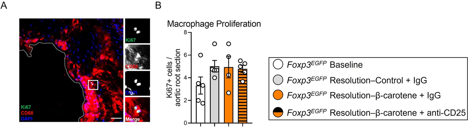
Four-week-old male and female expressing enhanced green fluorescence protein (EGFP) under the control of the forkhead box P3 (Foxp3) promoter (Foxp3EGFP mice) were fed a purified Western diet deficient in vitamin A (WD-VAD) and injected with antisense oligonucleotide targeting the low-density lipoprotein receptor (ASO-LDLR) once a week for 16 wk to induce the development of atherosclerosis.
After 16 wk, a group of mice was harvested (baseline) and the rest of the mice were injected once with sense oligonucleotide (SO-LDLR) to block ASO-LDLR (resolution). Mice undergoing resolution were either kept on the same diet (resolution–control) or switched to a Western diet supplemented with 50 mg/kg of β-carotene (resolution–β-carotene) for three more weeks. An additional group of mice fed with β-carotene was injected twice before sacrifice with anti-CD25 monoclonal antibody to deplete Treg (resolution–β-carotene+anti-CD25). The rest of the resolution groups were injected with IgG isotype control antibody. To quantify macrophage egress and monocyte recruitment, we injected a dose of EdU at week 15 and fluorescently labeled beads 2 d before harvesting the mice, respectively (see ‘Methods’ for details). (A) Macrophages proliferating in the lesion were identified by the colocalization of Ki67 (green) and DAPI (blue) in CD68+ (red) cells. (B) Number of Ki67+ macrophages in the lesion. Size bars = 50 μm. N = 4–10 mice/group. Values are represented as means ± SEM. Statistical differences were evaluated using one-way ANOVA with Tukey’s multiple comparisons test. Differences between groups were considered significant with a p-value<0.05. * p<0.05.
Author response image 1

(A) mBMDMs were divided in M0 or M2 (exposed to IL-4 for 24 h), and then treated with either DMSO or retinoic acid for 6 h before harvesting for RNA seq analysis. Exploring the RNA seq dataset, we identified Cyp26b1 as a RA-sensitive gene in mBMDMs (PMID: 36754230). (B) Validation of Cyp26b1 antibody in mBMDMs exposed to retinoic acid confirms the suitability of this antibody for measuring retinoic acid signaling in our experimental settings.
Additional files
-
MDAR checklist
- https://cdn.elifesciences.org/articles/87430/elife-87430-mdarchecklist1-v2.pdf
-
Source data 1
Raw data for all figures.
- https://cdn.elifesciences.org/articles/87430/elife-87430-data1-v2.xlsx
-
Supplementary file 1
Composition of the experimental diets utilized in the study.
- https://cdn.elifesciences.org/articles/87430/elife-87430-supp1-v2.docx
Download links
A two-part list of links to download the article, or parts of the article, in various formats.
Downloads (link to download the article as PDF)
Open citations (links to open the citations from this article in various online reference manager services)
Cite this article (links to download the citations from this article in formats compatible with various reference manager tools)
β-Carotene accelerates the resolution of atherosclerosis in mice
eLife 12:RP87430.
https://doi.org/10.7554/eLife.87430.3


